
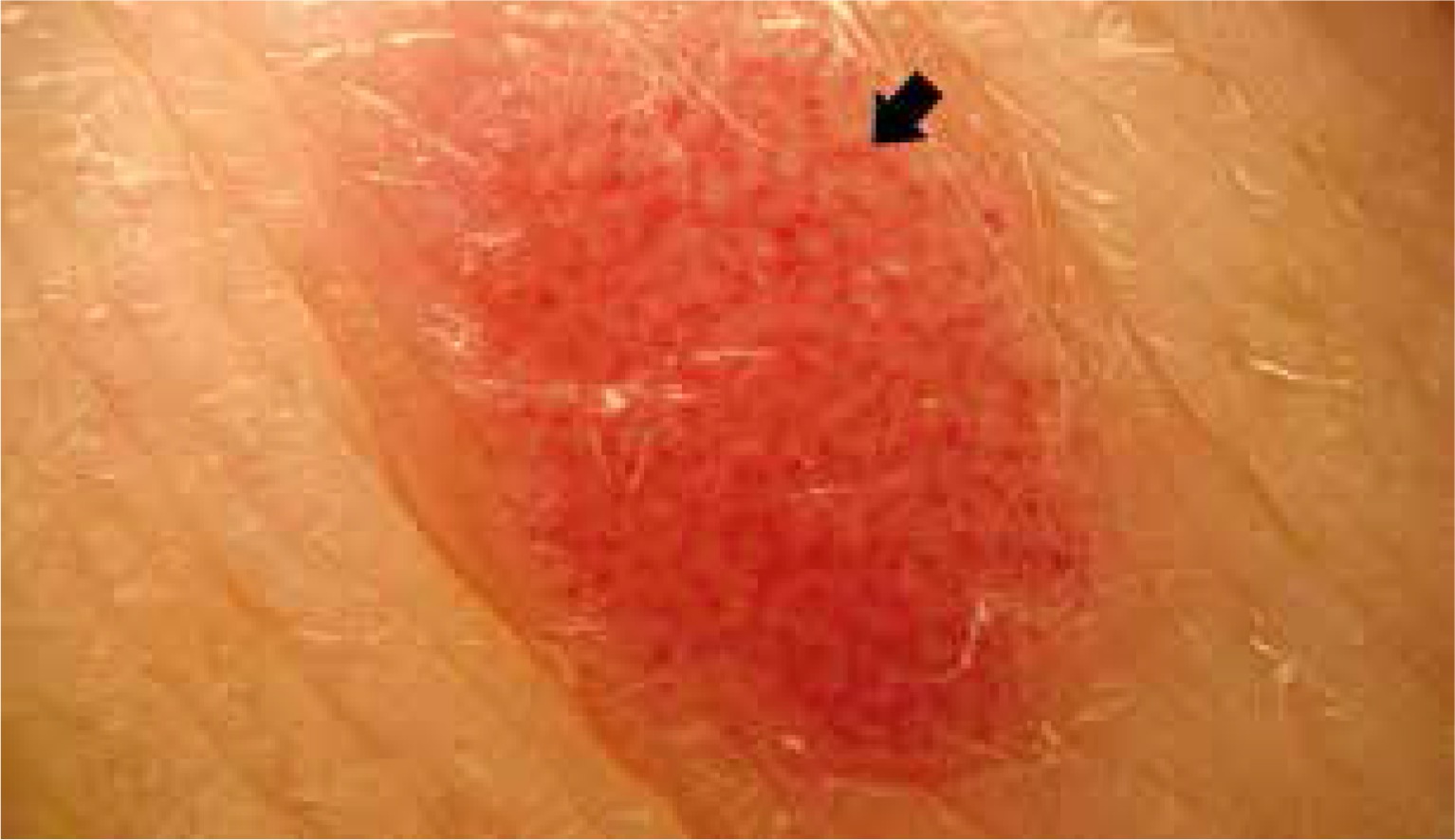
Acanthoma clarocellulare known as clear cell acanthoma (Degos tumour) is an uncommon, benign epidermal tumour first described by Degos in 1962. It mostly occurs in the skin of elderly patients [1]. Clinically clear cell acanthoma presents as a solitary, well-circumscribed red-pinkish nodule, with a diameter ranging from 5 mm to 2 cm. In most cases it grows slowly with no subjective symptoms. This lesion does not show a gender or ethnic predilection. Some authors consider that clear cell acanthoma could be a melanoma simulator. The clinical differential diagnosis includes: basal cell carcinoma, Bowen’s disease, irritated seborrheic keratosis, verruca vulgaris, pyogenic granuloma and even amelanotic melanoma. The pathogenesis of clear cell acanthoma is unknown. The higher incidence in legs may suggest a reactive, inflammatory nature, probably induced by stasis dermatitis. Another hypothesis indicates derivation of acanthoma clarocellulare from the eccrine apparatus.
Dermoscopic features of clear cell acanthoma were first described by Blumm in 2001.
In dermoscopy, clear cell acanthoma has its typical and specific presentation: many linear or curvilinear glomerular vessels in necklace-like arrangement (Figure 1). Only a few cases evaluated by dermoscopy showed an incomplete vascular pattern without typical linear or glomerular vessels [2]. Zargari et al. indicate the possibility of uncommon dermoscopic features as areas of haemorrhage, orange crusts or peripheral collarette of translucent scales. Some cases show crystalline structures or white lines [3]. Sometimes there is a scaly surface.
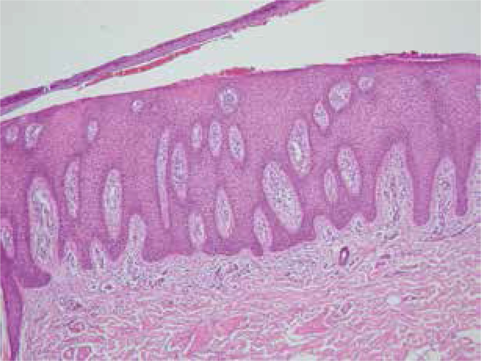
In histopathology evaluation, clear cell acanthoma often reveals itself in epidermis especially in the centre of the lesion with characteristic big, clear cells abound in glycogen. There are many dilated blood vessels in the dermal papillae (Figure 2). In both epidermis and superficial skin layer, there is the inflammatory infiltrate mainly made up of granulocytes.
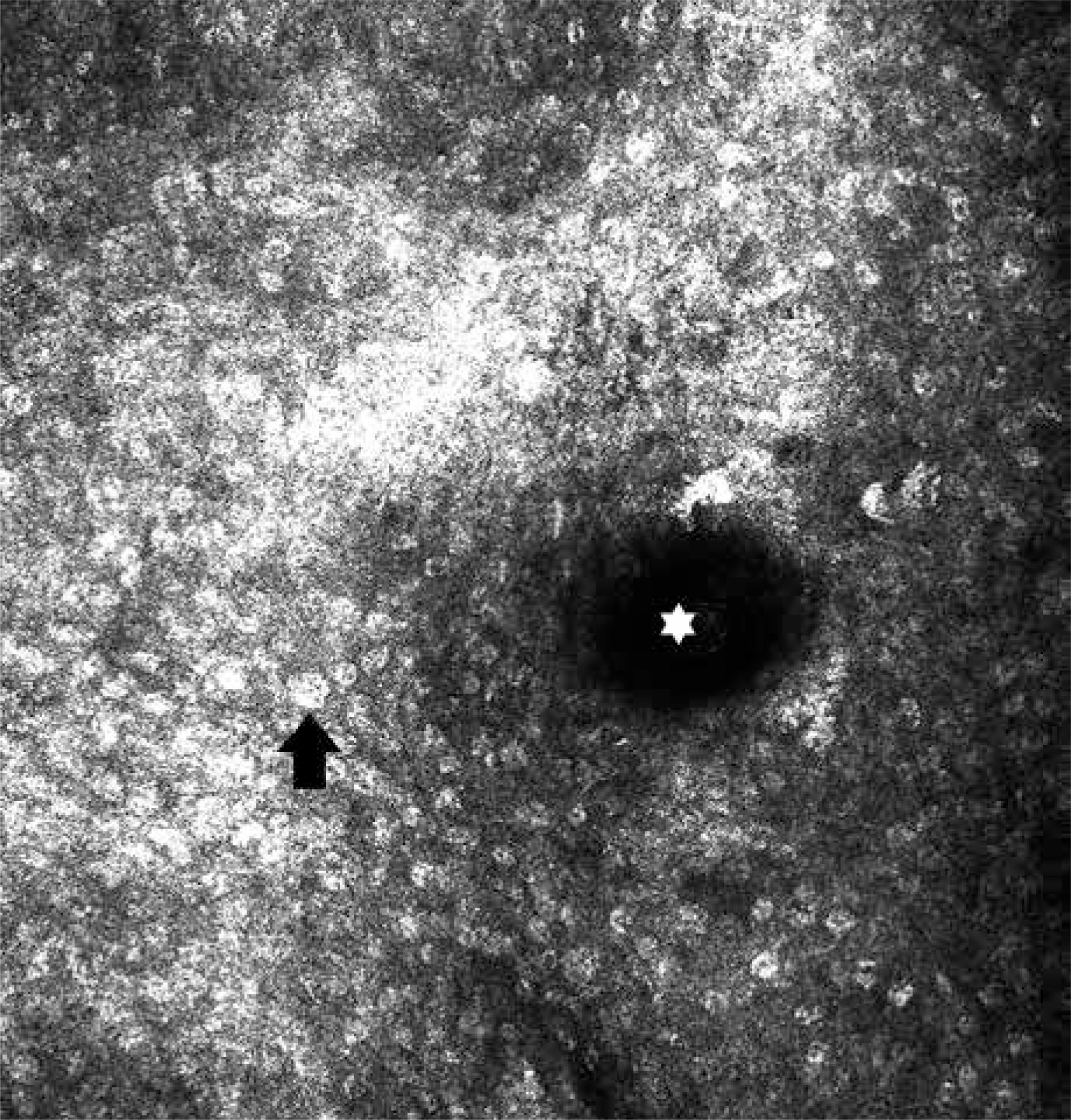
Reflectance confocal microscopy is a non-invasive optical imaging technique, very useful in the diagnosis of clear cell acanthoma. There are a few typical features, such as sharp lateral circumscription or epidermal disarray related to parakeratosis (Figure 3). In most cases there are large, acanthotic cells distributed over the entire surface of the lesion [4]. Clear cell acanthoma in reflectance confocal microscopy shows dilated blood vessels expanding dermal papillae, visible even in the spinous layer of the epidermis. Sometimes inflammatory cell infiltrate is present [5].
Figure 3
Clear cell acanthoma in reflectance confocal microscopy. Acanthotic cell (arrow) and dilated blood vessel (asterisk)
Clear cell acanthoma is an uncommon benign tumour of the skin frequently presented as a solitary nodule. The pathogenesis of clear cell acanthoma is unknown, although the reactive, inflammatory nature is considered. Some authors suggest reactive origin induced by stasis dermatitis. In some cases, the eccrine apparatus may be the place of growth of clear cell acanthoma. The diagnosis of the typical clear cell acanthoma is usually simple, especially through dermoscopy. Dermoscopic pattern of clear cell acanthoma as a necklace-like arranged capillaries was first time featured by Blumm in 2001. In some cases the typical vascular pattern can be incomplete. Even more, some inflammatory dermatoses, such as psoriasis, eczema or pityriasis lichenoides may present a similar vascular pattern. Reflectance confocal microscopy could improve the accuracy of diagnosis of clear cell acanthoma and help us to avoid misdiagnosis [5, 6]. Clear cell acanthoma is a totally benign tumour, so after the confident diagnosis, surgical treatment is not indicated for small lesions. In bigger ones, simple surgical excision is curative [7].


