
INTRODUCTION
Depression and obesity are conditions that are undoubtedly linked – they increase the risk of other diseases, significantly reduce the quality of everyday life, and pose a real threat to patients’ lives. Adipose tissue is now considered an endocrine organ that secretes bio-logically active substances, including leptin, resistin, and adiponectin [1]. In turn, numerous studies show that depression is accompanied by chronic inflammation, elevated levels of Th1 response markers (interleukin [IL]-6, IL-1, tumor necrosis factor alpha [TNF-α]), Th1/Th2 imbalance, and hypothalamic-pituitary-adrenal (HPA) axis dysfunction [2]. Pro-inflammatory interleukins (IL-1, IL-6) and anti-inflammatory IL-10 play an important role in the etiology of recurrent depression [3]. An excess of macronutrients in adipose tissue stimulates the release of inflammatory mediators, disrupts the functioning of the HPA axis, and reduces the production of adiponectin, which promotes the development of inflammation and oxidative stress [4]. A positive correlation has also been demonstrated between high body mass index (BMI) and activation of the anterior cingulate cortex and right inferior frontal gyrus – areas whose activity is also impaired in depression [5]. The inflammatory theory of depression, combined with the monoaminergic theory and hormonal and epigenetic factors, provides a coherent framework for explaining the pathophysiology of the disease. Excess adipose tissue, by stimulating inflammatory processes and weakening anti-inflammatory mechanisms, appears to significantly increase the risk of depression.
The inflammatory theory of depression
The inflammatory theory of depression – in combination with the monoaminergic theory and hormonal factors – provides a comprehensive explanation of the pathophysiological basis of this disorder [6]. It assumes that an imbalance in the immune system, particularly between the Th1 and Th2 lymphocyte populations, plays a significant role in the development of depression. Some scientific studies indicate that the elevated Th1/Th2 ratio observed in patients with depression and the accompanying increase in pro-inflammatory cytokine concentrations may contribute to the development of the disease [7, 8]. Patients with depression have also been found to have increased activity of natural killer (NK) cells, which secrete interferon gamma (INF-γ). These cells activate macrophages and enhance the production of pro-inflammatory cytokines. However, an alternative meta-analysis showed reduced INF-γ concentrations in patients with major depressive episodes [9]. Cytokines such as IL-1, IL-6, and TNF-α are key mediators of inflammation that can affect brain function and contribute to depressive symptoms [7]. In addition, some studies have shown an increased risk of suicide in patients with significantly elevated levels of IL-6 and TNF-α [10]. Studies in animal models have also shown that IL-10 deficiency in microglia can lead to depressive behavior and cognitive impairment [11]. Patients with severe depression had significantly lower serum IL-10 levels compared to healthy individuals. Conversely, the restoration of normal IL-10 levels was associated with clinical improvement [11, 12].
The HPA axis and pro-inflammatory cytokines
Elevated levels of pro-inflammatory cytokines are observed as a cause of excessive cortisol secretion [13]. This occurs through two mechanisms: direct activation of the HPA axis, and indirectly as a result of a reduced expression of glucocorticoid receptors (CAR). The latter phenomenon leads to increased tissue resistance to glucocorticoids. Studies have shown elevated concentrations of glucocorticoids (GCS) in both plasma and urine, as well as increased secretion in response to heightened levels of corticotropin-releasing hormone (CRH) and adrenocorticotropic hormone (ACTH) [14]. In summary, the currently accepted model of “glucocorticoid resistance” assumes that the increase in inflammation is due to reduced tissue sensitivity to the anti-inflammatory effects of cortisol. On the other hand, there is ample evidence that cortisol may also exhibit immunostimulatory properties and directly contribute to the inflammation observed in depression [15]. Patients with depression have also been shown to have elevated levels of pro- inflammatory cytokines co-occurring with increased concentrations of acute phase proteins [16]. An example is C-reactive protein (CRP), the most commonly used marker of inflammation, the secretion of which is stimulated by IL-6. Although previous studies have not conclusively confirmed a causal relationship between depression and elevated CRP levels, most of them have shown that people with depression have higher concentrations of this protein [17, 18].
Inflammation and the kynurenine pathway
The inflammatory process also contributes to the dysfunction of the kynurenine pathway, which is responsible for converting tryptophan into serotonin and melatonin – substances with anti-inflammatory effects [19]. Inflammatory factors produced by the Th1 and NK lymphocytes (in particular IL-1, INF-γ, and TNF-α) activate the enzyme indoleamine 2,3-dioxygenase (IDO), which catalyzes the conversion of tryptophan into kynurenine, limiting the availability of tryptophan for the synthesis of serotonin and melatonin [19]. A decrease in serotonin levels can lead to the development of depressive disorders [20]. It has also been shown that kynurenine causes depressive and anxiety symptoms in animals [21]. A correlation has been proven between increased IDO activity, higher levels of pro-inflammatory factors, and the severity of depression symptoms [22].
Chronic inflammation and depression
The credibility of the inflammatory theory of depression is confirmed by research showing that adding nonsteroidal anti-inflammatory drugs (NSAIDs) to antidepressant therapy improved treatment efficacy in patients with depressive disorders [23, 24]. In addition, in vitro studies on the effects of antidepressants – both serotonin reuptake inhibitors (SSRIs) and tricyclic anti-depressants – have shown a reduction in the INF-γ to IL-10 ratio, which may indicate their anti-inflammatory effect [25]. However, further research is needed to fully understand the mechanism of action of these substances. Therefore, patients suffering from diseases involving chronic inflammation, such as autoimmune diseases, obesity, atherosclerosis, or asthma, may be more prone to developing depression [26, 27].
Adipokines
Adipose tissue is a source of over 600 potentially secretory proteins that are involved in the regulation of insulin, appetite, fat distribution, energy expenditure, endothelial function, blood pressure, hemostasis, and inflammatory processes [28]. The best-known adipo-kines are adiponectin, leptin, and resistin. Disturbances in their concentrations in the course of obesity promote chronic, low-grade inflammation and the development of metabolic pathologies, and at the same time may be one of the pathogenetic mechanisms of depression [29, 30].
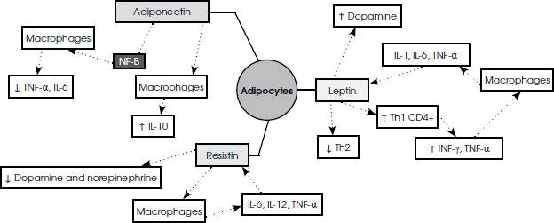
Figure I
Schematic diagram of the connections between adipokines secreted by adipocytes and pro-inflammatory and neurohormonal factors. The figure illustrates the interaction of key adipokines with elements of the immune system and neurotransmitters, which has potential clinical implications in the pathogenesis of psychiatric disorders
Adiponectin
Adiponectin is a monomeric glycoprotein with a molecular weight of 30 kDa, showing significant structural similarity to the complement component C1q. For this reason, it was originally referred to as “complement- related adipocyte protein” (Acrp30) [31]. Acrp30 is a protein that is very abundant in plasma and is secreted exclusively by adipose tissue. However, it has been shown that its concentration in plasma is significantly lower in obese individuals than in individuals of normal weight [32]. Moreover, it has also been found that the expression of cDNA encoding this protein is significantly reduced in the adipose tissue of obese mice and humans, and that it is present only in mature adipocytes [33].
Adiponectin plays a central role in regulating body weight and increasing energy expenditure [34]. It has also been proven to reduce insulin resistance by reducing triglyceride concentrations in muscles and the liver, and its reduced levels are associated with impaired insulin sensitivity [35]. Adiponectin mainly has anti- inflammatory effects [36]. In turn, pro-inflammatory cyto-kines secreted by adipocytes, such as TNF-α and IL-6, inhibit its synthesis [37]. Many studies have confirmed that serum adiponectin concentrations decrease significantly with increasing body weight and the development of obesity [38]. In lean individuals, high adiponectin concentrations and low leptin and resistin levels maintain metabolic homeostasis, promote normal muscle and liver activity, and have antihyperglycemic effects. Conversely, adiponectin concentrations are low in obese indivi-duals [37]. Adiponectin has been shown to cross the blood-brain barrier, and its AdipoR1 and AdipoR2 receptors have been identified in the hypothalamus, brain stem, hippocampus, and cerebral cortex, among other places [39]. Decreased plasma adiponectin levels correlate with the occurrence of major depressive episodes. A decrease in its levels, and thus its anti-inflammatory properties, may predispose patients to an increased inflammatory response in limbic structures [40]. Adiponectin may affect mood and inflammatory responses, which are often exacerbated in patients with depression. Studies suggest that lower adiponectin concentrations may be associated with a higher risk of depression, as a deficiency of it promotes inflammation in the body and dysfunction of the central nervous system [41].
Adiponectin has been shown to stimulate brain cell proliferation and alleviate depressive behaviour. Reduced levels of it may decrease the effectiveness of physical activity in stimulating neurogenesis in the hippocampus, possibly through activation of the ADNR1/AMPK signalling pathway [42]. Animal studies have confirmed that reduced adiponectin levels lead to depressive beha-vior, anhedonia, learned helplessness, and hypothalamic- pituitary-adrenal axis disorders [43]. Administration of an adiponectin-neutralizing antibody into the ventri-cular system of the brain induced depressive behavior in mice, while central administration of adiponectin produced antidepressant effects in both obese and normal- weight mice [43]. In addition, adiponectin promotes neurogenesis, which is reduced in people with depression [44, 45]. Modifiable factors such as physical activity, weight reduction, a diet rich in omega-3 polyunsaturated fatty acids, and certain medications (e.g., statins or thiazolidinediones) have been shown to increase adiponectin levels [46].
A study conducted on mice subjected to high stress levels showed that adiponectin has a rapid antidepressant effect, which is associated with increased glutamatergic activity in the medial prefrontal cortex [47]. The findings of another study were also interesting: serum adiponectin levels were found to be significantly lower in elderly people with severe depressive disorder compared to healthy people of the same age, with low adiponectin levels persisting even after taking into account and controlling for factors such as BMI, cognitive test scores (CAMCOG, MMSE), depression severity, and education level, suggesting that reduced adiponectin levels are independently associated with depression [48]. The meta-analysis by Carvalho et al. [49], although mainly concerned with leptin, also examined adiponectin and found no significant difference in its levels between people with depression and those without depression in different BMI categories. The lack of association may be due to the limitations of BMI, which is an approximate measure of obesity and does not take into account the distribution of body fat. Some studies report that higher adiponectin levels are associated with lower severity of depressive symptoms, possibly due to its anti-inflammatory effects [50]. These inconsistencies may be due to differences in depression subtypes and severity; for example, Jeong et al. [51] reported elevated levels only in individuals with subclinical depression, while other studies included a variety of depressive phenotypes [52].
Leptin
Leptin is a relatively recently discovered protein hormone encoded by the LEP gene, also known as the obesity gene (Ob gene) [53]. The main source of leptin is white adipose tissue adipocytes, although it is also produced in smaller amounts in brown adipose tissue, muscles, gastrointestinal tract cells, bone marrow, the nervous system, the placenta, and other organs and tissues [54]. Plasma leptin concentration depends on several factors – it increases in proportion to the amount of adipose tissue and also depends on the size of adipocytes and gender [55]. In women, blood leptin levels are about three times higher than in men, which is due to the greater amount of subcutaneous adipose tissue, composed of larger fat cells [56]. In addition, it has been shown that female sex hormones, insulin, and pro-inflammatory cytokines stimulate the production and secretion of leptin, while starvation, catecholamines, growth hormone, and male sex hormones inhibit its synthesis [37].
Leptin binds to the Lep-R (Ob-R) receptor, which occurs in several isoforms and belongs to the family of type I cytokine receptors. The long form (ObR-L) is mainly present in the hypothalamus and, to a lesser extent, in other tissues and organs, including the adrenal glands, adipose tissue, gonads, the osteoarticular system, the pancreas, the liver, and immune system cells [55]. The presence of receptors in many locations explains the pleiotropic action of leptin – this hormone is involved not only in the regulation of energy balance, but also in the control of immune response, angiogenesis, hematopoiesis, and ossification processes [37]. The primary function of leptin is to regulate feelings of hunger and satiety. After binding to a receptor in the arcuate nucleus of the hypothalamus, numerous signalling pathways are activated, leading to the inhibition of the synthesis of neuropeptide Y and agouti-related protein (AGRP), which increase feelings of hunger. At the same time, POMC neurons are activated, producing melanotropin (MSH) and endorphins, which further reduce appetite [56]. Paradoxically, obese individuals have high leptin concentrations coexisting with tissue resistance to its action, which contributes to further weight gain. Leptin deficiency or Lep-R receptor dysfunction, on the other hand, leads to severe obesity in young people, sexual maturation disorders, and impaired cellular immunity [57].
Leptin has pro-inflammatory properties [58]. It participates in the regulation of both specific and non-specific immune responses, and high concentrations of it are observed in the early stages of the inflammatory response. Cytokines such as IL-1, IL-6, and TNF-α stimu-late the synthesis and secretion of this adipokine, while leptin itself promotes the proliferation and differentiation of CD4+ Th1 lymphocytes and prevents their apoptosis. Th1 cells produce INF-γ and TNF-α, which in turn stimulate macrophages to secrete cytokines that enhance leptin production, resulting in a positive feedback mecha-nism that amplifies the inflammatory response. Activated macrophages release numerous substances, including prostaglandins, leukotrienes, and nitric oxide. Leptin has also been shown to influence neutrophil activation and chemotaxis, the expression of adhesion molecules, the severity of oxidative stress, and the proliferation and cytotoxicity of NK cells. In addition, leptin inhibits the production of Th2 lymphocytes [58]. An imbalance between Th1 and Th2 lymphocytes may promote the development of numerous inflammatory and autoimmune diseases, such as rheumatoid arthritis, non-alcoholic fatty liver disease, and atherosclerosis [59]. A chronic, generalized inflammatory response can lead to the dysfunction of Lep-R receptors in the brain, causing leptin resistance and hyperleptinemia, which in turn exacerbate inflammation [60].
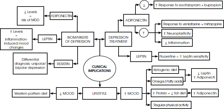
Figure II
Schematic mind map illustrating the relationship between biomarkers of depression, their clinical implications in treatment, and the impact of lifestyle changes on mood
Depressive disorders are associated with immune dysregulation, a predominance of Th1 responses, and excessive synthesis of IL-1, IL-2, IL-6, INF-γ, and TNF-α [61]. This is accompanied by an accumulation of oxygen free radicals and nitric oxide [2]. Leptin has a similar pro- inflammatory effect, which may suggest a link between the severity of depressive symptoms and hyperleptinemia. Depression can be seen as the body’s response to chronic stress, in which there is overactivation of the HPA axis and hypercortisolemia. Glucocorticoids have been shown to increase appetite, which stimulates adipocytes to produce leptin. Under normal conditions, leptin inhibits the further synthesis of glucocorticoids [62]. However, in the course of hyperleptinemia and Lep-R receptor dysfunction, this feedback mechanism does not work [63]. As a result, excessive stimulation of adipocytes by glucocorticoids persists, which increases leptin production and exacerbates generalized inflammation. Furthermore, leptin has been shown to affect hippocampal plasticity and dopamine secretion [64], which further links it to the neurobiological mechanisms of depression.
Nevertheless, a meta-analysis has been conducted which showed that leptin is not a reliable biomarker for depression or other mood disorders. Elevated levels of it in some patients probably reflect coexisting metabolic disorders and leptin resistance rather than a direct pathophysiological mechanism of depression [65]. Another study showed that leptin dysregulation (i.e., resistance) may be a mechanism linking obesity and MDD with atypical features [66]. Furthermore, in obese women after breast cancer treatment, increased anxiety and depressive symptoms were directly associated with higher leptin levels, highlighting the reciprocal influence of mental health and adipocytokines in the context of obesity-related health risks [67]. In contrast, another study found no significant differences in serum leptin levels between patients with major depressive disorder and a healthy control group, contrasting with the simultaneous discovery of reduced levels of the neurotrophic factor EGF, suggesting that EGF, rather than leptin, may be a better risk indicator in the pathogenesis of MDD in this population [68].
Conclusions from cohort studies suggest that the risk of developing depressive symptoms is associated not with leptin concentration itself, but with its low biological signaling, which is measured by a significant interaction between leptin levels and abdominal obesity (waist circumference); this risk is particularly increased when high leptin and large waist circumference co-occur (indicating leptin resistance) [69].
Resistin
Resistin is an adipokine that is primarily produced by inflammatory cells in peripheral blood – monocytes and macrophages – but is also found in other tissues, such as adipocytes, bone marrow, the lungs, placenta, pancreatic islet cells, and cerebrospinal fluid [70]. The expression of the gene encoding resistin is stimulated by the cytokines TNF-α and IL-6 [71]. Resistin itself, in turn, has a reciprocal effect on macrophages, stimulating them to secrete IL-6, IL-12, and TNF-α [72]. CAP-1 (adenylate cyclase- associated protein-1) is considered to be the receptor for human resistin. The binding of resistin to this receptor leads to an increased expression of NF-κB, cAMP, and protein kinase A (PKA), which triggers a pro-inflammatory response [72]. In vitro studies have shown that resistin increases the expression of VCAM-1 and ICAM-1 adhesion molecules, enhances MCP-1 production, and activates endothelial cells, ultimately resulting in the release of endothelin-1 [73].
Resistin also inhibits the release of dopamine and norepinephrine from the hypothalamus [74]. Meanwhile, the course of depression is associated with a decrease in the concentration of neurotransmitters such as dopamine in the cerebral cortex and striatum, and dopaminergic transmission in the subiculum and ventral striatum is impaired [20]. Reduced dopaminergic activity leads to problems with motivation, pleasure, and reward system functioning. Norepinephrine, the secretion of which from the hypothalamus is also inhibited by resistin [74], plays a key role in the pathophysiology of depression [75]. A decrease in noradrenergic activity results in difficulties with concentration and cognitive functions, while increasing its level is one of the goals of antidepressant pharmacotherapy. It has also been suggested that norepinephrine is important in improving social relationships, which are often impaired in people with depression [75].
A correlation has been demonstrated between atypi-cal symptoms of depression and resistin levels, but no such correlation has been found for typical symptoms. People with depression have elevated TNF-α receptor concentrations, and increased levels of this cytokine are associated with the number of depressive episodes and the risk of suicide [20]. Resistin reduces dopamine secretion from the hypothalamus [74], while TNF-α stimulates the expression of the resistin gene [71, 74]. In addition, the expression of this gene is stimulated by IL-6, elevated concentrations of which, according to meta-analysis results, correlate with the occurrence and severity of depression [20]. An increase in the number of adipocytes leads to an increased release of resistin. Elevated concentrations of this adipokine promote the redirection of fat storage towards its accumulation in skeletal muscles and the liver, which in turn promotes insulin resistance. It has been proven that high levels of resistin correlate with insulin resistance [76] and that there is a positive correlation between BMI and resistin concentration [77]. It can therefore be concluded that an increased amount of adipose tissue secreting resistin may lead to a reduced release of dopamine and norepinephrine from the hypothalamus, which may be associated with depressive symptoms.
Clinical implications
Adipokines as biomarkers of depression
Given the evidence pointing to a potential link between reduced adiponectin levels and an increased risk of major depression, it seems reasonable to consider its use as a biomarker for this disease [78]. Adiponectin has also been shown to have prognostic significance in assessing response to antidepressant treatment. Although no correlation has been found between its levels and the initial severity of depression, adiponectin may act as a factor modifying the effectiveness of therapy. In addition, adiponectin is considered a predictor of antidepressant response to drugs with different mechanisms of action (SSRIs, SNRIs, ketamine) [79]. Leptin may be associated with mood and behavioral changes induced by inflammation, which is more commonly observed in women [80]. However, the association between P-leptin and depressive symptoms ceased to be statistically significant when BMI and antidepressant use were included in the analysis. This suggests that these factors limit the usefulness of leptin as a clinical biomarker of depression [81].
Since obesity is characterized by adipokine secretion disorders – with increased leptin and decreased adiponectin levels – the adiponectin/leptin ratio (AL ratio) may be an effective indicator for assessing adipose tissue dysfunction. A study by Senkus et al. [87] showed that a 12-month diet and exercise program aimed at weight reduction improves the AL ratio. Resistin, which intensifies inflammatory processes and affects neurotransmission, has been identified as a potential biomarker for differentiating between people with depression and healthy individuals, although the results of studies are heterogeneous [50]. Furthermore, resistin concentration was significantly lower in patients with unipolar depression compared to those with bipolar depression. This indicates that resistin may be a good marker in the differential dia-gnosis of depression subtypes [82].
In addition, the assessment of body fat (using bioelectrical impedance analysis or dual-energy X-ray absorptiometry – DEXA) may indirectly indicate the amount and activity of adipokines through a very strong correlation with their main source of production: adipose tissue. These relatively simple, minimally invasive methods would enable early identification of high-risk patients and the personalization of treatment based on metabolic and immunological profiles, which could potentially improve prognosis and reduce the risk of recurrence and hospitalization [83].
Treatment of depression and adipokines
Studies have shown that low adiponectin levels predicted a better response to the combination of escitalopram and bupropion compared to treatment with venlafaxine and mirtazapine. In turn, high adiponectin levels were associated with better efficacy of the venlafaxine + mirtazapine combination than escitalopram + bupro-pion [88]. It has also been found that an increase in adiponectin concentration after ketamine administration improves neuroplasticity and reduces inflammation, which is a desirable antidepressant mechanism and may contribute to the therapeutic effect of this drug. Interestingly, it has been shown that the lower the baseline concentration of adiponectin in the blood, the stronger the response to ketamine [41]. Higher adiponectin concentrations were observed in patients who experienced improvement in depressive symptoms (statistically significant both before and after electroconvulsive therapy). This suggests that higher adiponectin levels may be an indicator of a better response to treatment. A positive correlation between adiponectin and BDNF concentrations (before treatment) was also found, indicating that higher initial adiponectin concentrations may promote better neurodegeneration [82].
The concept that leptin resistance may be a therapeutic target in depression in obese individuals is supported by a study in which fluoxetine restored leptin sensitivity [84]. In addition, it is suspected that the response to depression treatment is related to the polymorphism of the leptin receptor gene: rs12564738 and rs12029311 [85]. Resistin concentrations decreased in patients who achieved remission after antidepressant treatment, regardless of the drug used (amitriptyline or paroxetine) No changes in resistin concentration were observed in patients who did not achieve remission [86]. One compound of potential importance in the brain – acting as an antioxidant, neuromodulator, and neuroprotector – is carnosine [89]. Carnosine supplementation has been shown to normalize the concentration of certain adipokines, including resistin [90]. Supplementation with anserine/carnosine, L-carnosine, or β-alanine may therefore potentially alleviate the symptoms of depression [91].
Lifestyle and environmental factors and adipokines
Studies have shown that a 6-month ketogenic diet can have a beneficial effect on adipokine levels by lowering leptin and increasing adiponectin and may also reduce symptoms of depression [92]. Regulating adipokine secretion therefore appears to be a promising therapeutic approach in the treatment of depression. In addition, plant and marine sources of omega-3 fatty acids (n-3 PUFA) have been shown to increase adiponectin and decrease leptin levels in patients with type 2 diabetes [93]. Meta- analyses also confirm the beneficial effect of omega-3 acids on the treatment of depression, especially those containing ≥ 60% eicosapentaenoic acid (EPA) at a dose of ≤ 1 g/day, which may alleviate depressive symptoms [94]. It is believed that diets characterized by reduced fat content may contribute to the beneficial modulation of resistin levels. Furthermore, enriching low-fat diets with a higher protein supply may result in increased concentrations of adiponectin. However, it should be noted that due to the variety and limited strength of the available evidence, it is impossible to draw definitive conclusions [95].
Dietary interventions based on calorie restriction have led to a reduction in depressive symptoms [96]. A Western- style diet, rich in processed foods and sugar, is associated with a deterioration in emotional states [97]. In contrast, a Mediterranean-type diet, rich in fruits, vegetables, nuts, and whole grains, is widely recognized as beneficial for mood [98]. Improved mood after weight loss may also be linked to psychosocial factors, such as improved body image satisfaction and active participation in treatment, rather than weight loss alone [99]. In addition, it has been proven that people who practice various sports disciplines have increased levels of anti-inflammatory cytokines and decreased levels of pro-inflammatory cytokines [100], a more favorable adipokine profile, and lower levels of oxi-dative stress [101]. A study has been conducted which shows that exercise at the right intensity can increase adiponectin levels, mainly in overweight individuals, and that intense exercise lowers leptin levels [102].








