
Dissecting cellulitis of the scalp is a rare, chronic inflammatory dermatosis that mainly affects young men. It manifests as single or multiple pustules, nodules and abscesses, which interconnect and form draining sinus tracts. The most frequent area which is affected is the vertex region and then the occipital region. This dermatosis can lead to scarring alopecia [1].
Dissecting cellulitis may be associated with hidradenitis suppurativa, acne conglobata and pilonidal cysts. Co-occurrence of these disorders is identified as follicular occlusion triad or tetrad. It can concurrently present with arthritis and spondyloarthropathy. Treatment of dissecting cellulitis can be challenging, depending on the severity of the disease.
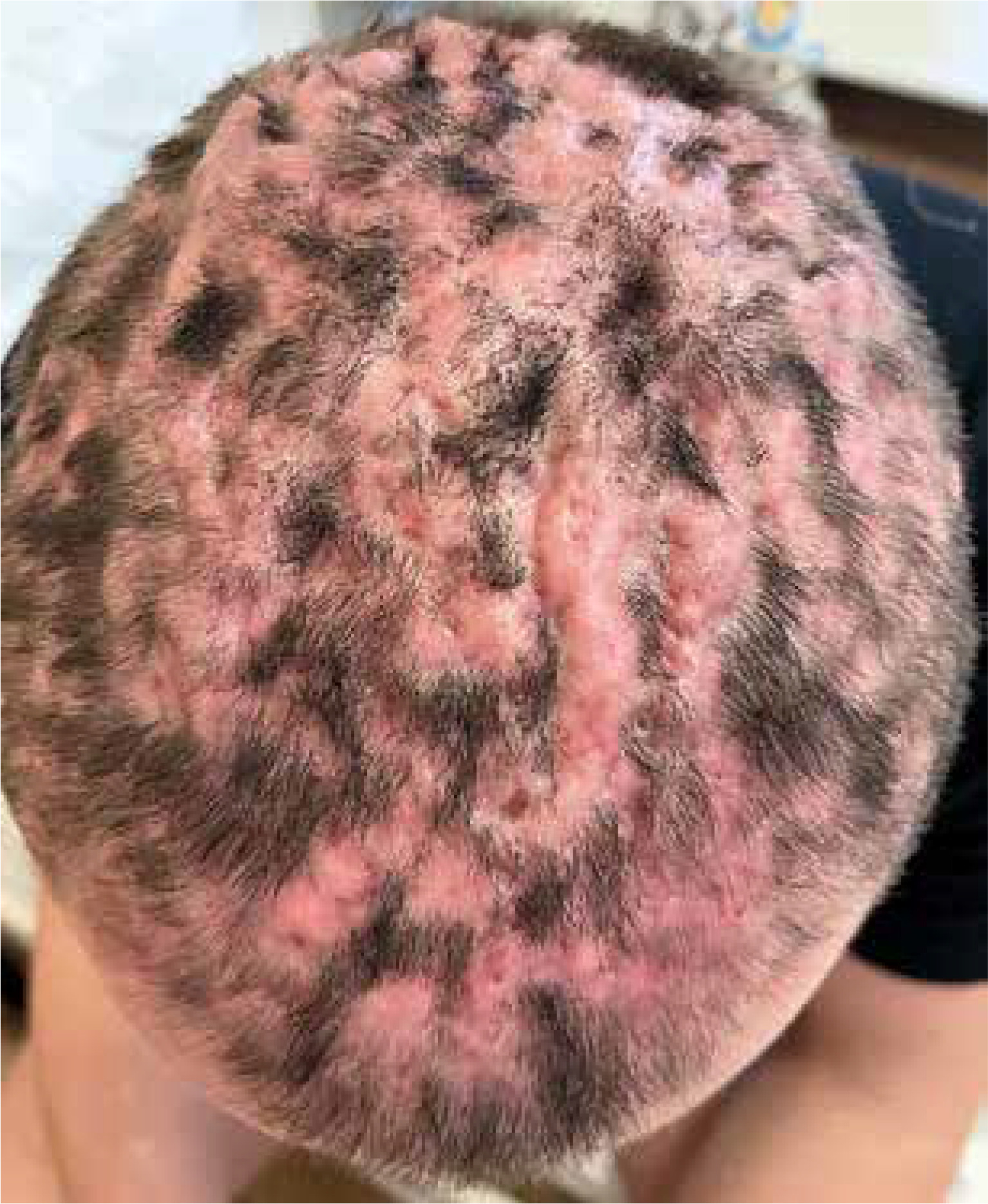
A 24-year-old male patient was admitted to the Dermatology Department with 1-year history of painful skin nodules in the scalp region, with abscesses and draining, purulent sinus tracts forming. Nodules were initially located in the vertex region of the head, then also affecting the occipital area and gradually spreading to the entire skin of the scalp (Figure 1). On physical examination, inflammatory nodules with purulent and blood exudation were observed on the entire skin of the scalp. Additionally, the patient complained of multi-joint pain for 1.5 months. Before admission, he was treated in an outpatient clinic by a dermatologist without any significant improvement with multiple antibiotics and topical disinfecting agents [2]. Prior to the hospitalization he was treated for acne conglobata with oral isotretinoin in maximum daily dose of 50 mg for 7 months with a good therapeutic effect. Furthermore, the patient underwent surgical treatment of the inflamed pilonidal cyst 4 years prior to admission.
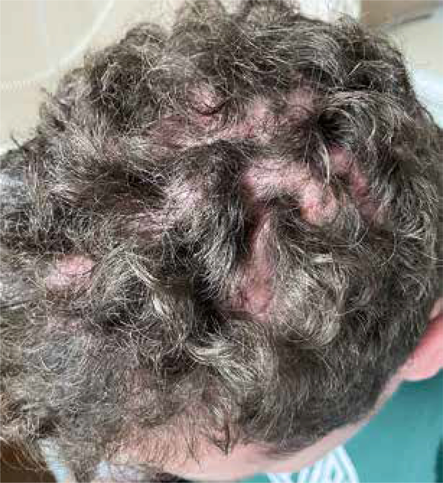
During the hospitalization, laboratory tests showed increased inflammatory markers (C-reactive protein 130 mg/dl). The histopathological examination of the altered skin demonstrated keratosis pilaris, lymphocyte infiltration within skin appendages, proliferation and widening of the lymphatic vessels in the subepidermal area. Based on the clinical picture and trichoscopic examination, dissecting cellulitis was diagnosed. Additionally, because the patient complained of multiple joint pain (knee joints, shoulders, elbows and wrists), he was consulted by the rheumatologist, who linked presented symptoms with SAPHO (synovitis, acne, pustulosis, hyperostosis, osteitis) syndrome. Patient was treated for 7 days with cefuroxime (1.5 g/day), with following clindamycin (600 mg/day) and rifampicin (600 mg/day), as well as acitretin (25 mg/day) and methylprednisolone (from the maximal dose of 16 mg/day with a gradual reduction in 6 weeks) with average improvement. After 6 weeks of therapy, local injections of triamcinolone (15 mg/ml) were added to the systemic treatment. There was a significant improvement of the skin of the scalp. After 4 injections of steroids, used at intervals of 3–4 weeks, hair regrowth was observed (Figure 2). Additionally, during glucocorticosteroid therapy, joint paint was reduced.
Figure 2
Skin of the scalp after 5 months of treatment (7 days with cefuroxime (1.5 g/day), with following clindamycin (600 mg/day) and rifampicin (600 mg/day), acitretin (25 mg/day) and methylprednisolone from the maximal dose of 16 mg/day with a gradual reduction in 6 weeks as well as 4 sessions of local triamcinolone (15 mg/ml) injections performed in 3–4 weeks’ interval)
We present this case as an example of the follicular occlusion triad. There was a co-occurrence of three dermatoses. The patient has suffered from the dissecting cellulitis for a year. Moreover in the past, he had been diagnosed with acne conglobata and pilonidal cyst. Obtaining the desired therapeutic effect with hair regrowth and reduced joint paint was possible due to the use of multi-directional treatments.


