
INTRODUCTION
Eccentric foci of hyperpigmentation, later termed the “Bolognia sign”, were first described by Bolognia et al. in 1994 as “small dark dots” within melanocytic nevi [1]. This feature is clinically important because eccentric peripheral hyperpigmentation may also occur in melanoma. The typical clinical presentation consists of small, round, brown-to-black hyperpigmented foci measuring up to 3 mm in diameter and located at the periphery of a melanocytic nevus [1, 2].
In approximately 70% of cases, these small dark dots result from increased melanin content in epidermal melanocytes and/or keratinocytes, usually accompanied by an increased number of superficial dermal melanophages. In approximately 3% of cases in the original series, an eccentrically located small dark dot represented a focus of melanoma arising within a pre-existing nevus [1]. Because eccentric foci of hyperpigmentation may also be found in combined nevi, Clark nevi, and other types of nevi, the term “Bolognia sign” has been used to describe eccentric foci of hyperpigmentation within melanocytic nevi [2–4].
CASE REPORT
In May 2021, a 2-year-old girl presented to a private dermatology practice with a pigmented nevus on the back. She had been referred by a pediatric surgeon for surgical excision with concern of possible malignancy.
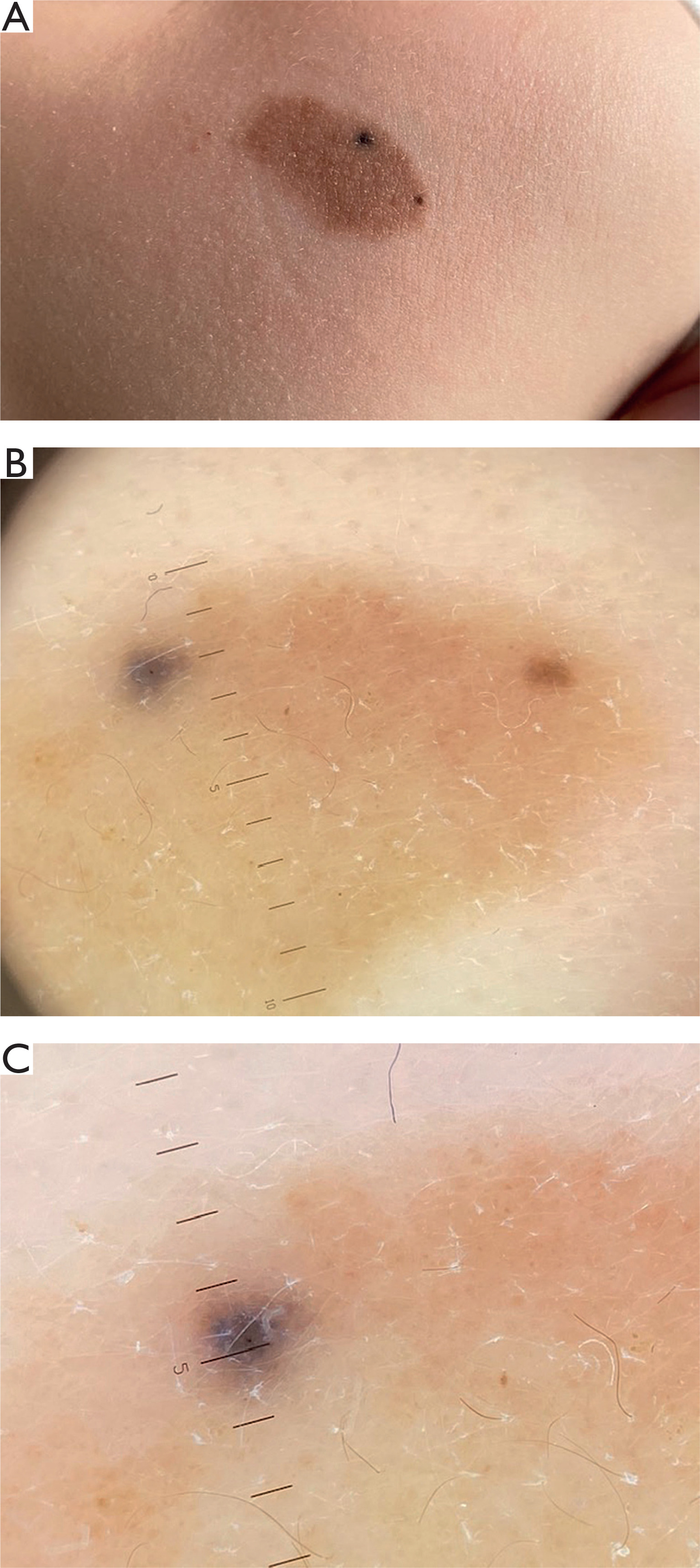
The lesion consisted of a brown macule with two blue-black dots located peripherally (fig. 1 A). Initial dermoscopy showed light-brown pigmentation with two areas of homogeneous blue-gray pigmentation in the upper part of the lesion. The nevus was clinically and dermoscopically diagnosed as a melanocytic nevus with eccentric foci of hyperpigmentation (figs. 1 B, C).
Figure 1
Visit 1. A – Iirregular brown macule with two different colored dots. B, C – The dots are different in color, the left is more bluish whilst the right is more brown. Dermoscopy examination with Heine Delta 30 PRO
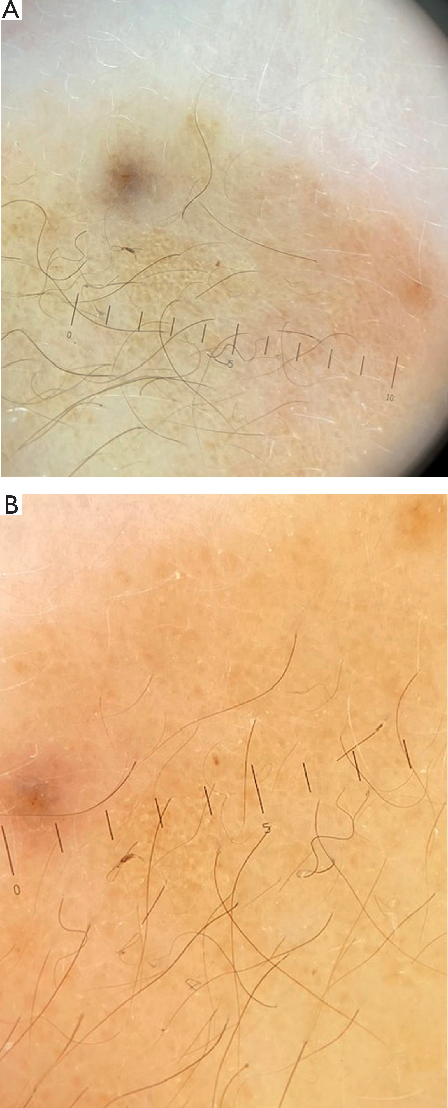
Follow-up dermoscopy performed 6 months after the initial examination showed a decrease in the intensity of pigmentation within the hyperpigmentated foci. The two homogeneous foci were light brown and less intensely pigmented than at baseline (figs. 2 A, B).
Figure 2
Visit 2 after 6 months. A – Decrease in the intensity of pigmentation dots. B, C – Two homogeneous dots were light brown and less intensely pigmented than at the baseline. Dermoscopy examination with Heine Delta 30 PRO
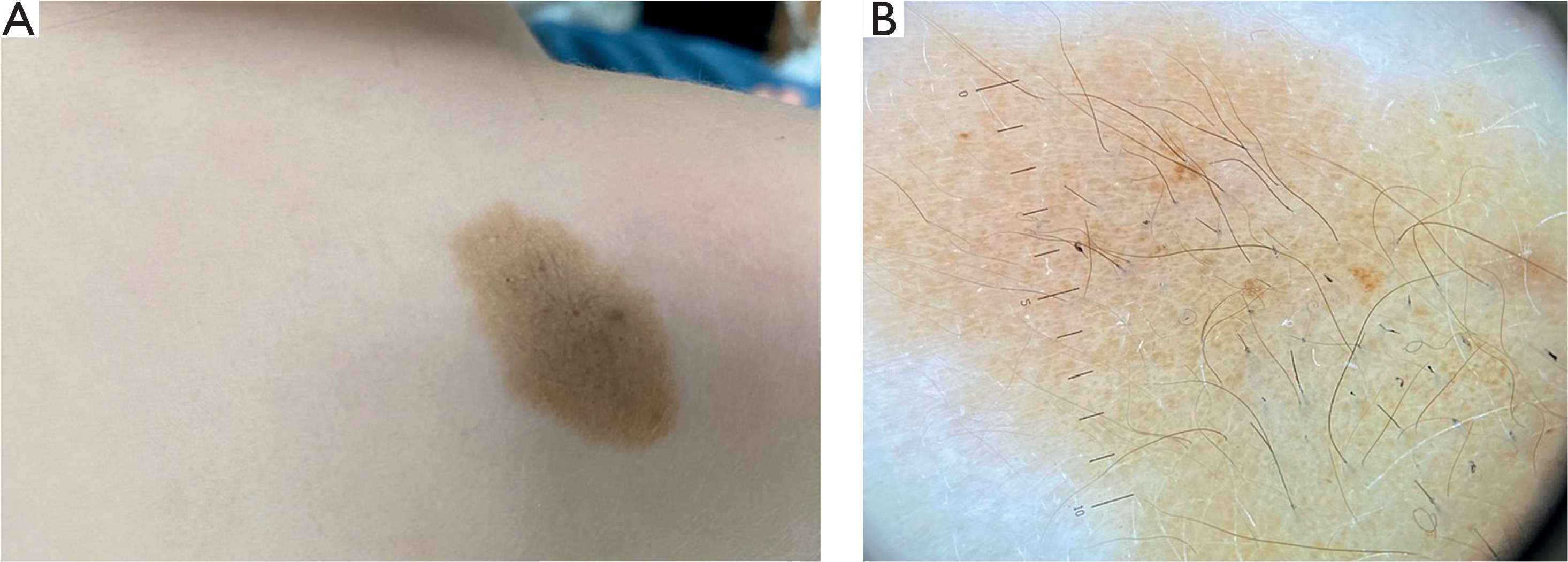
At the third visit, 12 months after baseline, the two homogeneous foci were no longer detectable clinically or dermoscopically (figs. 3 A, B). Hair growth was also observed around the nevus. The pigmentation pattern was typical of a congenital melanocytic nevus.
Figure 3
Visit 3 a year after the initial visit when the girl age was 3 years. A – The macule of the nevus in light brown color. B – On dermoscopy the light brown background with hairs is visible without no different in color dots within. Dermoscopy examination with Heine Delta 30 PRO
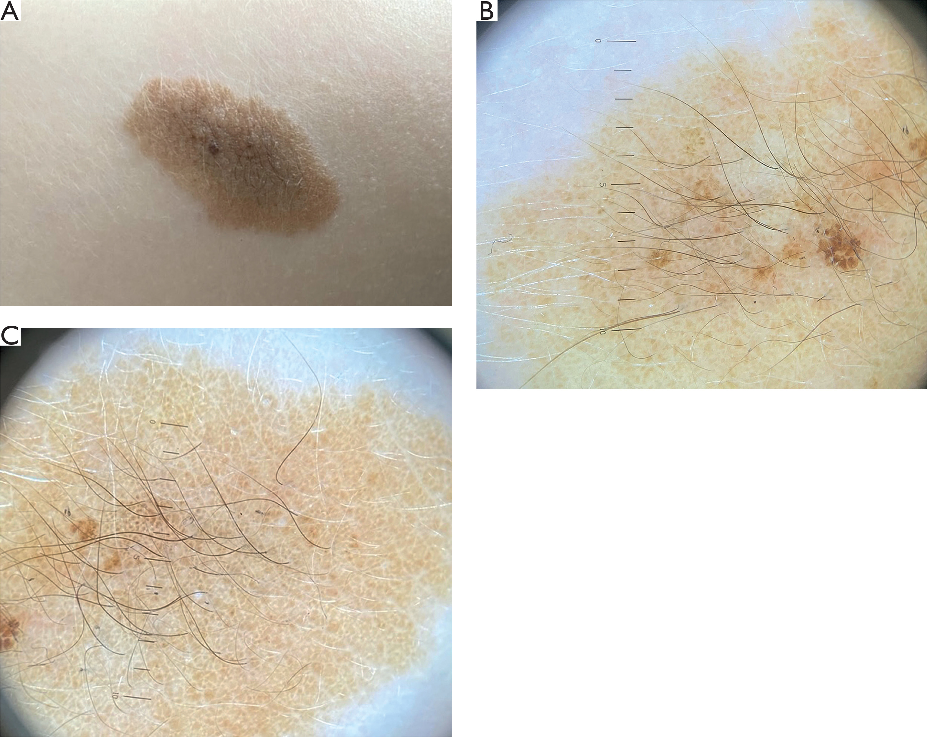
At the fourth visit, 24 months after baseline, clinical and dermoscopic examination showed regular pattern of a congenital melanocytic nevus (figs. 4 A–C).
Figure 4
Visit 4 when the girl was 4-yo. A – The nevus forms a brown plaque. B – Dermoscopy shows the light brown cobblestone pattern characteristic for the nevus in youngsters with small dots of darker brown color in the middle. Dermoscopy examination with Heine Delta 30 PRO. C – Cobblestone pattern of the nevus with darker vellus hairs growing on its brown background
CONCLUSIONS
We report the case of a 2-year-old girl with a melanocytic nevus with eccentric foci of hyperpigmentation and describe the morphological and dermoscopic changes observed during 24 months of follow-up.
We emphasize the importance of dermoscopic follow-up in the management of melanocytic nevi with eccentric foci of hyperpigmentation in children. Based on our observation, eccentric foci of hyperpigmentation within a melanocytic nevus may represent a particular, transient phenotypic stage in the development of a congenital melanocytic nevus. Regular dermoscopic follow-up of appropriately selected lesions may help avoid unnecessary surgical excisions.
These conclusions should not be extrapolated to adults. In adults, eccentric foci of hyperpigmentation are important simulators of early melanoma in Clark's nevi [3]. Eccentric peripheral hyperpigmentation has been reported in 25.3% of melanomas and in only 4.5% of benign melanocytic lesions [5]. Because this pattern may raise concern for melanoma developing in association with an atypical nevus, the surgical excision is recommended in adults [6]. By contrast, dermoscopic follow-up may represent an alternative to surgical treatment in appropriately selected pediatric cases.












