
INTRODUCTION
Nevus sebaceus of Jadassohn (NSJ) is one of the most distinctive organoid hamartomas of the skin, involving the epidermis and its adnexal derivatives – follicular, sebaceous, and apocrine structures. Classically, it presents at birth as a smooth, yellowish plaque, most commonly located on the scalp or face, and gradually becomes verrucous during puberty and adulthood. Although benign, NSJ may harbor or give rise to secondary adnexal tumors, such as trichoblastoma or syringocystadenoma papilliferum [1].
Histologically, NSJ represents a microcosm of cutaneous adnexal architecture. Alsaad et al. described its evolutionary phases: early lesions with primitive follicular germs and scattered sebaceous lobules, whereas mature lesions demonstrate epidermal papillomatosis, prominent sebaceous hyperplasia, and increased numbers of apocrine structures in the dermis [1]. The lesion thus reflects the close anatomic and embryologic relationship within the pilosebaceous–apocrine unit, in which the hair follicle, sebaceous gland, and apocrine gland develop from a shared epithelial germ [1].
Within this developmental framework, aberrations may result in unusual histologic configurations. Although apocrine glands and sebaceous lobules are expected components of NSJ, the coexistence of sebocytes and calcifications within the lumina of apocrine glands appears not to have been previously reported. This finding raises questions regarding possible morphogenetic interactions and degenerative processes within hamartomatous adnexal proliferations.
OBJECTIVE
To report an apparently novel histopathologic finding in nevus sebaceus, characterized by the coexistence of sebocytes and calcifications within the lumina of apocrine glands, and to discuss its potential developmental, mechanical, and degenerative mechanisms in the context of organoid hamartomatous growth.
CASE REPORT
A 50-year-old man with stage 3 chronic kidney disease presented with a solitary, asymptomatic, elevated, linear, yellowish-brown lesion on the left side of the forehead. The lesion had been yellowish at birth and became verrucous around puberty. It remained relatively stable for many years; however, during the preceding 2 years, the patient had noticed a gradual increase in its size. There was no history of pain, pruritus, discharge, or ulceration, although the lesion caused cosmetic concern.
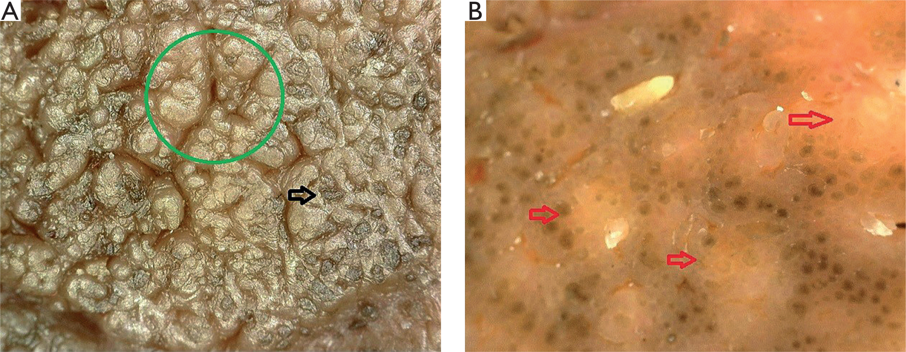
Cutaneous examination revealed a 3 × 1.5 cm linear, yellowish-brown plaque with a papillomatous, verrucous surface (fig. 1). Dermoscopy showed exophytic papillary projections forming a cobblestone or cerebriform pattern, follicular plugging, and yellowish-brown areas suggestive of sebaceous differentiation (fig. 2). The clinical differential diagnoses included nevus sebaceus and verrucous epidermal nevus, with nevus sebaceus considered the most likely diagnosis.
Figure 1
Clinical image showing a linear, verrucous, yellowish--brown plaque on the left forehead. According to the patient, a flat yellowish lesion had been present at the same site since birth
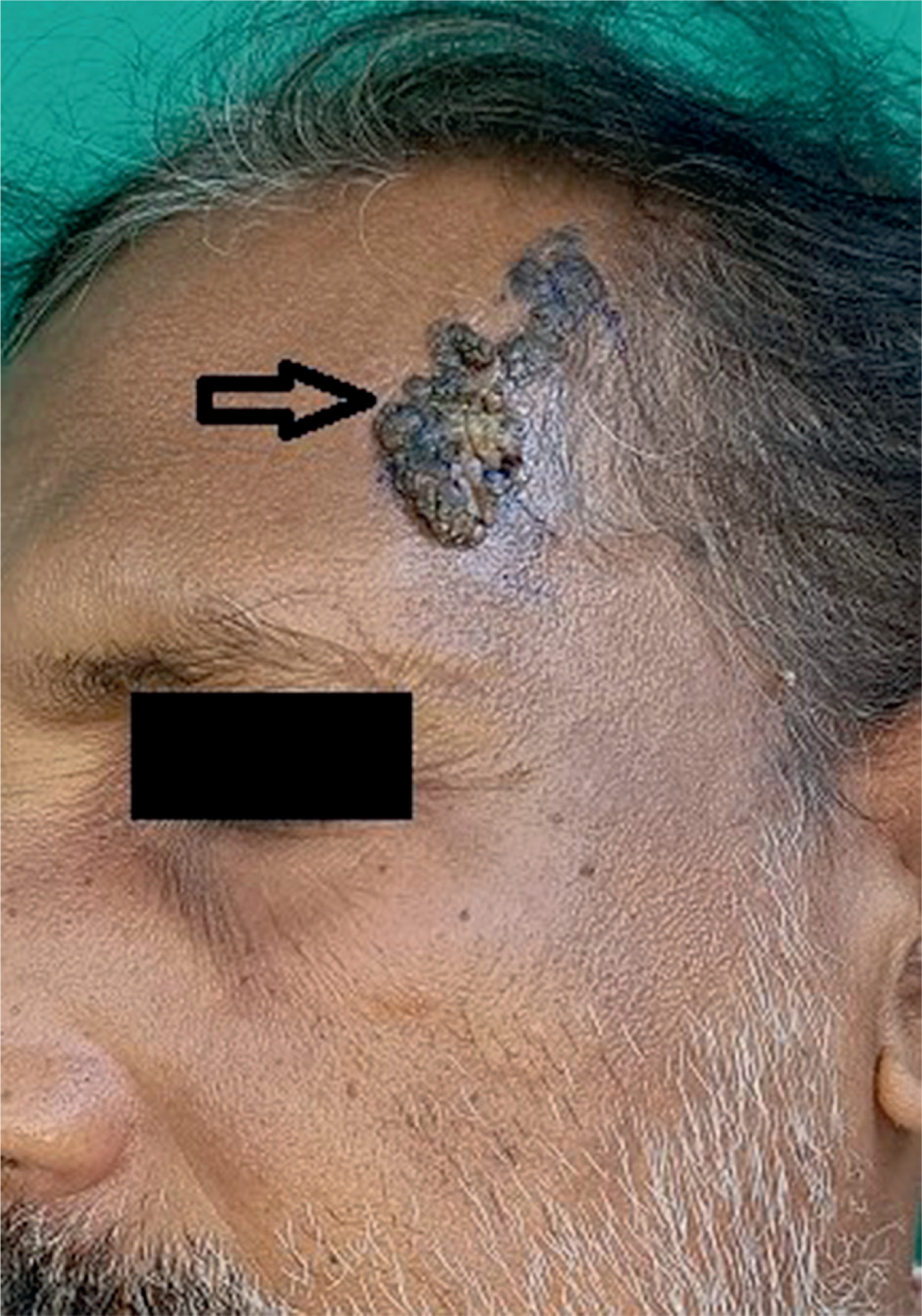
Figure 2
Dermoscopic examination using a Dino-Lite device at ×100 magnification. Nonpolarized dermoscopy shows a cerebriform pattern (green circle) and follicular plugging (black arrow) (A). Polarized dermoscopy shows yellowish areas suggestive of sebaceous differentiation (red arrows) (B)
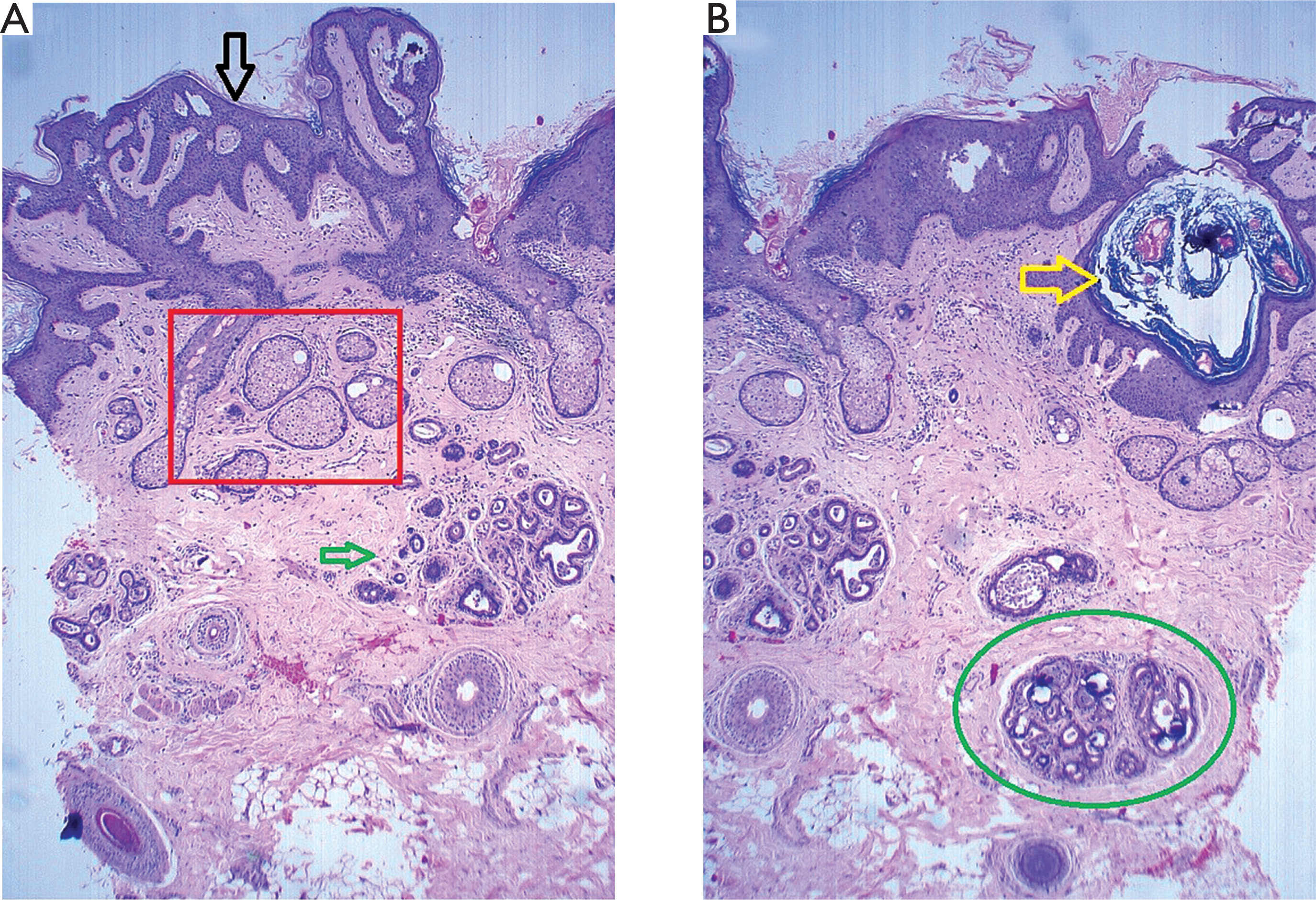
His complete blood counts were within normal limits. Biochemical evaluation showed normal serum calcium (9.4 mg/dl; reference range 8.6–10 mg/dl), serum phosphate (3.5 mg/dl; reference range 2.5– 4.5 mg/dl), parathyroid hormone (44 pg/ml; reference range 15–65 pg/ml), and alkaline phosphatase (67 IU/l; reference range 44–147 IU/l). To exclude a secondary neoplasms, a 4-mm punch biopsy was performed. Hematoxylin and eosin-stained sections revealed epidermal papillomatosis and irregular acanthosis. Numerous mature sebaceous lobules were observed in the upper dermis, many of which opened directly into the overlying epidermis (fig. 3 A). Increased numbers of apocrine glands containing intraluminal calcifications were noted in the mid-dermis (fig. 3 B).
Figure 3
Low-power view showing an irregularly hyperplastic epidermis (black arrow), hyperplastic sebaceous glands opening directly into the epidermis (red rectangle), and prominent apocrine glands (green arrow) (H&E, ×4) (A). A dilated follicular infundibulum with keratin plugging and irregular branching is visible (yellow arrow). The circled area contains apocrine glands with amorphous basophilic calcifications (green circle) (H&E, ×4) (B)
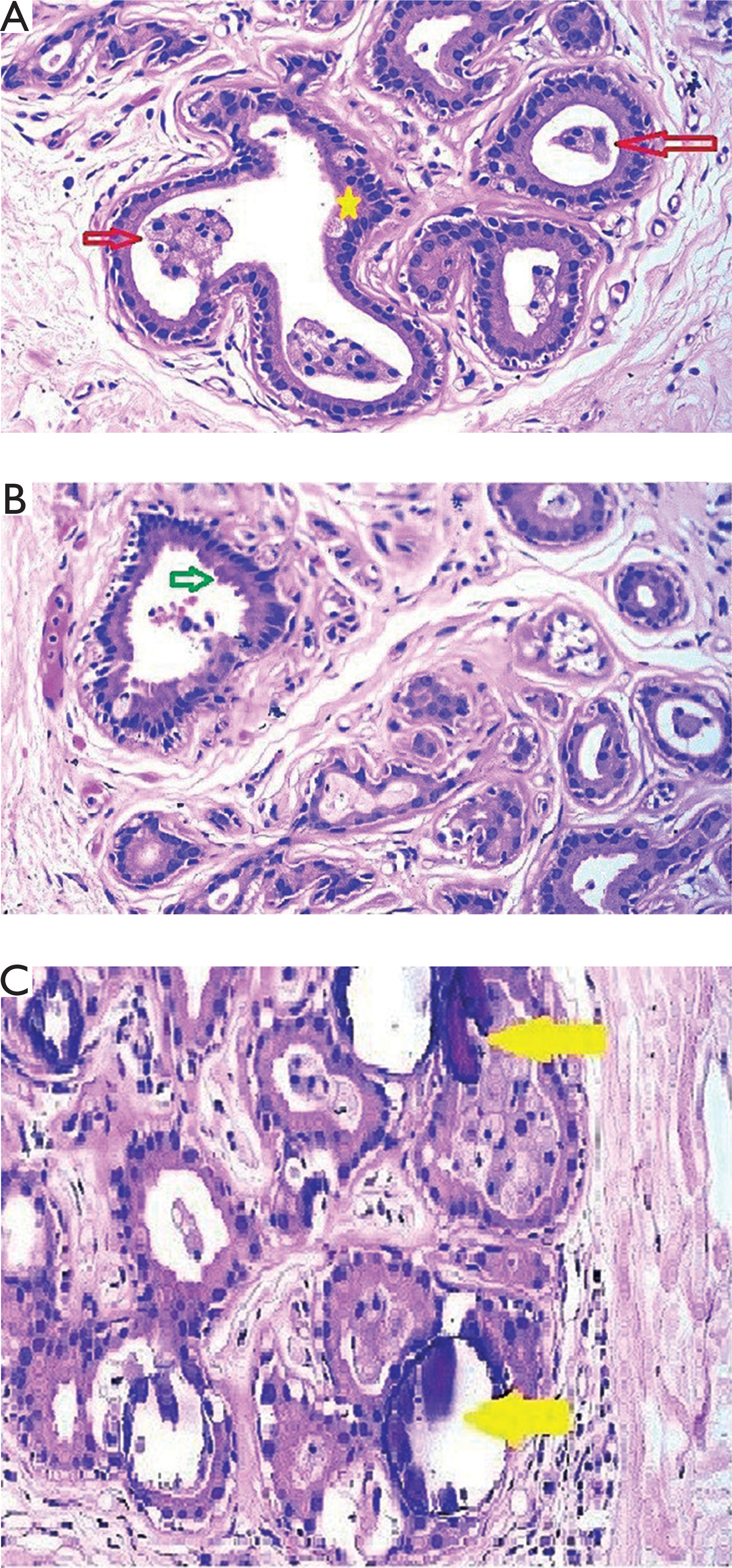
A striking finding was the presence of clusters of sebocytes with vacuolated cytoplasm and scalloped nuclei within the lumina of the apocrine glands and along the luminal aspect of the glandular epithelium (fig. 4 A). The apocrine glands showed decapitation secretion (fig. 4 B). In addition, multiple amorphous basophilic calcifications were present within the glandular lumina (fig. 4 C).
Figure 4
Higher-power view showing clusters of mature sebocytes within the lumina of apocrine glands (red arrows) and along the luminal aspect of the glandular epithelium (yellow asterisk) (H&E ×40) (A). Apical snouting consistent with decapitation secretion is visible (green arrow), supporting apocrine differentiation (H&E ×40) (B). Foci of amorphous basophilic material consistent with calcification are present within the lumina of apocrine glands (yellow arrows) (H&E ×40) (C)
The biopsy specimen included the full thickness of the dermis to a depth of approximately 3.5 mm, allowing evaluation of the sebaceous, follicular, apocrine, and eccrine structures. Multiple deeper sections were examined and confirmed these findings without any additional abnormalities. There were no histopathological features of secondary neoplasms, such as trichoblastoma or syringocystadenoma papilliferum.
The histopathological findings were consistent with nevus sebaceus showing sebocytes and calcifications within apocrine glands. The patient was informed of the benign nature of the lesion and referred for surgical excision and reconstruction. The lesion was completely excised, followed by reconstructive surgery, with a satisfactory postoperative outcome. However, the excised specimen was not submitted for histopathological examination, and therefore the unusual intraluminal findings observed in the punch biopsy could not be confirmed in the excision specimen. The patient was subsequently lost to follow-up.
DISCUSSION
Nevus sebaceus exemplifies an organoid hamartoma in which multiple adnexal structures coexist at varying degrees of maturation and architectural disorganization. The pilosebaceous–apocrine unit represents a developmental continuum [1]. During embryogenesis, as described by Alsaad et al., the hair follicle and sebaceous gland develop from a downward proliferation of basaloid epidermal cells into the dermis. Apocrine glands arise later from the upper portion of the developing follicle and open into the follicular canal [1].
This close developmental relationship may explain why apocrine structures are frequently encountered in NSJ. Their proliferation within a sebaceous milieu may be regarded as exaggeration of normal embryologic association rather than an entirely aberrant phenomenon. However, the presence of sebocytes within apocrine lumina, as observed in the present case, represents a more unusual degree of adnexal intermingling and may reflect developmental intermixing, mechanical displacement, or postmaturational degenerative change.
The occurrence of sebaceous structures in unexpected locations has precedents in dermatopathology. Mehregan and Pinkus described transfollicular extrusion of sebaceous glands as a possible processing artifact caused by displacement of fragile sebaceous lobules during microtomy [2]. Similar findings involving sebaceous or pilosebaceous structures within the blister cavities of pemphigus vulgaris have subsequently been reported and attributed either to mechanical displacement during tissue processing or to acantholytic loosening of the adnexal epithelium [3–5]. Related adnexal changes have also been described in pemphigus vulgaris and bullous pemphigoid [6, 7].
Although these previous reports primarily concerned immunobullous disorders, they demonstrate that sebaceous structures may become displaced into adjacent epithelial spaces through mechanical or biological processes. In the present case, there was no acantholysis or prominent inflammatory infiltrate. Therefore, developmental intermixing arising from the shared epithelial origin of the pilosebaceous–apocrine unit represents one possible explanation. Alternatively, the fragility of sebaceous lobules may have permitted displacement of sebocytes into adjacent apocrine lumina during tissue processing.
Calcification within apocrine glands is rare but has previously been described in nevus sebaceus. Jang et al. reported psammomatous calcified spherules within apocrine glands and proposed a dystrophic mechanism associated with cellular degeneration and subsequent calcium deposition [8]. Choi et al. also reported calcification within the apocrine glands of nevus sebaceus [9]. Thus, apocrine calcification itself is not a novel finding; the unusual feature of the present case is its coexistence with sebocytes within the same glandular lumina.
The spatial association between sebocytes and calcifications raises the possibility that degenerating sebocytes or apocrine epithelial debris may have served as a nidus for local dystrophic mineralization. However, this proposed relationship remains speculative and cannot be established from a single histopathological observation. Moreover, because the patient had stage 3 chronic kidney disease, a possible contribution of altered mineral metabolism should also be considered. The patient was under regular treatment and follow-ups for his chronic kidney disease. His serum calcium, phosphate, parathyroid hormone, and alkaline phosphatase levels were all within the normal reference ranges, making a clinically significant disturbance of mineral metabolism less likely. However, as calcium-phosphate homeostasis and tissue calcification are dynamic processes, a subtle systemic contribution cannot be completely excluded. Therefore, although the localized intraluminal distribution favours dystrophic calcification, the proposed mechanism remains speculative.
Microtomy-related artifacts may arise when the paraffin block is insufficiently hardened, sections are trimmed too rapidly, or the blade encounters tissues of uneven consistency [10]. Fragile, lipid-rich sebocytes may be particularly susceptible to mechanical displacement. In the present case, the presence of sebocytes in similar locations across multiple deeper sections and their apparent continuity with adjacent sebaceous lobules argue against a simple microtomy artifact. Nevertheless, these findings do not entirely exclude processing-related displacement. Unfortunately, the excision specimen was not examined histopathologically, precluding confirmation of the unusual intraluminal findings and representing an important limitation of this report, as a tissue-processing artefact cannot be completely excluded.
Management options for nevus sebaceus include clinical observation, surgical excision, and selected destructive or laser-based procedures, particularly when treatment is undertaken for cosmetic reasons. Surgical excision is appropriate when there is diagnostic uncertainty, a significant clinical change, histopathological atypia, or concern regarding a secondary neoplasm. It also enables examination of the entire lesion, which cannot be achieved using a limited punch biopsy. Most secondary neoplasms arising in nevus sebaceus are benign, and malignant transformation is uncommon; therefore, routine prophylactic excision remains controversial [1, 11]. Management should be individualized according to the patient’s age, clinical findings, cosmetic concerns, and preferences. In the present case, the recent enlargement of the lesion, the patient’s cosmetic concerns, and the unusual histopathological findings supported the decision to perform complete surgical excision and reconstruction.
The coexistence of sebocytes and calcifications within apocrine glands represents an unusual histopathological pattern in nevus sebaceus. Possible explanations include developmental intermixing resulting from the shared origin of the pilosebaceous–apocrine unit, mechanical displacement of fragile sebaceous cells, and local dystrophic calcification secondary to cellular degeneration. In summary, the proposed developmental, mechanical, and degenerative mechanisms remain speculative and their relative contributions cannot be conclusively determined from a single case.
CONCLUSIONS
This case describes an apparently novel histopathological finding in nevus sebaceous: the coexistence of sebocytes and calcifications within the lumina of apocrine glands. This unusual pattern may reflect developmental intermixing within the pilosebaceous–apocrine unit, mechanical displacement of sebaceous cells, and local dystrophic calcification.
Although the exact mechanism remains uncertain, this observation expands the recognized histopathological spectrum of nevus sebaceus and highlights the architectural complexity of adnexal differentiation within this organoid hamartoma.












