
Introduction
Ovarian masses are common findings in general gynecology [1]. Epithelial neoplasms of the ovary account for 60% of ovarian masses. Ovarian cystadenomas are common benign epithelial neoplasms which have a very good prognosis. Serous and mucinous are the two most frequent types of cystadenomas [2]. These tumors are not usually symptomatic. Abdominal pain and abdominal distension are the two most frequent symptoms and usually appear when these tumors become large [3]. Abdominal and pelvic ultrasonography (US), magnetic resonance imaging (MRI) and computed tomography (CT) are useful tools in making the initial diagnosis of an ovarian mass [4]. In this report, we discuss a case of large serous cystadenoma, which was diagnosed in a 68-year-old female patient.
Case report
A 68-year-old postmenopausal woman presented to our department with complaints of massive abdominal distention which started gradually 6 months ago. The patient also complained of difficulty in breathing and ambulation. There were no other gastrointestinal, urinary, or gynecological symptoms. The woman had a significant past medical history of hypercholesterolemia and her past surgical history included one cesarean section 40 years ago. Her family history is negative for malignant ovarian and breast cancer in first-degree relatives.
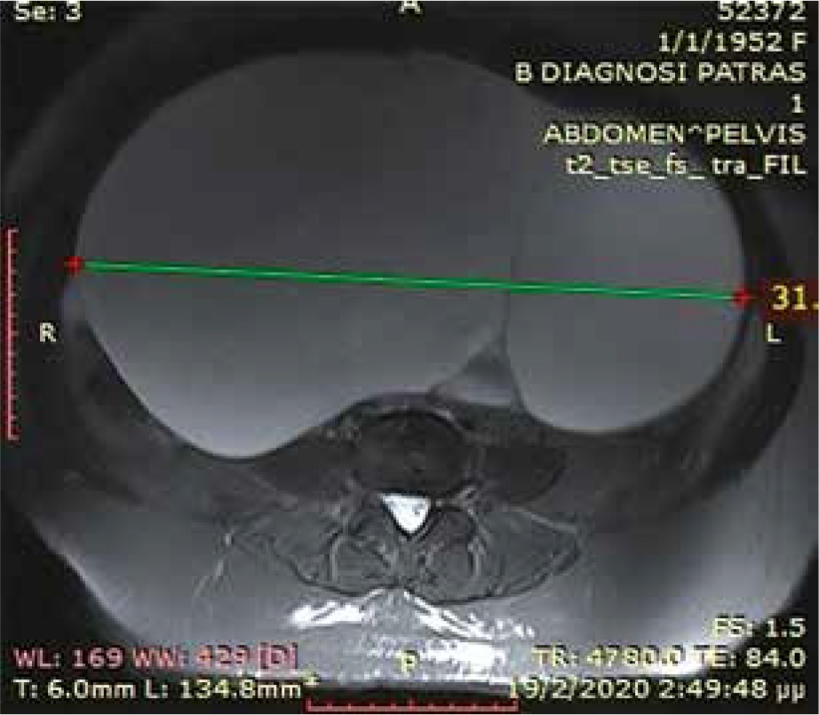
On physical examination, the patient’s abdomen was grossly distended, hard and tense on palpation. Blood analysis revealed hemoglobin of 11.4 g/dl, 9170 leukocytes and 235000 platelets. All tumor markers were within normal limits. However CA-125 was slightly elevated (42.4 U/ml). An abdominal and pelvis magnetic resonance imaging (MRI) was performed. The MRI revealed a huge pelvi-abdominal mass measuring 34 × 31 × 27 cm, which originated from the right ovary (Fig. 1).
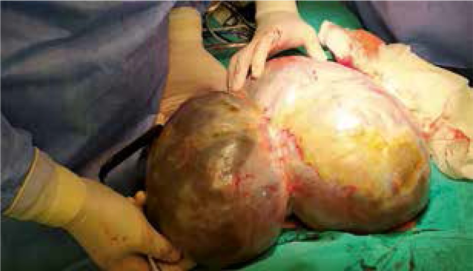
Because of elevation of serum CA-125 levels, ovarian malignancy was included in differential diagnosis. The patient was operated for exploratory laparotomy with removal of right ovarian mass with ovarian cystectomy (Fig. 2). The patient had no intra-operative or postoperative complications. On day 5 of surgery, patient was discharged home in good condition. Her final histopathology report was suggestive of serous cystadenoma of right ovary.
Discussion
Ovarian serous cystadenomas, also known as serous cystadenoma, are the most common ovarian neoplasms. They are benign lesions and represent 16% of all ovarian epithelial neoplasms [5]. Its prevalence peaks between 60-70 years of the human lifespan. Serous ovarian cystadenocarcinomas account for ~25% of serous tumors [6]. Tumor markers, such as CA-125 can be a useful tool that helps to distinguish between benign and malignant ovarian masses [7]. CA-125 levels of less than 35 U/mL are now accepted as normal. Elevated levels of CA-125 are more strongly associated with serous, rather than mucinous tumors [8]. It is now widely accepted that the tumor marker CA-125 is a predictive and prognostic factor in CA-125 positive ovarian cancers. Serum CA-125 level is a strong prognostic factor for overall survival and progression free survival in ovarian cancer. There is an inverse relationship between serum CA-125 levels and survival in ovarian cancer. That means that a decreasing level generally indicates a positive response to cancer therapy while an increasing level indicates tumor recurrence and poor survival [9]. However, a few prospective studies indicated the inadequate sensitivity of CA-125 in the setting of ovarian cancer screening in asymptomatic populations [10-12]. Some authors have described elevated levels of these markers in patients with benign tumors [7]. The combination of normal findings at serum CA-125 assay, imaging, and clinical findings exclude the possibility of ovarian cancer [13]. Abdominal and pelvic ultrasonography (US) seems to be useful in making the initial diagnosis of an ovarian mass. Furthermore, magnetic resonance imaging and computed tomography are helpful in confirming the diagnosis of these masses [4].
Surgery is the treatment of choice for serous cystadenomas. Ovarian cystectomy or unilateral salpingo-oophorectomy is commonly considered in huge tumors. Clinical recurrence of serous cystadenomas is not very common and reflects either incomplete resection or a new primary tumor [2, 14]. Until now, there has been no randomized controlled trial for the laparoscopic management of ovarian cysts > 20 cm, so laparotomy remained the ideal method for the excision of the giant ovarian cysts [15].
Ovarian masses will continue to be a risk for woman, as the majority of patients will not be diagnosed at an early stage. It is suggested that every female patient with pain in any region of the abdomen or pelvis be at least evaluated by imaging techniques.
Conclusions
Giant ovarian mass is a rare finding in general gynecology. Physicians must maintain heightened awareness and index of suspicion when approaching a woman with pain in any region of the abdomen or pelvis. Further investigation with abdominal and pelvic ultrasonography and magnetic resonance imaging or computed tomography is necessary. Although tumor markers such as CA-125 can be a useful tool to distinguish between benign and malignant ovarian masses, they are not very specific and sensitive for malignancy.


