
An 81-year-old female with coronary disease, arterial hypertension, paroxysmal atrial fibrillation, and DDDR implantation 6 years ago due to sick sinus syndrome (SSS) was admitted to the cardiology ward with symptoms of stable angina pectoris to have invasive diagnostics performed. Selective coronarography revealed calcific ostial stenosis of the right coronary artery (RCA) and proximal stenosis of the left anterior descending artery (LAD). As well as the above-mentioned findings, it exposed the tip of an RV electrode with healing tissue around its spiral coil, which was indenting into the lumen of the middle part of the LAD causing stenosis and potential blood flow restriction (Figures 1 A–C). Further assessment of the LAD and the RCA stenoses using intravascular ultrasound (IVUS) and subsequent PCI of any lesion that would be considered significant were scheduled for the following month. During the second coronarography the exact mutual RV lead and the LAD’s position was determined by IVUS, which also showed no signs of damage to the artery wall (Figure 1 D). Considering the morphology of the LAD, the localization of indention in the distal part, the opinion of a cardiac surgeon, and our experience, we decided that the patient would not benefit from electrode removal or any other invasive treatment of the indented area. Therefore, we withdrew from invasive diagnostics and implemented conservative therapy with ambulatory observation. The stenosis of the ostial RCA was assessed in IVUS as nonsignificant (MLA = 6.5 mm2). However, the proximal, significant stenosis of LAD (MLA = 2.7 mm2) was treated with shockwave intravascular lithotripsy and DES stenting.
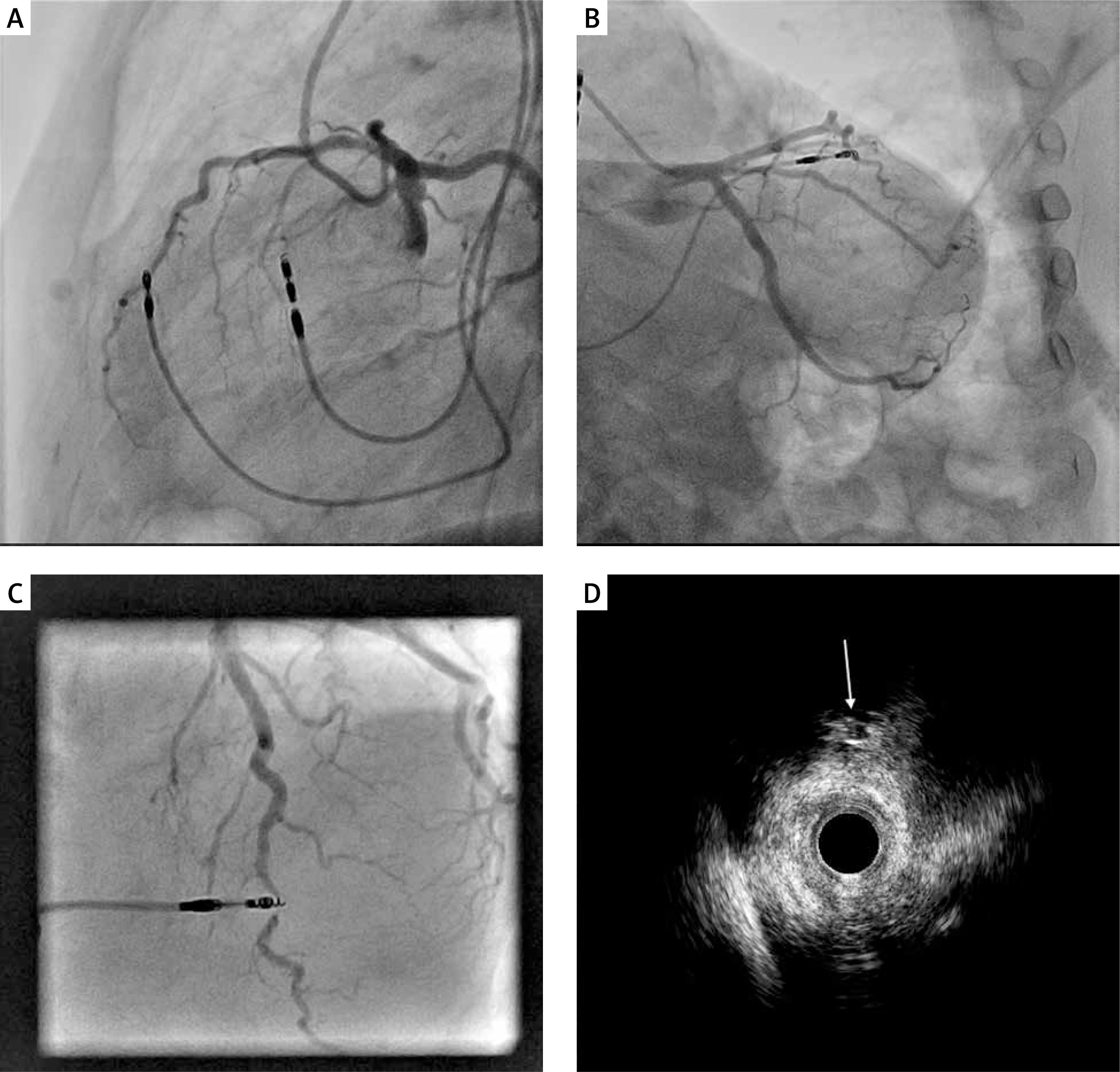
Figure 1
A–C – Angiographic images in 3 different projections presenting interference between the tip of the electrode and the left anterior descending artery. D – Intravascular ultrasound image of the left anterior descending artery. An arrow pointing at the tip of the electrode
Cardiovascular implantable electronic devices cause various complications, and 0.2% of such procedures are complicated by cardiac tamponade, which is considered a rare incident [1]. In comparison to the above, damage of a coronary artery, with only a few cases reported [2], is an extremely rare complication. Narrowing it down to LAD injuries, there have been 4 cases reported [2, 3]. Nonetheless, many of the implantable leads remain in a very close relationship to the coronary arteries. The mean distance between them is 4.7 mm for the antero-septal junction fixation presented in this case [4]. Inferring from the study above and the analysed case, the optimal lead implantation technique for an RV electrode would be fixation on the true RV septum, and hence avoidance of antero-septal junction fixation. Two-dimensional fluoroscopy without contrast is the imaging method used in a lead fixation procedure that does not allow to determine possible coronary artery intrusion and is very rarely followed by computed tomography (CT) or coronary angiography. However, fluoroscopy performed in right anterior oblique (RAO) and left anterior oblique (LAO) projections confirms the optimal lead positioning on the true RV septum. Considering the above, it is possible that restriction of blood flow after lead fixation is in fact much more common but not clinically significant. It is essential to raise awareness of the reported complication due to the possibility that coronary artery damage might be more common in the future considering the growing use of left bundle branch area pacing (LBBAP), a method with the propensity to damage septal perforators and cause coronary artery spasm due to the transseptal route of the pacing lead [5].


