
Bone cyst is a benign osteolytic cyst that develops within bone. There are mainly two types of bone cysts: the common form, simple bone cyst; and aneurysmal bone cyst (ABC), the uncommon form. The latter is rare and affects about one in a million people in a given year [1]. The most common location is on the long bone of legs, upper arms, spine, and pelvis [1]. The rib is an uncommon location of ABC. We report here two cases of ABC of the ribs.
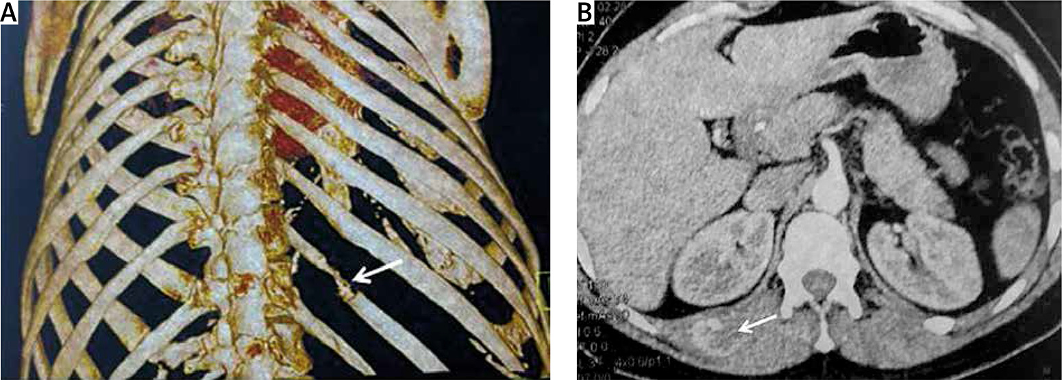
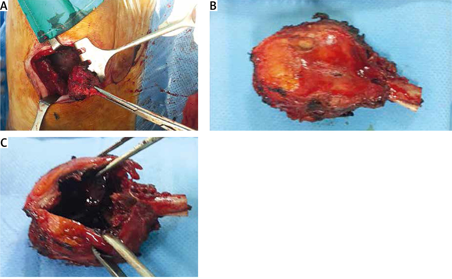
Case 1: A 59-year-old male patient was admitted in March 2018 with a complaint of an increasing swelling of the left base of chest wall since 1 year before. There was no fever or respiratory associated symptoms. On physical examination, the left anterior chest wall in the 8th rib was slightly bulging, with no mass palpation. Plain and lateral chest radiographs revealed a chest shadow (Figure 1 A). Computed tomography (CT) scan of the chest revealed a regular-shaped 66 × 47 × 48 mm bone cyst in the anterior 8th rib (Figure 1 B). Magnetic resonance imaging (MRI) and ultrasound were performed and confirmed the diagnosis of bone cyst but with a suspicion of the hydatid origin of the cyst, while laboratory assessments were all within normal limits. Complete removal surgery was performed through a left posterolateral thoracotomy (Figure 2). An anatomopathologist confirmed the diagnosis of ABC with a free surgical margin. Five year follow-up revealed no recurrence.
Case 2: A 57-year-old female patient presented in September 2015 with a 2-year history of right posterior chest swelling with pain. On physical examination, the right posterior chest wall in the region of the 9th rib was bulging. The respiratory sounds were normal. Chest X-rays (Figure 3 A) and computed tomography (CT) scan showed a posterior irregular shaped bone cyst of the 9th rib. Laboratory data were all within normal values. A right sided posterolateral thoracotomy was performed with complete removal (Figure 4). A microscopic examination showed a blood-filled space, surrounded by a fibrous wall; granulation and giant cells were found in the cyst wall (Figures 5 A, B). The surgical margin was free and no malignant lesion was observed. Follow-up during 3 years was uneventful.
Figure 3
Radiological findings in case 2. A – Chest radiograph showing left paracardiac shadow. B – CT scan revealing an osteolytic lesion of the anterior bow of the 8th rib. C, D – MRI exposed the expandable character of the osteolytic lesion with multiloculated blood-filled cavities with a “honeycomb” appearance
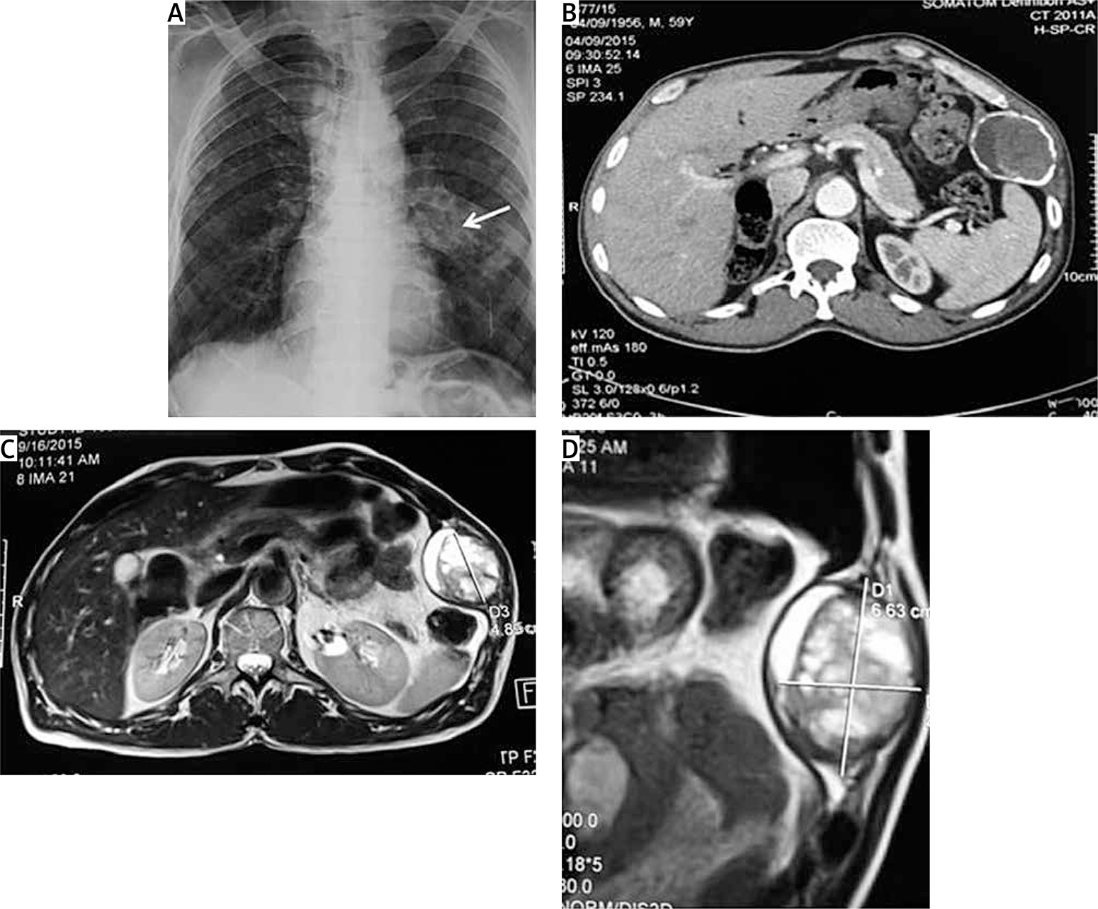
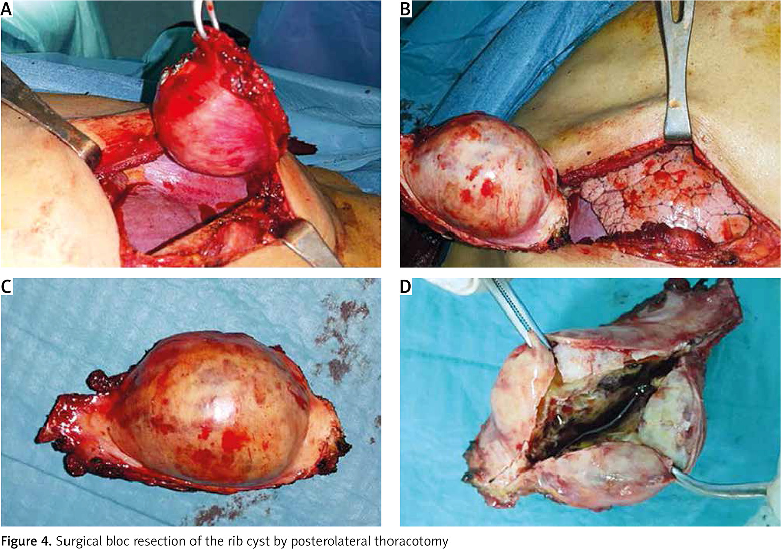
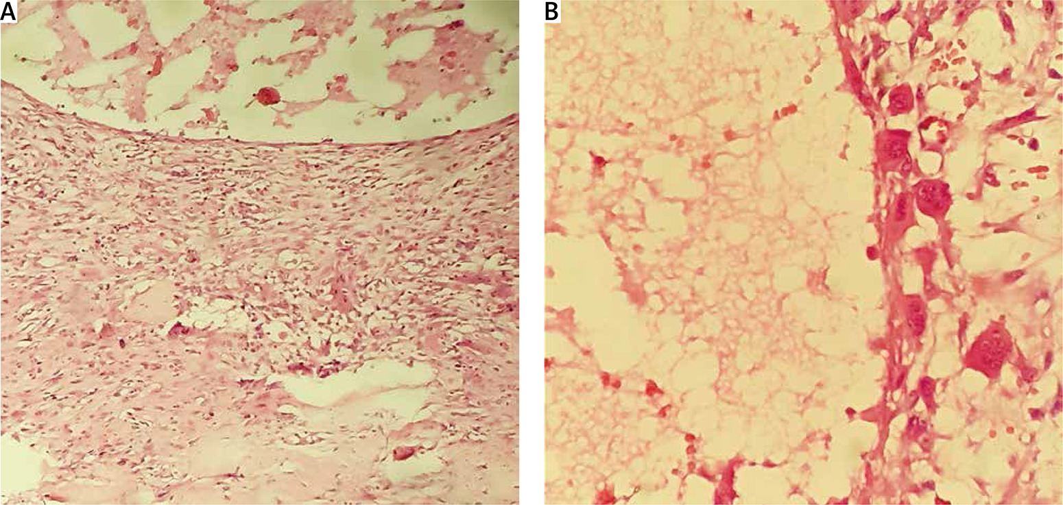
Figure 5
Cystic feature with hemorrhagic content in fibrous tissue containing some osteoclast active cells in a dystrophic bone tissue (A – H + E, 40×, B – H + E, 100×)
ABC located in the rib is particularly rare, about 2.7% of all ABC [1, 2]. It may occur in every rib [2]. About 29% of reported cases have been found incidentally by a routine chest X-ray but it is usually associated with pain, swelling, a palpable lump, dyspnea, paraplegia, or pathological fractures [1]. The etiology of ABC is unknown but arteriovenous malformation is a widely accepted theory. The radiological data of costal ABC are not sufficiently specific to establish a definitive diagnosis; CT scan or MRI has great value to demonstrate the characteristic findings of the condition, namely an expansile lytic bony lesion with multiloculated blood-filled cavities with a ‘soap bubble’ or ‘honeycomb’ appearance [3]. However, it is often difficult to obtain a precise preoperative diagnosis. The usual differential diagnosis in young children is between an ABC and simple bone cyst, and in young adults between ABC and giant cell tumor. In pathology ABC has a uniform pattern: large cystic spaces filled with blood and separated by fibrous septa, alternating with solid areas. Primary lesions arise with no known cause. Secondary ABCs arise from another preceding lesion, and these are presumed to occur as the result of an intralesional hemorrhage within the primary lesion. Primary ABCs such as in our cases are uncommon; they represent only 5–7% of all bone tumors [1, 4]. Approximately one-third are the secondary type [5]. Therefore surgical treatment is recommended, but other methods of treatment are available such as curopsy, embolization, cryotherapy, sclerotherapy or radiotherapy. Local extension can cause pathologic fractures, paralysis due to spinal cord compression, compression of the vital organs and malignant transformation. To date, complete surgery and large excision allow the lowest risk of local recurrence [2]. However, few authors have reported recurrence after a total excision [6, 7]. Radiotherapy should be considered only for inoperable cases, keeping in mind that transformation into a sarcoma due to irradiation is possible [3, 8].
In conclusion, ABC of the rib is a rare, benign condition which is considered in the differential diagnosis of chest wall tumors. Surgery by bloc resection of the mass and affected portion of the rib can be curative and provide good results due to potential risk of recurrence.


