There are 43 cases describing aortic trombosis and COVID-19, among which only 2 cases reported the anatomic localization to be the ascending aorta [1]. We present a patient with SARS-CoV-2 and thrombus in the ascending aorta. Clinically the patient also had significant coronary artery disease, which required treatment by cardiac surgeons.
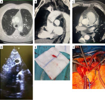
A 75-year-old patient was admitted to the COVID department with positive antigen test results, fever, shortness of breath. He had no comorbidities but had a history of coronary artery disease. No vaccination history against COVID-19 was recorded. Computed tomography showed typical inflammatory changes of approximately 40–50% in the lungs consistent with COVID-19 infection (Figure 1 A). In the ascending aorta, a floating thrombus measuring 18 × 12 mm was found, which was a remarkable discovery (Figure 1 B). Computed tomography (CT) scan also showed calcification in all coronary arteries (Figure 1 C). Therapeutic anticoagulation with heparin was immediately started. After Heart-Team discussion, the decision of computed tomography of coronary arteries was made, which showed significant coronary artery disease in the right coronary artery (RCA) and left descending artery (LAD). CTA showed all the pathologies perfectly, and there was no reason to perform transoesophageal echocardiography. The next decision was to transfer the patient to a cardiac surgery ward for coronary artery bypass grafting (CABG) and removal of the thrombus from the aorta. The patient was consulted by an interventional radiologist to consider minimally invasive techniques, but there was a risk of leaving a clot and the patient should have open chest surgery for CABG. Preoperative transthoracic ultrasound showed good function of heart chambers without valve diseases and confirmed a thrombus in the ascending aorta (Figure 1 D). Surgery was performed by medial sternotomy. The cardiopulmonary bypass was connected between the right atrium and the aortic arch. If the view of the aortic arch had not been sufficient for cannulation, the femoral artery would have been the second method of cannulation. After 38 min of circulation, distal anastomoses (LAD, RCA) and systematic hypothermia to 28°C was performed. During circulatory arrest the aorta was opened and the thrombus was removed; the time of circulatory arrest was 7 min (Figures 1 E, F). The decision to perform Deep Hypothermic Circulatory Arrest Surgery was made due to minimized risk of closing the thrombus by cross clamp. The aortic wall was clear, without suspicion of dissection or endocarditis. The total time of cardiopulmonary circulation was 122 min. After the surgery, the patient was transferred to the intensive care unit. He was extubated on day 1 and moved to the cardiosurgery department on day 3. During hospitalization, one episode of atrial fibrillation was treated with Cordarone, and sinus rhythm returned the next day. Patient was discharged home after 10 days of hospitalization with full recovery.
Figure 1
A – Chest computed tomography, B – floating thrombus in ascending aorta in CT, C – calcification in coronary artery, D – thrombus in ascending aorta in TTE, E – removed thrombus, F – perioperative photo

We present a patient with a very rare complication of coronavirus infection. A minimally invasive technique should be used if the coronary artery disease was not for intervention [2]. This is the first case with additional coronary artery bypass grafting and thrombus removal with deep hypothermic circulatory arrest.


