
Catheter-induced coronary artery dissection (CICAD) is a rare but very serious complication of invasive diagnostics and percutaneous treatment of coronary artery disease. The incidence of CICAD is < 0.1% [1]. CICAD is usually treated with stent deployment, less often conservatively and only a small percentage of patients require surgical intervention [1, 2].
In this article, we present the case of a patient with CICAD who, after initial conservative therapy, underwent intravascular ultrasound-guided percutaneous coronary intervention (IVUS-guided PCI).
A 58-year-old patient with chronic coronary syndrome was referred to the clinic from a district hospital, after scheduled coronary angiography complicated by iatrogenic dissection of the left coronary artery (LCA) (Figure 1 A). On admission, the patient was in a good general condition and had no chest pain. There were no significant abnormalities in electrocardiography or echocardiography. High-sensitivity troponin concentration was 1210 ng/l (n < 15 ng/l). The decision of a conservative approach was made. However, after a few days, symptoms of angina pectoris (CCS III) occurred. Computed tomography (CT) revealed a left main (LM) coronary artery dissection causing narrowing of the left coronary artery ostium (ratio of the false lumen to the true lumen was 3 : 1). The decision to perform IVUS-guided PCI was made by the Heart Team.
Figure 1
A – Angiographic imaging: dissection of the left coronary artery (LCA) (red arrow), B – Angiographic imaging: guidewires inserted into the left anterior descending artery (LAD) (blue arrow) and circumflex artery (Cx) (red arrow), C – Intravascular ultrasound (IVUS) imaging: after stent deployment the true lumen has a larger dimension than the false lumen (red arrow), D – Angiographic imaging: after 6 weeks – good effect of the PCI of the left main (LM) coronary artery, E – Computed tomography: after 5 months – good effect of the PCI of the left main (LM) coronary artery (red arrow)
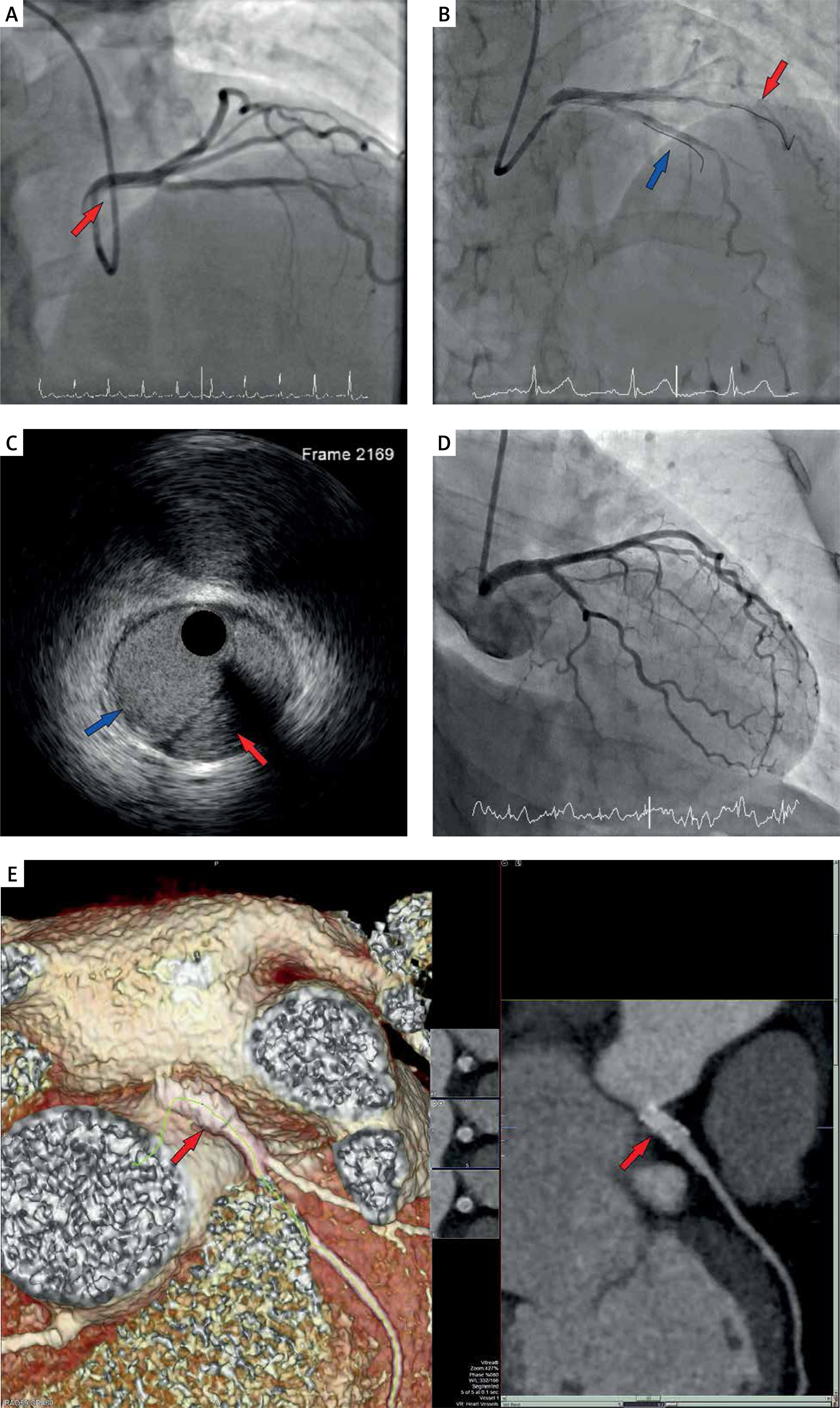
Coronary angiography showed the dissection of the LM and its branches. Under IVUS control, the guidewires were inserted into the true lumen (approximately 1 mm in diameter) (Figure 1 B) and a stent (Xience Sierra 4.0 12 mm) was implanted into the LM to close the dissection entry site. IVUS revealed adequate expansion and apposition of the stent (Figure 1 C).
During the following days, the patient was in a good general condition and had no symptoms of angina. Controlled CT done 5 days after PCI showed the patent stent and the dissections of the initial section of the LAD and Cx. Seven days after PCI the patient was discharged. Dual antiplatelet therapy (aspirin and ticagrelor) for 12 months was recommended. Coronary angiography performed after 6 weeks showed a good effect of the PCI of the LM, a small linear dissection in the LAD and a partially healed dissection narrowing the lumen by 50% in the middle of the Cx (Figure 1 D). After another 5 months, a CT was performed showing coronary arteries without any signs of dissection (Figure 1 E).
Treatment of CICAD depends on the patient’s condition and angiographic image. This example shows that when a conservative approach is chosen, very careful observation of the patient is essential and sometimes an invasive strategy has to be implemented. The most important and often the most difficult part of the procedure is insertion of the guidewires into the true lumen. IVUS is used to confirm the correct location of the guidewires [3, 4]. The best treatment of CICAD is stent deployment along the entire length of the dissection [3], but it is not always possible. In the above-described case, the implantation of the stent in the dissection entry turned out to be effective therapy.


