eISSN: 2449-8238
ISSN: 2392-1099
Clinical and Experimental Hepatology
Current issue Archive Manuscripts accepted About the journal Editorial board Abstracting and indexing Subscription Contact Instructions for authors Ethical standards and procedures
Editorial System
Submit your Manuscript
SCImago Journal & Country Rank
3/2022
vol. 8
 
Share:
Share:
Original paper

Cellular and mitochondrial taurine depletion in bile duct ligated rats: a justification for taurine supplementation in cholestasis/cirrhosis

Asma Najibi
1
,
Heresh Rezaei
2
,
Ram Kumar Manthari
3
,
Hossein Niknahad
1, 2
,
Akram Jamshidzadeh
1, 2
,
Omid Farshad
1
,
Feng Yan
4
,
Yanqin Ma
4
,
Dongmei Xu
4
,
Zhongwei Tang
4
,
Mohammad Mehdi Ommati
5
,
Reza Heidari
1

1.
Pharmaceutical Sciences Research Center, Shiraz University of Medical Sciences, Shiraz, Iran
2.
Department of Pharmacology and Toxicology, School of Pharmacy, Shiraz University of Medical Sciences, Shiraz, Iran
3.
Department of Biotechnology, GITAM Institute of Science, Gandhi Institute of Technology and Management, Visakhapatnam, Andhra Pradesh, India
4.
Department of Life Sciences, Shanxi Agricultural University, Shanxi, Taigu, China
5.
College of Veterinary Medicine, Shanxi Agricultural University, Jinzhong, China
Clin Exp HEPATOL 2022; 8, 3: 195-210
Online publish date: 2022/09/21
Article file
- Cellular (1).pdf  [0.25 MB]
Get citation
 
PlumX metrics:
 

Introduction

Taurine (β-amino-ethane sulfonic acid; TAU) is the most plentiful free amino acid in the human body [1, 2]. This amino acid is abundantly found in almost all cell types [1, 2]. Although TAU does not contribute to the protein’s structure, several physiological roles have been attributed to this amino acid [1, 2]. TAU plays a significant role in bile acid conjugation and acts as an essential osmolyte in various biological systems [1-5]. Despite the ubiquitous physiological functions of TAU, its action in the pathophysiology of diseases is largely disputed. On the other hand, it has been found that TAU deficiency can seriously impair the function of several organs such as the skeletal muscle and heart [6-9]. Therefore, it is essential to investigate the role of TAU deficiency in the pathogenesis of the human disease.

Body TAU can be synthesized in the liver or supplied from the diet. The source of body TAU is essentially species-dependent [10, 11]. Some species, such as foxes and felines, entirely depend on the dietary TAU [10, 11]. In humans, the hepatic TAU synthesizing capability is negligible, and this compound is primarily supplied from the diet [12, 13]. Seafood (e.g., oysters and muscles) is a well-known dietary source of TAU [14]. TAU is taken up by various cell types via specific transporters (TauT) [15]. The accumulation of TAU in different organs is widely variable [15]. Tissues such as the heart and skeletal muscle contain very high concentrations of TAU [15]. At the subcellular levels, TAU is mostly compartmentalized in mitochondria [12, 16]. Several lines of evidence support the pivotal role of TAU in mitochondrial function [17-20]. It has been found that TAU can regulate mitochondrial energy metabolism, attenuate mitochondria-facilitated reactive oxygen species (ROS) formation, and prevent mitochondria-mediated apoptosis and cell death [17-24]. Interestingly, it has been found that TAU is also incorporated in mitochondrial tRNA structure [12, 17, 25-27]. Thus, the synthesis of mitochondrial proteins (e.g., respiratory chain complexes) is impaired in TAU deficiency [12, 17, 25-27]. Decreased integrity of mitochondrial respiratory chain complexes is repeatedly mentioned in TAU deficiency conditions [22, 28]. These events may impair ATP metabolism, cellular energy crisis, cell death, and organ injury [22, 28].

Cholestasis is a severe clinical complication induced by xenobiotics or liver disease [29, 30]. The liver is the main organ influenced by cholestasis [31-34]. Prolonged cholestasis can lead to tissue fibrosis, cirrhosis, and fulminant hepatic failure [29, 30]. However, organs other than the liver may also be affected during cholestasis [31-33]. It is well known that cholestatic liver disease is related to brain injury, skeletal muscle damage, cardiovascular dysfunction, lung injury, renal impairment, intestinal damage and permeability, and poor function of reproductive organs [35-42]. Several lines of evidence indicate that bile duct ligation (BDL) is a suitable experimental tool to investigate cholestasis-induced organ injury [33, 43-48]. Severe liver histopathological changes, cardiac dysfunction, skeletal muscle atrophy, muscle mass loss (sarcopenia), brain and lung injury, hepatic encephalopathy, intestinal and renal damage (cholemic nephropathy), and poor reproductive organs function are appropriately induced in the BDL model of cholestasis [33, 36, 42-47]. On the other hand, various investigations, including our research on BDL animals, indicate the essential role of mitochondrial impairment in the pathogenesis of cholestasis-associated complications [23, 33, 38, 41, 49-66].

Based on the above literature, the current investigation was designed to evaluate tissue and mitochondrial TAU levels in various organs of BDL rats. The obtained data could help to identify factors involved in the pathogenesis of cholestasis/cirrhosis-induced organ injury and, eventually, the development of therapeutic options in this disease.

Material and methods

Reagents

Trichloroacetic acid, potassium chloride, sucrose, 3-(N-morpholino) propanesulfonic acid (MOPS), iodoacetic acid, ethylenediamine tetra-acetic acid (EDTA), potassium hydroxide, phosphoric acid, acetonitrile HPLC grade, 2-[4-(2-hydroxyethyl)piperazin-1-yl]ethane sulfonic acid (HEPES), sodium chloride, 2amino2-hydroxymethyl-propane-1,3-diol-hydrochloride (Tris-HCl), sucrose, trypsin, bovine serum albumin (BSA), trichloroacetic acid, sodium carbonate, and D-mannitol were obtained from Merck (Darmstadt, Germany). Ketamine and xylazine were purchased from Bioveta (Czech Republic). Anhydride calcium, methyl tetrazolium, dinitrofluorobenzene (DNFB), dimethyl sulfoxide, and taurine (2-aminoethanesulfonic acid) were purchased from Sigma-Aldrich (St. Louis, MO, USA).

Animals

Forty-eight healthy mature male and female Sprague Dawley (SD) rats (250-300 g) were obtained from Shiraz University of Medical Sciences, Shiraz, Iran. Animals were housed in polystyrene cages over wood-chip bedding. There was an environmental temperature of 24 ±1°C and a 12 l photoschedule, along with ≈ 40% relative humidity. Rats had free access to a regular rodents’ chow diet (RoyanFeed, Isfahan, Iran) and tap water [67]. An ethics committee approved all animal experiments in Shiraz University of Medical Sciences, Shiraz, Iran (#95-01-36-11587) and the ARRIVE guidelines (Animal Research: Reporting of In Vivo Experiments).

Experimental setup

Bile duct ligation is an appropriate animal model to investigate the adverse effect of cholestasis in the liver [42, 62, 68-70]. For BDL surgery, rats were anesthetized (a mixture of 8 mg/kg of xylazine and 60 mg/kg of ketamine, intraperitoneally [IP]). Then, a midline incision through the linea alba was made, and the common bile duct was localized and doubly ligated [42, 62, 68-70]. The sham operation involved laparotomy and bile duct localization without ligation.

Sample collection

Eight animals per group (sham-operated or BDL animals) were anesthetized (thiopental 80 mg/kg) at 14 and 42 days after the BDL operation. Tissue samples including brain, heart, liver, kidney, skeletal muscle, lung, intestine, ovary, testis, and blood samples were collected. Equal amounts of tissue samples (5% w : v) were homogenized in a solution containing 70 mM mannitol, 220 mM sucrose, 2 mM HEPES, 0.5 mM EGTA, and 0.1% essentially fatty acid-free bovine serum albumin (pH = 7.4) [63]. One milliliter of each blood sample was centrifuged (4000 g, 15 min, 4°C) and used for serum biochemical analysis. One milliliter of tissue homogenate was used for TAU evaluation, and the rest of the samples were used for mitochondria isolation. Liver tissue samples were also histopathologically analyzed (H&E staining for regular histopathological alterations and trichrome staining for tissue fibrosis) to confirm the occurrence of cholestasis/cirrhosis in the current model.

Mitochondria isolation protocol

Mitochondria were isolated from different tissues with high mitochondrial content (brain, heart, liver, skeletal muscle, and kidney) based on the differential centrifugation protocol [33, 71-75]. The right and left hind legs’ gastrocnemius muscle was used for skeletal muscle mitochondria isolation. Samples of heart tissue and skeletal muscle were minced in isolation buffer (70 mM mannitol, 220 mM sucrose, 2 mM HEPES, 0.5 mM EGTA, and 0.1% essentially fatty acid-free bovine serum albumin, pH = 7.4) containing trypsin (0.1% w : v), and incubated on ice for 15 minutes [38, 71].Then, samples were centrifuged (10,000 g, 10 min, 4°C), and the supernatant was discarded. The pellet (heart and skeletal muscle tissue) was homogenized in the isolation buffer at a 10 : 1 ratio of isolation buffer to tissue (v : w) and homogenized [71, 76, 77]. Other tissues were washed and minced in an ice-cold (4°C) isolation buffer medium. The minced tissues were transported into a fresh buffer (10 : 1 ratio) medium and homogenized. Then, the mitochondria-rich fraction was isolated by the differential centrifugation method [53, 67, 71, 78, 79]. First, tissue sample homogenates were centrifuged at 1000 g for 20 min (4°C) to pellet intact cells and RBCs. Then, the supernatant was centrifuged (10,000 g, 4°C, 20 min) to pellet the mitochondrial fraction. The crude mitochondrial fraction was further centrifuged at least three times (12,000 g, 4°C, 20 min) [80, 81]. Protein levels were measured using bovine serum albumin as a standard based on the Bradford method.

Tissue and mitochondrial taurine content

Samples (1 ml of the 5 mg protein/ml of isolated mitochondrial preparations or 1 ml of 10% w : v of tissue homogenate) were treated with 100 µl of TCA (50% w : v), vortexed well (30 s), and incubated at room temperature for 10 minutes. Afterward, tubes were vortexed again and centrifuged (15,000 g, 20 min). Then, the supernatant was collected in 10 ml tubes and treated with 2 ml of carbonate buffer (0.1 M, pH = 9.0), 0.5 ml of DMSO, and 100 µl of DNFB. Samples were protected from light and mixed well (vortexed for 30 s) then incubated at 40°C for 15 min. After the incubation period, samples were centrifuged (16,000 g, 20 min) and protected from light. Samples (25 µl) were injected into an HPLC apparatus consisting of a C18 column (250 × 4.6 mm, Alltech Econosphere, 3 µm) and a UV detector (set at λ = 360 nm) [82]. Mobile phases were composed of buffer A (phosphate buffer, 0.01 M, pH = 3.0) and buffer B (HPLC grade acetonitrile). The gradient program begins at 10% B, ramps to 25% Bat 10 minutes, then ramps to 50% B at 15 minutes, and was held at 50% B until 20 minutes. Next, the flow was back to 10% buffer B until the run time ended (30 min). The flow rate was 1 ml/min [82].

Statistical analysis

Data are given as mean ± SD. The Shapiro-Wilk test was applied to evaluate data normality. Afterward, the comparison of data sets was carried out by the one-way analysis of variance (ANOVA). Tukey’s multiple comparison was used as the post hoc test.

Results

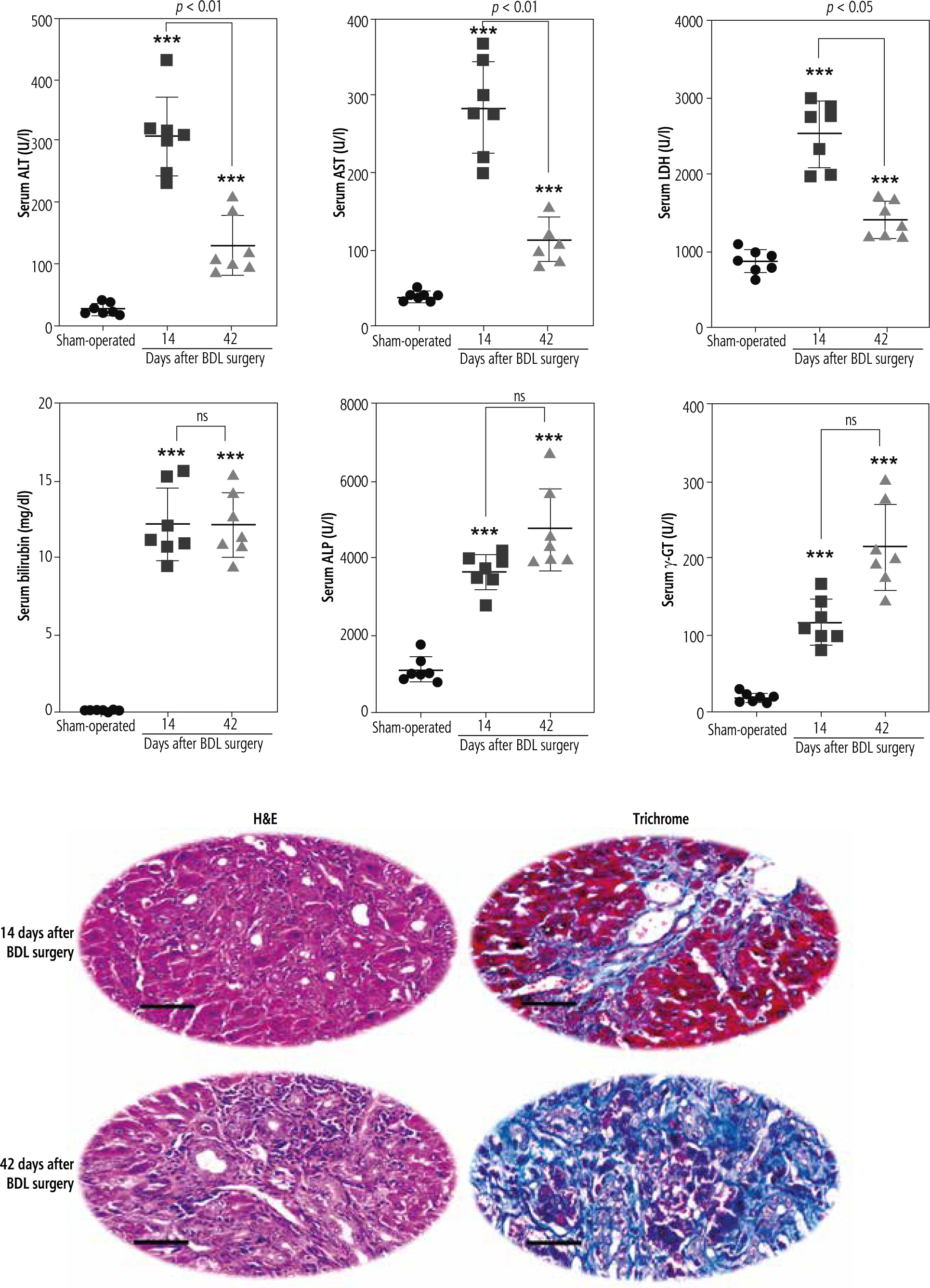
Assessment of serum biomarkers revealed a significant increase in alanine aminotransferase (ALT), aspartate aminotransferase (AST), and lactate dehydrogenase (LDH) in BDL animals compared with the sham-operated group (Fig. 1). Moreover, serum bilirubin, alkaline phosphatase (ALP), and γ-glutamyltranspeptidase (γ-GT) drastically increased at different time intervals after the BDL operation, although there was no significant difference between 14- and 42-day groups. It should be mentioned that the level of biomarkers such as ALP, γ-GT, and bilirubin is constantly high in the BDL model of cholestasis due to permanent ligation of the bile duct. Furthermore, tissue histopathological alterations, including bile duct proliferation, necrosis, and inflammatory cell infiltration, as well as significant collagen deposition (trichrome stain), were detected in the liver of BDL animals (Fig. 1). These data indicate the appropriate induction of cholestasis/cirrhosis in the current study.

Fig. 1

Liver tissue histopathological alterations and serum biomarkers indicate appropriate induction of cholestasis/cirrhosis in bile duct ligated (BDL) rats. Bile duct proliferation and inflammatory cell infiltration were detected in the liver of cholestatic animals (H&E stain). Scale bar = 100 µm. Moreover, significant collagen deposition (blue area in Masson-trichrome stain) indicates liver fibrotic areas. Data are presented as mean ± SD (n = 7). ***Indicates significantly different from the sham-operated group (p < 0.001). ns – not significant

/f/fulltexts/CEH/47739/CEH-8-47739-g001_min.jpg

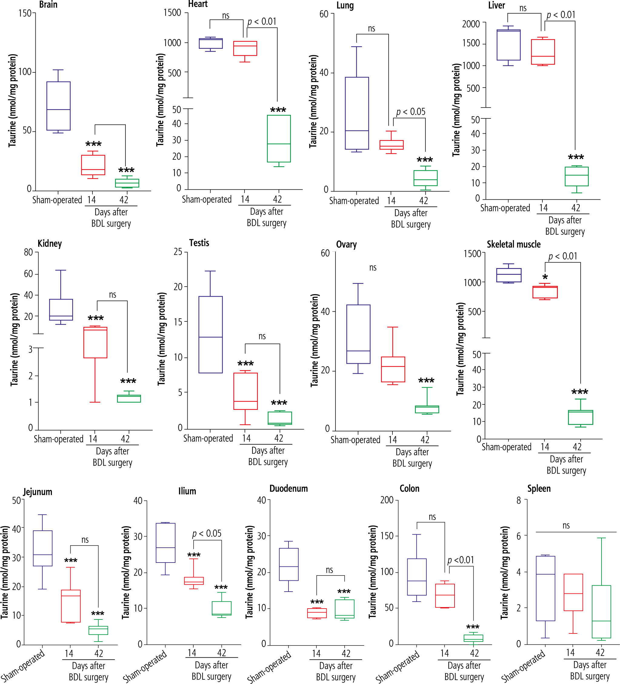
TAU levels were assessed in various tissues of cholestatic rats 14 and 42 days after the BDL operation. Significant depletion in TAU content was evident in most tissues 14 days after BDL. On the other hand, the TAU content of all tissues was drastically decreased 42 days after the BDL surgery. As TAU depletion was not significant at 14 days after BDL operation, this parameter was not time-dependent in tissues such as the liver, ovary, lung, heart, and colon (Fig. 2).

Fig. 2

Tissue taurine levels in bile duct ligated (BDL) rats. Data are shown as box and whiskers (min to max). Asterisks indicate significantly different as compared with the sham-operated group (*p < 0.05 and ***p < 0.001). ns – not significant

/f/fulltexts/CEH/47739/CEH-8-47739-g002_min.jpg

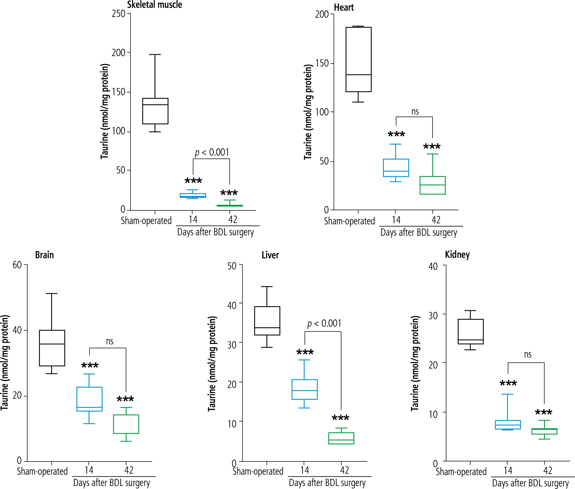
Mitochondrial TAU content of tissues, including skeletal muscle, brain, heart, liver, and kidney, significantly decreased at both intervals of 14 and 42 days after the BDL operation. The decrease in TAU levels of mitochondria was not time-dependent in most tissues (heart, brain, and kidney) assessed in the current study (Fig. 3).

Fig. 3

Mitochondrial taurine levels in cholestatic/cirrhotic animals. Data are shown as box and whiskers (min to max). Asterisks indicate significantly different as compared with the sham-operated group (***p < 0.001). ns – not significant

/f/fulltexts/CEH/47739/CEH-8-47739-g003_min.jpg

Discussion

Taurine is a very safe amino acid abundantly found in the human body. Various experimental models have highlighted the physiological and pharmacological roles for TAU [1, 2]. However, the role of TAU deficiency in the pathogenesis of many human diseases is far from clear. The data obtained from the current investigation revealed a significant decrease in TAU content of several tissues as well as mitochondria in BDL rats as a reliable animal model of cholestasis/cirrhosis. These findings indicate a pivotal role for tissue and mitochondrial TAU in the pathogenesis of cholestasis/cirrhosis-induced organ injury. As TAU plays a viable function as an osmolyte in various tissues [1, 2], and most importantly is crucial for mitochondrial function and energy metabolism [12, 17, 22, 25-28], significant alteration in its levels could play pathogenic roles in cholestasis/cirrhosis-linked complications.

The effects of TAU on mitochondrial indices are an exciting feature of this amino acid [12, 17, 25-27]. TAU is localized in mitochondria via specific transporters [83, 84]. Moreover, some studies also found that TAU could be synthesized in the mitochondrial matrix [85]. These data indicate the importance of TAU in mitochondrial function. Therefore, disruption of the fundamental processes such as energy metabolism could accompany cellular TAU depletion. Based on these data, investigating the TAU level in cholestasis/cirrhosis could enhance our understanding of the mechanism of organ injury in these pathological conditions and provide viable therapeutic options.

Oxidative stress is a well-known phenomenon in various tissues of cholestatic animals [34, 62, 86-89]. Oxidative stress could damage multiple cellular targets, including proteins, lipids, and essential organelles such as mitochondria [31, 35, 88, 90-97]. It is well known that oxidative stress is a general phenomenon in cholestasis [34, 87, 88]. On the other hand, mitochondria are the major sources of intracellular ROS [98]. Interestingly, various investigations have mentioned the role of TAU in preventing mitochondria-facilitated ROS formation and oxidative stress [19, 20, 22]. The role of TAU in mitochondrial tRNA structure and function is an interesting feature of this amino acid [15, 26, 83, 99-102]. It is well known that the proper modification of mitochondrial tRNA structure by TAU leads to appropriate synthesis and function of mitochondrial proteins (e.g., mitochondrial respiratory chain complexes) [15, 26, 83, 99-102]. Thus, the impact of TAU in the appropriate synthesis and function of mitochondria respiratory chain complexes is a fundamental role of this compound in preventing mitochondria-mediated oxidative stress [19, 20]. In our recent studies on cholestatic animals, we repeatedly found that TAU supplementation could improve mitochondrial function and blunt oxidative stress in various organs [23, 103, 104]. Based on these data, TAU depletion in various organs during cholestasis/cirrhosis is directly connected to the occurrence of oxidative stress and its linked complications.

Some studies indicate that TAU deficiency mediates apoptosis and cell death through a mitochondria-dependent pathway [28]. Interestingly, it has been found that TAU deficiency could lead to the induction of mitochondrial permeability transition pore (mPT) [105]. Jong et al. revealed that mitochondrial TAU content is directly associated with increased cell apoptosis [105]. mPT induction could cause the release of cell death mediators (e.g., cytochrome c) from this organelle [105]. The release of other cell death mediators such as apoptosis-inducing factor (AIF) also has been reported in different tissues of cholestatic animals [53]. However, more studies are needed to identify a connection between mitochondrial TAU deficiency and the release of such mediators from mitochondria.

It has been well documented that collagen deposition and fibrosis are important events in the liver, kidney, lung, and heart during cholestasis/cirrhosis [47, 106-110]. Tissue fibrosis results from a complex process connected to oxidative stress [111]. On the other hand, mitochondria are significant sources of intracellular ROS [98]. Therefore, mitochondrial impairment could play a pathogenic role in tissue fibrosis during cholestasis. It has been reported that protecting cellular mitochondria could significantly prevent liver injury and fibrosis in an experimental model of cirrhosis [112]. Pérez et al. found that mitochondria-mediated cell death could play a crucial role in liver injury during cirrhosis [112]. These investigations highlight the importance of mitochondrial impairment in chronic liver disease, leading to fibrotic lesions. The antifibrotic properties of TAU have also been frequently mentioned in various tissues [113-117]. Previous investigations indicated that liver mitochondrial function was severely impaired in the BDL model of hepatic fibrosis [33, 118-120]. On the other hand, it has been found that TAU could significantly enhance mitochondrial function and prevent mitochondria-facilitated ROS formation and oxidative stress [103, 121]. Therefore, we might be able to hypothesize that a part of the antifibrotic effects of TAU could be mediated through mitochondrial-dependent mechanisms.

In the following parts, the role of TAU deficiency and its potential link with organ injury reported in cholestasis/cirrhosis (liver, brain, heart, kidney, skeletal muscle, and intestinal damage) is discussed in the context of the fundamental role of this amino acid in mitochondrial function and mitigating oxidative stress.

It has been found that TAU had significant hepatoprotective properties in both experimental models and human cases of cholestasis/cirrhosis [122-124]. For example, the effect of TAU on portal hypertension is an exciting feature of this amino acid [122-124]. The impacts of TAU on the morphology of the liver and biomechanical properties of the portal vein seem to be involved in its effects on portal hypertension in cirrhotic patients [122-124]. It is well known that the effects of TAU in mitigating oxidative stress and its associated complications in the liver play a pivotal role in its hepatoprotective properties [70, 103, 121, 125-135]. On the other hand, the effects of TAU on hepatocytes’ mitochondrial function have also been repeatedly investigated [33, 103, 118-121, 136-138]. It is well established that TAU could enhance mitochondria energy metabolism, prevent mitochondrial permeabilization, and blunt mitochondria-mediated cell death in experimental models of hepatic injury [33, 103, 118-121, 136, 137]. These data indicate that TAU is an excellent and safe compound for managing liver dysfunction as the main complication in cholestasis/cirrhosis. The current study found that liver tissue and hepatocytes’ mitochondrial TAU content were significantly depleted in cholestatic/cirrhotic animals (Figs. 2 and 3). Therefore, TAU supplementation could be a viable therapeutic option to blunt liver injury during cholestasis/cirrhosis.

Brain injury is a critical complication in cholestasis/cirrhosis [139, 140]. Several mechanisms have been proposed for cholestasis/cirrhosis-induced brain injury [139-142]. The first, and probably the most important one, is the disruption of the urea cycle in the liver and the accumulation of ammonia in plasma and brain tissue [139]. Ammonia is a neurotoxin, and it has been repeatedly mentioned that this agent is responsible for neuronal damage, cognitive dysfunction, brain edema, and coma in cirrhotic patients [139]. Some other compounds, such as bilirubin and manganese (Mn), also accumulate in different tissues such as the brain during cholestasis [139]. The mentioned compounds are well-known neurotoxicants [139]. Oxidative stress is a common feature of cholestasis/cirrhosis-induced brain injury [139, 143, 144]. Also, mitochondrial injury seems to play a crucial role in the pathogenesis of these complications [139]. All bile constituents such as Mn, bilirubin, and bile acids have adverse effects on mitochondrial function and energy metabolism [139, 145, 146]. In the current study, we discovered a significant decline in the TAU content of the brain tissue and isolated mitochondria (Figs. 2 and 3). On the other hand, a plethora of evidence indicates that this amino acid dramatically alleviates oxidative stress and enhances mitochondrial function in various experimental models of brain injury [147-149]. Our previous studies also found that TAU could improve brain mitochondrial function, mitigate biomarkers of oxidative stress, and enhance animals’ locomotor activity [70, 76, 103]. These data suggest that cholestatic/cirrhotic patients could benefit from the neuroprotective properties of TAU.

Sarcopenia or muscle mass loss is a serious complication in cirrhotic patients, leading to severe disability [51, 150-152]. Cirrhosis-induced sarcopenia could also significantly influence the outcome of therapeutic interventions such as liver transplantation [150, 153]. Several studies have mentioned that mitochondrial impairment and oxidative stress are crucial mechanisms involved in the pathogenesis of sarcopenia-induced muscle injury [51]. The effects of TAU on muscle function are another critical feature of this amino acid [24, 154-158]. TAU is found at very high concentrations in the skeletal muscle [155, 157]. It has been found that the effect of TAU on mitochondrial function and energy metabolism is an essential feature of this amino acid in the skeletal muscle [9, 20, 24, 159]. Therefore, decreased muscle TAU level is associated with muscle dysfunction [9, 24]. Some studies have revealed that TAU transporter knock-out animals exhibited a significant decrease in skeletal muscle mass and function [9, 160]. Interestingly, changes in mitochondrial morphology of the skeletal muscle have also been detected in the ultrastructural analysis of the tissue samples from TAU deficient models [8, 9]. On the other hand, several studies indicate that TAU could significantly enhance muscle performance [24, 69, 161-165]. These events indicate that TAU plays a crucial role in skeletal muscle function. A big part of TAU effects on the skeletal muscle is mediated through this amino acid’s impact on mitochondrial function and energy metabolism [24, 69, 161, 165]. In the current study, we detected that TAU levels in skeletal muscle tissue and mitochondria were significantly decreased at different time intervals after BDL surgery (Figs. 2 and 3); thus, TAU deficiency-associated mitochondrial impairment could play a critical role in muscle injury induced by cholestasis/cirrhosis. Therefore, the administration of this amino acid could be a viable strategy for managing cirrhosis-associated muscle dysfunction and sarcopenia.

Cardiac contraction abnormalities, decreased cardiac output, and heart failure could occur in cholestasis/cirrhosis [166-170]. Cardiac arrhythmia is also a com-mon pathological finding in cirrhotic patients [171, 172]. Previous studies also indicate oxidative stress, inflammation, and mitochondrial impairment in the heart tissue during cirrhosis [23, 173-175]. On the other hand, several studies suggest that TAU deficiency is associated with cardiac abnormalities [7, 176]. It has been found that pathological conditions such as cardiac atrophy and heart failure accompanied TAU deficiency [7, 176]. Recent studies also mentioned that essential pathways involved in tissue energy metabolism, such as fatty acid oxidation, are suppressed in the cardiac muscle under TAU deficiency conditions [177]. Interestingly, in TAU transporter knockout models, ultrastructural changes in cardiomyocytes’ mitochondria have been detected [7]. Recently we found that TAU could significantly improve mitochondrial function and blunted oxidative stress in the heart tissue of cirrhotic animals [178]. Moreover, it has been repeatedly reported that TAU could normalize various types of arrhythmia [179]. The effects of TAU on cirrhosis-induced arrhythmia could be an interesting subject for future investigations. The current study detected that TAU levels dropped significantly in cardiac tissue and isolated mitochondria (Figs. 2 and 3). All these data support the positive impact of TAU on cardiac function. Therefore, compensating for TAU deficiency could play an essential role in blunting adverse cardiac events in cholestatic/cirrhotic patients.

The adverse effect of cholestasis/cirrhosis on renal function is another essential subject widely investigated [93, 180-184]. At the early stages of bile duct obstruction, several potentially cytotoxic molecules such as bilirubin and bile acids are suspected to be responsible for direct renal injury [38, 71, 184]. Cholemic nephropathy is a phenomenon developed as an early response to cholestasis [38]. Bile cast formation, tubular damage, tissue necrosis, and inflammatory cell infiltration have been detected in cholemic nephropathy [38, 45, 181]. Renal fibrosis could also occur in cholestasis [38]. On the other hand, cirrhosis-induced renal injury is mainly developed as a hepatorenal syndrome [185]. In addition to histopathological changes detected in cholemic nephropathy, progressive tissue fibrosis, severe alteration in renal vasculature, hemodynamic changes, and renal failure are common in hepatorenal syndrome [185]. Mechanistically, oxidative stress and its related events seem to play a fundamental role in the pathogenesis of cholemic nephropathy and hepatorenal syndrome [38, 71, 90, 186, 187]. Additionally, several investigations have revealed the importance of mitochondrial impairment in the mechanism of cholestasis-induced renal injury [60, 188]. Mitochondrial impairment in the kidney could lead to an energy crisis and subsequent impairment of high energy demand processes such as reabsorption of chemicals in the renal tubules [189, 190]. This phenomenon is known as the Fanconi syndrome [90]. There are reports of the Fanconi syndrome linked with cholestasis/cirrhosis [92, 191-193]. Several studies suggested the therapeutic role of antioxidants against cholestasis-induced renal injury [39, 125, 194, 195]. Recently, we found that TAU administration to cholestatic rats could significantly alleviate cholemic nephropathy in BDL animals [104]. The effects of TAU on mitochondrial parameters and oxidative stress seem to be the primary mechanism for its renoprotective properties in cholestatic rats [104]. On the other hand, the effects of TAU on the renal blood flow, osmoregulation, glomeruli filtration rate, and ion absorption and secretion are mentioned as the physiological roles of this amino acid in the renal system [196, 197]. Several studies have revealed the positive effects of TAU on various types of renal disorders and xenobiotics-induced renal injury [126, 127, 197-204]. In the current study, we found that tissue and mitochondrial TAU levels were dramatically decreased in the kidney of BDL rats (Figs. 2 and 3). Therefore, TAU deficiency could play a significant role in the pathogenesis of oxidative stress, mitochondrial dysfunction, and renal injury in cholestasis/cirrhosis. Based on these data, TAU supplementation could serve as a therapeutic option to mitigate cholestasis/cirrhosis-linked renal impairment.

The role of TAU in reproductive system function, in both males and females, is also the subject of many investigations in this field [205-214]. On the other hand, it has been found that reproductive organs are severely damaged during cholestasis [59]. Oxidative stress and mitochondrial impairment seem to play a pivotal role in the mechanisms of reproductive system injury in cholestasis [59]. Meanwhile, it is well established that the effects of TAU in counteracting oxidative stress and its related complications are a major mechanism for the protective properties of this amino acid in reproductive organs [214, 215]. It has also been reported that TAU could significantly improve parameters such as sperm motility, sperm antioxidant levels, ATP content, and sperm capacitation [205, 213, 216-220]. TAU also could regulate the synthesis and release of important hormones such as testosterone and luteinizing hormone (LH) [213]. All these data highlight the crucial role of TAU in the reproductive system. In the current study, we found that testis and ovary TAU levels were significantly depleted in cholestatic rats (Fig. 2). Therefore, TAU depletion could be linked with poor reproductive factors in males and females. In this context, TAU supplementation could be considered as a strategy to protect reproductive organs in cholestasis/cirrhosis.

Several parameters could be involved in the mechanism of tissue and mitochondrial TAU depletion in cholestasis/cirrhosis. First, and most notably in the current model, the TAU synthesis is disrupted in the liver during cholestasis due to severe hepatic injury (Fig. 4). This could be the leading cause of perturbed TAU levels in rodent experimental models, as the liver is the main organ synthesizing this amino acid. However, humans have relied on dietary TAU, and the amount of TAU synthesis in the liver is negligible. Therefore, other factors such as disturbed absorbance of this amino acid could also lower TAU levels in cholestasis/cirrhosis. In the current study, the intestinal (duodenum, ileum, and jejunum) TAU level was significantly depleted in BDL rats. It is well known that TAU is a vital osmolyte that preserves enterocyte integrity [221, 222]. Hence, a depleted TAU level in the intestine could disturb the absorbance of many nutrients, including TAU itself (Fig. 4). These data indicate that TAU supplementation could protect intestinal tissue and prevent disturbances in the process of absorption of vital compounds into the bloodstream. On the other hand, these data suggest that future studies on the clinical administration of TAU to cholestatic/cirrhotic patients should be through the parenteral route because of intestinal damage in these patients.

Fig. 4

Taurine is synthesized in the liver of some species, such as rats and dogs, by utilizing methionine and cysteine amino acids. Some species such as foxes and felines cannot synthesize taurine endogenously and completely depend on the dietary sources of this amino acid. The taurine synthesis capability of the human liver is also negligible. The current study found that taurine level in various tissues and their isolated mitochondria was significantly depleted by cholestasis/cirrhosis. Taurine synthesis capability synthesis of the liver or its absorption through the intestinal brush border could be affected by cholestasis/cirrhosis. Lower taurine levels could lead to deleted amino acid stores in various organs and cause organ injury. These data could support the importance of strategies such as taurine supplementation in cholestatic/cirrhotic patients

/f/fulltexts/CEH/47739/CEH-8-47739-g004_min.jpg

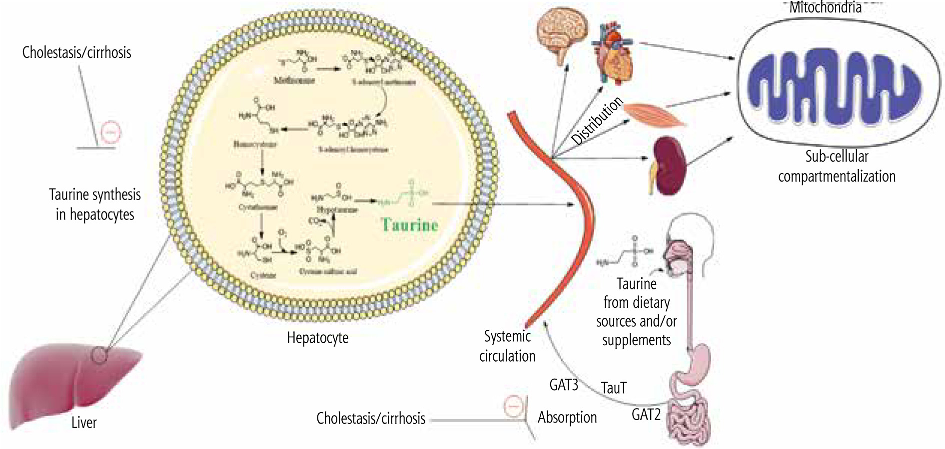
Another potential mechanism for decreased TAU levels of tissues and mitochondria in cholestasis/cirrhosis could be related to the changes in the expression and/or activity of TAU transporters (TauT). The cellular and mitochondrial uptake of TAU is mediated via specific TauT [83, 84]. Hence, monitoring the changes of these transporters during cholestasis/cirrhosis could give a better insight into the mechanisms of TAU depletion in various tissues (Fig. 4).

Cases of TAU deficiency have also been previously reported in association with different pathologies. It has been found that situations such as long-term paraenteral nutrition or acute exposure to cytotoxic agents (e.g., cancer chemotherapy regimens) could lead to TAU deficiency [223-225]. Today, the importance of TAU in regulating the physiological function of different systems is entirely approved by many experimental and clinical data. The current study found that tissue and mitochondrial TAU levels were significantly depleted during cholestasis/cirrhosis. Therefore, TAU supplementation could preserve various organs in a more functional state.

The safety of TAU and its application in critically ill patients (e.g., cirrhotic patients) is another subject that should be considered before the application of this amino acid in clinical settings. Fortunately, there are many studies on the application of TAU against various human diseases in clinic [226-228]. It has been found that TAU could be administered to humans at very high doses (e.g., 6-12 g/day) without any considerable side effect [228]. Interestingly, some studies demonstrated that TAU could be administered at high doses in disorders such as hepatic encephalopathy and cirrhotic patients (e.g., for controlling portal hypertension) [122]. These data indicate that TAU could be safely administered in clinical cases of cholestasis/cirrhosis. However, determining the long-term effects of TAU therapy in cholestatic/cirrhotic patients requires further studies.

The data obtained from the present research could provide an insight into the relevance of TAU deficiency in the pathogenesis of cholestasis/cirrhosis-induced organ damage and suggest a viable therapeutic option in patients. More studies, including clinical trials, could reveal the importance of TAU therapy in managing cholestasis/cirrhosis-induced complications.

Disclosure

The authors declare no conflict of interest.

References

1 

Schaffer SW, Lombardini JB, Huxtable RJ. Taurine 3: cellular and regulatory mechanisms. Springer Science & Business Media, 2013.

2 

Stapleton PP, Charles RP, Redmond HP, Bouchier-Hayes DJ. Taurine and human nutrition. Clin Nutr 1997; 16: 103-108.

3 

Huxtable RJ, Michalk D. Taurine in health and disease. Springer Science & Business Media, 2013.

4 

Yamori Y, Taguchi T, Hamada A, et al. Taurine in health and diseases: consistent evidence from experimental and epidemiological studies. J Biomed Sci 2010; 17 Suppl 1: S6.

5 

Pasantes-Morales H, Quesada O, Morán J. Taurine: an osmolyte in mammalian tissues. In: Schaffer S, Lombardini JB, Huxtable RJ (Eds.). Taurine 3: cellular and regulatory mechanisms. Springer US, Boston, MA 1998; 209-217.

6 

Ito T, Hanahata Y, Kine K, et al. Tissue taurine depletion inducesprofibrotic pattern of gene expression and causes aging-related cardiac fibrosis in heart in mice. Biol Pharm Bull 2018; 41: 1561-1566.

7 

Ito T, Kimura Y, Uozumi Y, et al. Taurine depletion caused by knocking out the taurine transporter gene leads to cardiomyopathy with cardiac atrophy. J Mol Cell Cardiol 2008; 44: 927-937.

8 

Ito T, Oishi S, Takai M, et al. Cardiac and skeletal muscle abnormality in taurine transporter-knockout mice. J Biomed Sci 2010; 17: S20.

9 

Ito T, Yoshikawa N, Inui T, et al. Tissue depletion of taurine accelerates skeletal muscle senescence and leads to early death in mice. PLoS One 2014; 9: e107409.

10 

Moise NS, Pacioretty LM, Kallfelz FA, et al. Dietary taurine deficiency and dilated cardiomyopathy in the fox. Am Heart J 1991; 121: 541-547.

11 

Pion PD, Kittleson MD, Skiles ML, et al. Dilated cardiomyopathy associated with taurine deficiency in the domestic cat: relationship to diet and myocardial taurine content. Adv Exp Med Biol 1992; 315: 63-73.

12 

Hansen SH, Grunnet N. Taurine, glutathione and bioenergetics. In: Idrissi AE, L’Amoreaux WJ (Eds.). Springer, New York 2013; 3-12.

13 

Huxtable RJ. Taurine in nutrition and neurology. Vol. 139. Springer Science & Business Media, 2013.

14 

Bouckenooghe T, Remacle C, Reusens B. Is taurine a functional nutrient? Curr Opin Clin Nutr Metab Care 2006; 9: 728-733.

15 

Lambert IH, Kristensen DM, Holm JB, Mortensen OH. Physiological role of taurine–from organism to organelle. Acta Physiologica 2015; 213: 191-212.

16 

El Idrissi A, Trenkner E. Taurine regulates mitochondrial calcium homeostasis. Adv Exp Med Biol 2003; 526: 527-536.

17 

Hansen SH, Andersen ML, Birkedal H, et al. The important role of taurine in oxidative metabolism. Adv Exp Med Biol 2006; 583: 129-135.

18 

Hansen SH, Andersen ML, Cornett C, et al. A role for taurine in mitochondrial function. J Biomed Sci 2010; 17: 1-8.

19 

Jong CJ, Azuma J, Schaffer S. Mechanism underlying the antioxidant activity of taurine: prevention of mitochondrial oxidant production. Amino Acids 2011; 42: 2223-2232.

20 

Schaffer S, Kim HW. Effects and mechanisms of taurine as a therapeutic agent. Biomol Ther (Seoul) 2018; 26: 225-241.

21 

Heidari R, Ghanbarinejad V, Ommati MM, et al. Mitochondria protecting amino acids: Application against a wide range of mitochondria-linked complications. PharmaNutrition 2018; 6: 180-190.

22 

Mohammadi H, Ommati MM, Farshad O, et al. Taurine and isolated mitochondria: a concentration-response study. Trend Pharm Sci 2019; 5: 197-206.

23 

Abdoli N, Sadeghian I, Mousavi K, et al. Suppression of cirrhosis-related renal injury by N-acetyl cysteine. Curr Res Pharmacol Drug Discov 2020; 1: 30-38.

24 

Ommati MM, Farshad O, Jamshidzadeh A, Heidari R. Taurine enhances skeletal muscle mitochondrial function in a rat model of resistance training. PharmaNutrition 2019; 9: 100161.

25 

Chen WQ, Jin H, Nguyen M, et al. Role of taurine in regulation of intracellular calcium level and neuroprotective function in cultured neurons. J Neurosci Res 2001; 66: 612-619.

26 

Fakruddin M, Wei FY, Suzuki T, et al. Defective mitochondrial tRNA taurine modification activates global proteostress and leads to mitochondrial disease. Cell Rep 2018; 22: 482-496.

27 

Hansen SH, Birkedal H, Wibrand F, Grunnet N. Taurine and regulation of mitochondrial metabolism. Adv Exp Med Biol 2015; 803: 397-405.

28 

Jong CJ, Ito T, Prentice H, et al. Role of mitochondria and endoplasmic reticulum in taurine-deficiency-mediated apoptosis. Nutrients 2017; 9: 795.

29 

Carey EJ, Lindor KD. Cholestatic liver disease. Springer, New York 2014.

30 

Jüngst C, Lammert F. Cholestatic liver disease. Dig Dis 2013; 31: 152-154.

31 

Orellana M, Rodrigo R, Thielemann L, Guajardo V. Bile duct ligation and oxidative stress in the rat: effects in liver and kidney. Comp Biochem Physiol 2000; 126: 105-111.

32 

O’Brien A, China L, Massey KA, et al. Bile duct-ligated mice exhibit multiple phenotypic similarities to acute decompensation patients despite histological differences. Liver Int 2016; 36: 837-846.

33 

Heidari R, Niknahad H, Sadeghi A, et al. Betaine treatment protects liver through regulating mitochondrial function and counteracting oxidative stress in acute and chronic animal models of hepatic injury. Biomed Pharmacother 2018; 103: 75-86.

34 

Copple BL, Jaeschke H, Klaassen CD. Oxidative stress and the pathogenesis of cholestasis. Semin Liver Dis 2010; 30: 195-204.

35 

Huang LT, Tiao MM, Tain YL, et al. Melatonin ameliorates bile duct ligation-induced systemic oxidative stress and spatial memory deficits in developing rats. Pediatr Res 2009; 65: 176-180.

36 

Shikata F, Sakaue T, Nakashiro KI, et al. Pathophysiology of lung injury induced by common bile duct ligation in mice. PLoS One 2014; 9: e94550.

37 

Bosoi CR, Oliveira MM, Ochoa-Sanchez R, et al. The bile duct ligated rat: A relevant model to study muscle mass loss in cirrhosis. Metab Brain Dis 2017; 32: 513-518.

38 

Heidari R, Mandegani L, Ghanbarinejad V, et al. Mitochondrial dysfunction as a mechanism involved in the pathogenesis of cirrhosis-associated cholemic nephropathy. Biomed Pharmacother 2019; 109: 271-280.

39 

Heidari R, Ghanbarinejad V, Mohammadi H, et al. Dithiothreitol supplementation mitigates hepatic and renal injury in bile duct ligated mice: Potential application in the treatment of cholestasis-associated complications. Biomed Pharmacother 2018; 99: 1022-1032.

40 

Martínez-Cecilia D, Reyes-Díaz M, Ruiz-Rabelo J, et al. Oxidative stress influence on renal dysfunction in patients with obstructive jaundice: a case and control prospective study. Redox Biol 2016; 8: 160-164.

41 

Ommati MM, Farshad O, Mousavi K, et al. Betaine supplementation mitigates intestinal damage and decreases serum bacterial endotoxin in cirrhotic rats. PharmaNutrition 2020; 12: 100179.

42 

Ommati MM, Farshad O, Niknahad H, et al. Oral administration of thiol-reducing agents mitigates gut barrier disintegrity and bacterial lipopolysaccharide translocation in a rat model of biliary obstruction. Curr Res Pharmacol Drug Discov 2020; 1: 10-18.

43 

Kountouras J, Billing BH, Scheuer PJ. Prolonged bile duct obstruction: a new experimental model for cirrhosis in the rat. Br J Exp Pathol 1984; 65: 305-311.

44 

Rodríguez-Garay EA. Cholestasis: human disease and experimental animal models. Ann Hepatol 2003; 2: 150-158.

45 

Fickert P, Krones E, Pollheimer MJ, et al. Bile acids trigger cholemic nephropathy in common bile-duct-ligated mice. Hepatology 2013; 58: 2056-2069.

46 

Tag CG, Sauer-Lehnen S, Weiskirchen S, et al. Bile duct ligation in mice: induction of inflammatory liver injury and fibrosis by obstructive cholestasis. J Vis Exp 2015; 96: 52438.

47 

Tag CG, Weiskirchen S, Hittatiya K, et al. Induction of experimental obstructive cholestasis in mice. Lab Anim 2015; 49: 70-80.

48 

Heidari R, Mohammadi H, Ghanbarinejad V, et al. Proline supplementation mitigates the early stage of liver injury in bile duct ligated rats. J Basic Clin Physiol Pharmacol 2019; 30: 91-101.

49 

Abdoli N, Sadeghian I, Mousavi K, et al. Suppression of cirrhosis-related renal injury by N-acetyl cysteine. Curr Res Pharmacol Drug Discov 2020; 1: 30-38.

50 

Dhanda S, Sunkaria A, Halder A, Sandhir R. Mitochondrial dysfunctions contribute to energy deficits in rodent model of hepatic encephalopathy. Metab Brain Dis 2018; 33: 209-223.

51 

Farshad O, Ommati MM, uuml, et al. Skeletal muscle mitochondrial impairment in cirrhosis-induced sarcopenia. Trend Pharm Sci 2020; 6: 189-204.

52 

Farshad O, Ommati MM, Yüzügülen J, et al. Carnosine mitigates biomarkers of oxidative stress, improves mitochondrial function, and alleviates histopathological alterations in the renal tissue of cholestatic rats. Pharm Sci 2021; 27: 32-45.

53 

Ghanbarinejad V, Jamshidzadeh A, Khalvati B, et al. Apoptosis-inducing factor plays a role in the pathogenesis of hepatic and renal injury during cholestasis. Naunyn Schmiedebergs Arch Pharmacol 2021; 394: 1191-1203.

54 

Heidari R, Ahmadi A, Ommati MM, Niknahad H. Methylene blue improves mitochondrial function in the liver of cholestatic rats. Trend Pharm Sci 2020; 6: 73-86.

55 

Ommati MM, Attari H, Siavashpour A, et al. Mitigation of cholestasis-associated hepatic and renal injury by edaravone treatment: evaluation of its effects on oxidative stress and mitochondrial function. Liver Res 2020; 5: 181-193.

56 

Ommati MM, Farshad O, Azarpira N, et al. Silymarin mitigates bile duct obstruction-induced cholemic nephropathy. Naunyn Schmiedebergs Arch Pharmacol 2021; 394: 1301-1314.

57 

Ommati MM, Farshad O, Azarpira N, et al. Betaine alleviates cholestasis-associated renal injury by mitigating oxidative stress and enhancing mitochondrial function. Biologia 2021; 76: 351-365.

58 

Ommati MM, Farshad O, Mousavi K, et al. Agmatine alleviates hepatic and renal injury in a rat model of obstructive jaundice. PharmaNutrition 2020; 13: 100212.

59 

Ommati MM, Farshad O, Niknahad H, et al. Cholestasis-associated reproductive toxicity in male and female rats: The fundamental role of mitochondrial impairment and oxidative stress. Toxicol Lett 2019; 316: 60-72.

60 

Ommati MM, Mohammadi H, Mousavi K, et al. Metformin alleviates cholestasis-associated nephropathy through regulating oxidative stress and mitochondrial function. Liver Res 2020; 5: 171-180.

61 

Shen K, Feng X, Su R, et al. Epigallocatechin 3-gallate ameliorates bile duct ligation induced liver injury in mice by modulation of mitochondrial oxidative stress and inflammation. PLoS One 2015; 10: e0126278.

62 

Siavashpour A, Khalvati B, Azarpira N, et al. Poly (ADP-Ribose) polymerase-1 (PARP-1) overactivity plays a pathogenic role in bile acids-induced nephrotoxicity in cholestatic rats. Toxicol Lett 2020; 330: 144-158.

63 

Ghanbarinejad V, Ommati MM, Jia Z, et al. Disturbed mitochondrial redox state and tissue energy charge in cholestasis. J Biochem Mol Toxicol 2021; 35: e22846.

64 

Mousavi K, Niknahad H, Li H, et al. The activation of nuclear factor-E2-related factor 2 (Nrf2)/heme oxygenase-1 (HO-1) signaling blunts cholestasis-induced liver and kidney injury. Toxicol Res 2021; 10: 911-927.

65 

Ahmadi A, Niknahad H, Li H, et al. The inhibition of NFкB signaling and inflammatory response as a strategy for blunting bile acid-induced hepatic and renal toxicity. Toxicol Lett 2021; 349: 12-29.

66 

Ommati MM, Heidari R, Ghanbarinejad V, et al. Taurine treatment provides neuroprotection in a mouse model of manganism. Biol Trace Elem Res 2019; 190: 384-395.

67 

Emadi E, Abdoli N, Ghanbarinejad V, et al. The potential role of mitochondrial impairment in the pathogenesis of imatinib-induced renal injury. Heliyon 2019; 5: e01996.

68 

Moezi L, Heidari R, Amirghofran Z, et al. Enhanced anti-ulcer effect of pioglitazone on gastric ulcers in cirrhotic rats: the role of nitric oxide and IL-1b. Pharmacol Rep 2013; 65: 134-143.

69 

Heidari R, Jamshidzadeh A, Ghanbarinejad V, et al. Taurine supplementation abates cirrhosis-associated locomotor dysfunction. Clin Exp Hepatol 2018; 4: 72-82.

70 

Heidari R, Jamshidzadeh A, Niknahad H, et al. Effect of taurine on chronic and acute liver injury: Focus on blood and brain ammonia. Toxicol Report 2016; 3: 870-879.

71 

Heidari R, Niknahad H. The role and study of mitochondrial impairment and oxidative stress in cholestasis. In: Vinken M (Ed.). Experimental cholestasis research. Springer, New York, NY 2019; 117-132.

72 

Ommati MM, Heidari R, Manthari RK, et al. Paternal exposure to arsenic resulted in oxidative stress, autophagy, and mitochondrial impairments in the HPG axis of pubertal male offspring. Chemosphere 2019; 236: 124325.

73 

Ommati MM, Manthari RK, Tikka C, et al. Arsenic-induced autophagic alterations and mitochondrial impairments in hpg-s axis of mature male mice offspring (f1-generation): a persistent toxicity study. Toxicol Lett 2020; 326: 83-98.

74 

Ommati MM, Shi X, Li H, et al. The mechanisms of arsenic-induced ovotoxicity, ultrastructural alterations, and autophagic related paths: an enduring developmental study in folliculogenesis of mice. Ecotoxicol Environ Saf 2020; 204: 110973.

75 

Jamshidzadeh A, Niknahad H, Heidari R, et al. Propylthiouracil-induced mitochondrial dysfunction in liver and its relevance to drug-induced hepatotoxicity. Pharm Sci 2017; 23: 95-102.

76 

Niknahad H, Jamshidzadeh A, Heidari R, et al. Ammonia-induced mitochondrial dysfunction and energy metabolism disturbances in isolated brain and liver mitochondria, and the effect of taurine administration: relevance to hepatic encephalopathy treatment. Clin Exp Hepatol 2017; 3: 141-151.

77 

Jamshidzadeh A, Niknahad H, Heidari R, et al. Carnosine protects brain mitochondria under hyperammonemic conditions: relevance to hepatic encephalopathy treatment. PharmaNutrition 2017; 5: 58-63.

78 

Ommati MM, Manthari RK, Tikka C, et al. Arsenic-induced autophagic alterations and mitochondrial impairments in HPG-S axis of mature male mice offspring (F1-generation): a persistent toxicity study. Toxicol Lett 2020; 326: 83-98.

79 

Niknahad H, Jamshidzadeh A, Heidari R, et al. Paradoxical effect of methimazole on liver mitochondria: in vitro and in vivo. Toxicol Lett 2016; 259: 108-115.

80 

Ommati MM, Heidari R, Ghanbarinejad V, et al. The neuroprotective properties of carnosine in a mouse model of manganism is mediated via mitochondria regulating and antioxidative mechanisms. Nutr Neurosci 2020; 23: 731-743.

81 

Niknahad H, Jamshidzadeh A, Heidari R, et al. The postulated hepatotoxic metabolite of methimazole causes mitochondrial dysfunction and energy metabolism disturbances in liver. Pharm Sci 2016; 22: 217-226.

82 

Orth DL. HPLC determination of taurine in sports drinks. J Chem Educ 2001; 78: 791.

83 

Suzuki T, Suzuki T, Wada T, et al. Taurine as a constituent of mitochondrial tRNAs: new insights into the functions of taurine and human mitochondrial diseases. EMBO J 2002; 21: 6581-6589.

84 

Baliou S, Kyriakopoulos AM, Goulielmaki M, et al. Significance of taurine transporter (TauT) in homeostasis and its layers of regulation (Review). Mol Med Rep 2020; 22: 2163-2173.

85 

Ubuka T, Okada A, Nakamura H. Production of hypotaurine from l-cysteinesulfinate by rat liver mitochondria. Amino Acids 2008; 35: 53-58.

86 

Ommati MM, Amjadinia A, Mousavi K, et al. N-acetyl cysteine treatment mitigates biomarkers of oxidative stress in different tissues of bile duct ligated rats. Stress 2020; 24: 1-16.

87 

Aboutwerat A, Pemberton PW, Smith A, et al. Oxidant stress is a significant feature of primary biliary cirrhosis. Biochim Biophys Acta 2003; 1637: 142-150.

88 

Grattagliano I, Oliveira PJ, Vergani L, Portincasa P. Oxidative and nitrosative stress in chronic cholestasis. In: Liver pathophysiology. Elsevier, 2017; 225-237.

89 

Poli G. Pathogenesis of liver fibrosis: role of oxidative stress. Mol Aspects Med 2000; 21: 49-98.

90 

Heidari R. The footprints of mitochondrial impairment and cellular energy crisis in the pathogenesis of xenobiotics-induced nephrotoxicity, serum electrolytes imbalance, and Fanconi’s syndrome: a comprehensive review. Toxicology 2019; 423: 1-31.

91 

Assimakopoulos SF, Thomopoulos KC, Patsoukis N, et al. Evidence for intestinal oxidative stress in patients with obstructive jaundice. Eur J Clin Invest 2006; 36: 181-187.

92 

Bomzon A, Holt S, Moore K. Bile acids, oxidative stress, and renal function in biliary obstruction. Semin Nephrol 1997; 17: 549-562.

93 

Holt S, Marley R, Fernando B, et al. Acute cholestasis-induced renal failure: effects of antioxidants and ligands for the thromboxane A2 receptor. Kidney Int 1999; 55: 271-277.

94 

Shafaroodi H, Ebrahimi F, Moezi L, et al. Cholestasis induces apoptosis in mice cardiac cells: the possible role of nitric oxide and oxidative stress. Liver Int 2010; 30: 898-905.

95 

Sheen JM, Huang LT, Hsieh CS, et al. Bile duct ligation in developing rats: temporal progression of liver, kidney, and brain damage. J Pediatr Surg 2010; 45: 1650-1658.

96 

Sokol RJ, Devereaux M, Khandwala RA. Effect of dietary lipid and vitamin E on mitochondrial lipid peroxidation and hepatic injury in the bile duct-ligated rat. J Lipid Res 1991; 32: 1349-1357.

97 

Wang P, Gong G, Wei Z, Li Y. Ethyl pyruvate prevents intestinal inflammatory response and oxidative stress in a rat model of extrahepatic cholestasis. J Surg Res 2010; 160: 228-235.

98 

Brookes PS, Yoon Y, Robotham JL, et al. Calcium, ATP, and ROS: a mitochondrial love-hate triangle. Am J Physiol 2004; 287: C817-C833.

99 

Kirino Y, Goto YI, Campos Y, et al. Specific correlation between the wobble modification deficiency in mutant tRNAs and the clinical features of a human mitochondrial disease. Proc Natl Acad Sci U S A 2005; 102: 7127-7132.

100 

Kirino Y, Yasukawa T, Ohta S, et al. Codon-specific translational defect caused by a wobble modification deficiency in mutant tRNA from a human mitochondrial disease. Proc Natl Acad Sci U S A 2004; 101: 15070-15075.

101 

Rikimaru M, Ohsawa Y, Wolf AM, et al. Taurine ameliorates impaired the mitochondrial function and prevents stroke-like episodes in patients with MELAS. Intern Med 2012; 51: 3351-3357.

102 

Yasukawa T, Kirino Y, Ishii N, et al. Wobble modification deficiency in mutant tRNAs in patients with mitochondrial diseases. FEBS Lett 2005; 579: 2948-2952.

103 

Jamshidzadeh A, Heidari R, Abasvali M, et al. Taurine treatment preserves brain and liver mitochondrial function in a rat model of fulminant hepatic failure and hyperammonemia. Biomed Pharmacother 2017; 86: 514-520.

104 

Abdoli N, Sadeghian I, Azarpira N, et al. Taurine mitigates bile duct obstruction-associated cholemic nephropathy: effect on oxidative stress and mitochondrial parameters. Clin Exp Hepatol 2021; 7: 30-40.

105 

Jong CJ, Azuma J, Schaffer SW. Role of mitochondrial permeability transition in taurine deficiency-induced apoptosis. Exp Clin Cardiol 2011; 16: 125-128.

106 

Rajaram P, Little B, Norvell JP, et al. A 49-year-old man with cirrhosis and pulmonary fibrosis. Chest 2016; 149: e57-e60.

107 

Fickert P, Krones E, Pollheimer MJ, et al. Bile acids trigger cholemic nephropathy in common bile-duct-ligated mice. Hepatology 2013; 58: 2056-2069.

108 

Kim HM, Kim HK, Lee JH, et al. Myocardial structural and functional changes in patients with liver cirrhosis awaiting liver transplantation: a comprehensive cardiovascular magnetic resonance and echocardiographic study. J Cardiovasc Magn Reson 2020; 22: 25.

109 

Heidari R, Moezi L, Asadi B, et al. Hepatoprotective effect of boldine in a bile duct ligated rat model of cholestasis/cirrhosis. PharmaNutrition 2017; 5: 109-117.

110 

Jamshidzadeh A, Heidari R, Latifpour Z, et al. Carnosine ameliorates liver fibrosis and hyperammonemia in cirrhotic rats. Clin Res Hepatol Gastroenterol 2017; 41: 424-434.

111 

Rosenbloom J, Mendoza FA, Jimenez SA. Strategies for anti-fibrotic therapies. Biochim Biophys Acta 2013; 1832: 1088-1103.

112 

Pérez R, García-Fernández M, Díaz-Sánchez M, et al. Mitochondrial protection by low doses of insulin-like growth factor-Iin experimental cirrhosis. World J Gastroenterol 2008; 14: 2731-2739.

113 

Erman F, Balkan J, Cevikbaş U, et al. Betaine or taurine administration prevents fibrosis and lipid peroxidation induced by rat liver by ethanol plus carbon tetrachloride intoxication. Amino Acids 2004; 27: 199-205.

114 

Mas MR, Isik AT, Yamanel L, et al. Antioxidant treatment with taurine ameliorates chronic pancreatitis in an experimental rat model. Pancreas 2006; 33: 77-81.

115 

Miyazaki T, Karube M, Matsuzaki Y, et al. Taurine inhibits oxidative damage and prevents fibrosis in carbon tetrachloride-induced hepatic fibrosis. J Hepatol 2005; 43: 117-125.

116 

Oudit GY, Trivieri MG, Khaper N, et al. Taurine supplementation reduces oxidative stress and improves cardiovascular function in an iron-overload murine model. Circulation 2004; 109: 1877-1885.

117 

Refik Mas M, Comert B, Oncu K, et al. The effect of taurine treatment on oxidative stress in experimental liver fibrosis. Hepatol Res 2004; 28: 207-215.

118 

Heidari R, Ghanbarinejad V, Mohammadi H, et al. Mitochondria protection as a mechanism underlying the hepatoprotective effects of glycine in cholestatic mice. Biomed Pharmacother 2018; 97: 1086-1095.

119 

Krähenbühl S, Talos C, Lauterburg BH, Reichen J. Reduced antioxidative capacity in liver mitochondria from bile duct ligated rats. Hepatology 1995; 22: 607-612.

120 

Arduini A, Serviddio G, Tormos AM, et al. Mitochondrial dysfunction in cholestatic liver diseases. Front Biosci 2012; 4: 2233-2252.

121 

Heidari R, Abdoli N, Ommati MM, et al. Mitochondrial impairment induced by chenodeoxycholic acid: The protective effect of taurine and carnosine supplementation. Trend Pharm Sci 2018; 4.

122 

Schwarzer R, Kivaranovic D, Mandorfer M, et al. Randomised clinical study: the effects of oral taurine 6g/day vs placebo on portal hypertension. Aliment Pharmacol Ther 2018; 47: 86-94.

123 

Liang J, Deng X, Lin ZX, et al. Attenuation of portal hypertension by natural taurine in rats with liver cirrhosis. World J Gastroenterol 2009; 15: 4529-4537.

124 

Vilaseca M, Guixé-Muntet S, Fernández-Iglesias A, Gracia-Sancho J. Advances in therapeutic options for portal hypertension. Therap Adv Gastroenterol 2018; 11: 1756284818811294.

125 

Kerai MD, Waterfield CJ, Kenyon SH, et al. Taurine: protective properties against ethanol-induced hepatic steatosis and lipid peroxidation during chronic ethanol consumption in rats. Amino Acids 1998; 15: 53-76.

126 

Yao HT, Lin P, Chang YW, et al. Effect of taurine supplementation on cytochrome P450 2E1 and oxidative stress in the liver and kidneys of rats with streptozotocin-induced diabetes. Food Chem Toxicol 2009; 47: 1703-1709.

127 

Das J, Ghosh J, Manna P, Sil PC. Taurine protects acetaminophen-induced oxidative damage in mice kidney through APAP urinary excretion and CYP2E1 inactivation. Toxicology 2010; 269: 24-34.

128 

Das J, Ghosh J, Manna P, Sil PC. Acetaminophen induced acute liver failure via oxidative stress and JNK activation: Protective role of taurine by the suppression of cytochrome P450 2E1. Free Radical Res 2010; 44: 340-355.

129 

Miyazaki T, Matsuzaki Y. Taurine and liver diseases: a focus on the heterogeneous protective properties of taurine. Amino Acids 2012; 46: 101-110.

130 

Heidari R, Babaei H, Eghbal MA. Ameliorative effects of taurine against methimazole-induced cytotoxicity in isolated rat hepatocytes. Sci Pharm 2012; 80: 987-1000.

131 

Heidari R, Ommati MM, Alahyari S, et al. Amino acid-containing krebs-henseleit buffer protects rat liver in a long-term organ perfusion model. Pharm Sci 2018; 24: 168-179.

132 

Heidari R, Jamshidzadeh A, Keshavarz N, Azarpira N. Mitigation of methimazole-induced hepatic injury by taurine in mice. Sci Pharm 2015; 83: 143-158.

133 

Karamikhah R, Jamshidzadeh A, Azarpira N, et al. Propylthiouracil-induced liver injury in mice and the protective role of taurine. Pharm Sci 2015; 21: 94-101.

134 

Heidari R, Sadeghi N, Azarpira N, Niknahad H. Sulfasalazine-induced hepatic injury in an ex vivo model of isolated perfused rat liver and the protective role of taurine. Pharm Sci 2015; 21: 211-219.

135 

Heidari R, Jamshidzadeh A, Niknahad H, et al. The hepatoprotection provided by taurine and glycine against antineoplastic drugs induced liver injury in an ex vivo model of normothermic recirculating isolated perfused rat liver. Trends Pharm Sci 2016; 2: 59-76.

136 

Heidari R, Babaei H, Eghbal MA. Cytoprotective effects of taurine against toxicity induced by isoniazid and hydrazine in isolated rat hepatocytes. Arch Indust Hyg Toxicol 2013; 64: 201-210.

137 

Heidari R, Babaei H, Eghbal MA. Amodiaquine-induced toxicity in isolated rat hepatocytes and the cytoprotective effects of taurine and/or N-acetyl cysteine. Res Pharm Sci 2014; 9: 97-105.

138 

Eftekhari A, Ahmadian E, Azarmi Y, et al. The effects of cimetidine, N-acetylcysteine, and taurine on thioridazine metabolic activation and induction of oxidative stress in isolated rat hepatocytes. Pharm Chem J 2018; 51: 965-969.

139 

Heidari R. Brain mitochondria as potential therapeutic targets for managing hepatic encephalopathy. Life Sci 2019; 218: 65-80.

140 

Li H, Liu B, Gu C, et al. Relations of neuropeptide Y and heme oxygenase-1 expressions with fetal brain injury in rats with intrahepatic cholestasis of pregnancy 1. Acta Cir Bras 2019; 34: e201900401.

141 

Assimakopoulos SF, Konstantinou D, Georgiou C, Chroni E. Metabolism of polyamines and oxidative stress in the brain of cholestatic rats. Amino Acids 2010; 38: 973-974.

142 

Hollingsworth KG, Jones DEJ, Taylor R, et al. Impaired cerebral autoregulation in primary biliary cirrhosis: implications for the pathogenesis of cognitive decline. Liver Int 2010; 30: 878-885.

143 

Chroni E, Patsoukis N, Karageorgos N, et al. Brain oxidative stress induced by obstructive jaundice in rats. J Neuropathol Exp Neurol 2006; 65: 193-198.

144 

Görg B, Qvartskhava N, Bidmon HJ, et al. Oxidative stress markers in the brain of patients with cirrhosis and hepatic encephalopathy. Hepatology 2010; 52: 256-265.

145 

Ostrow JD, Pascolo L, Brites D, Tiribelli C. Molecular basis of bilirubin-induced neurotoxicity. Trends Mol Med 2004; 10: 65-70.

146 

Ghanbarinejad V, Ahmadi A, Niknahad H, et al. Carnosine mitigates manganese mitotoxicity in an in vitro model of isolated brain mitochondria. Adv Pharm Bull 2019; 9: 294-301.

147 

Seol SI, Kim HJ, Choi EB, et al. Taurine protects against postischemic brain injury via the antioxidant activity of taurine chloramine. Antioxidants 2021; 10: 372.

148 

Menzie J, Pan C, Prentice H, Wu JY. Taurine and central nervous system disorders. Amino Acids 2014; 46: 31-46.

149 

Jamshidzadeh A, Abdoli N, Niknahad H, et al. Taurine alleviates brain tissue markers of oxidative stress in a rat model of hepatic encephalopathy. Trend Pharm Sci 2017; 3: 181-192.

150 

Montano-Loza AJ. Clinical relevance of sarcopenia in patients with cirrhosis. World J Gastroenterol 2014; 20: 8061.

151 

Tandon P, Ismond KP, Riess K, et al. Exercise in cirrhosis: translating evidence and experience to practice. J Hepatol 2018; 69: 1164-1177.

152 

Ebadi M, Bhanji RA, Mazurak VC, Montano-Loza AJ. Sarcopenia in cirrhosis: From pathogenesis to interventions. J Gastroenterol 2019; 1-15.

153 

Montano-Loza AJ, Meza-Junco J, Prado CMM, et al. Muscle wasting is associated with mortality in patients with cirrhosis. Clin Gastroenterol Hepatol 2012; 10: 166-173.e161.

154 

Wen C, Li F, Zhang L, et al. Taurine is involved in energy metabolism in muscles, adipose tissue, and the liver. Mol Nutr Food Res 2019; 63: e1800536.

155 

Camerino DC, Tricarico D, Pierno S, et al. Taurine and skeletal muscle disorders. Neurochem Res 2004; 29: 135-142.

156 

Papet I, Rémond D, Dardevet D, et al. Chapter 21. Sulfur amino acids and skeletal muscle. In: Walrand S (Ed.). Nutrition and skeletal muscle. Academic Press, 2019; 335-363.

157 

Schaffer SW, Jong CJ, Ramila KC, Azuma J. Physiological roles of taurine in heart and muscle. J Biomed Sci 2010; 17: S2.

158 

Seidel U, Huebbe P, Rimbach G. Taurine: a regulator of cellular redox homeostasis and skeletal muscle function. Mol Nutr Food Res 2018; 1800569.

159 

Schuller-Levis GB, Park E. Taurine: new implications for an old amino acid. FEMS Microbiol Lett 2003; 226: 195-202.

160 

Warskulat U, Heller-Stilb B, Oermann E, et al. Phenotype of the taurine transporter knockout mouse. Methods Enzymol 2007; 428: 439-458.

161 

Ito T, Yoshikawa N, Schaffer SW, Azuma J. Tissue taurine depletion alters metabolic response to exercise and reduces running capacity in mice. J Amino Acids 2014; 2014: 964680.

162 

De Carvalho FG, Galan BSM, Santos PC, et al. Taurine: a potential ergogenic aid for preventing muscle damage and protein catabolism and decreasing oxidative stress produced by endurance exercise. Front Physiol 2017; 8: 710.

163 

Waldron M, Patterson SD, Jeffries O. Oral taurine improves critical power and severe-intensity exercise tolerance. Amino Acids 2019; 51: 1433-1441.

164 

De Luca A, Pierno S, Camerino DC. Taurine: the appeal of a safe amino acid for skeletal muscle disorders. J Transl Med 2015; 13: 243.

165 

Thirupathi A, Pinho RA, Baker JS, et al. Taurine reverses oxidative damages and restores the muscle function in overuse of exercised muscle. Front Physiol 2020; 11: 582449.

166 

Milani A, Zaccaria R, Bombardieri G, et al. Cirrhotic cardiomyopathy. Dig Liver Dis 2007; 39: 507-515.

167 

Zardi EM, Abbate A, Zardi DM, et al. Cirrhotic cardiomyopathy. J Am Coll Cardiol 2010; 56: 539-549.

168 

Wiese S, Hove JD, Bendtsen F, Møller S. Cirrhotic cardiomyopathy: pathogenesis and clinical relevance. Nat Rev Gastroenterol Hepatol 2014; 11: 177-186.

169 

Møller S, Hove JD, Dixen U, Bendtsen F. New insights into cirrhotic cardiomyopathy. Int J Cardiol 2013; 167: 1101-1108.

170 

Gaskari SA, Honar H, Lee SS. Therapy insight: cirrhotic cardiomyopathy. Gastroenterol Hepatol (N Y) 2006; 3: 329-337.

171 

Mozos I. Arrhythmia risk in liver cirrhosis. World J Hepatol 2015; 7: 662-672.

172 

Fede G, Privitera G, Tomaselli T, et al. Cardiovascular dysfunction in patients with liver cirrhosis. Ann Gastroenterol 2015; 28: 31-40.

173 

Ommati MM, Amjadinia A, Mousavi K, et al. N-acetyl cysteine treatment mitigates biomarkers of oxidative stress in different tissues of bile duct ligated rats. Stress 2020; 24: 213-228.

174 

Isaak A, Praktiknjo M, Jansen C, et al. Myocardial fibrosis and inflammation in liver cirrhosis: MRI study of the liver-heart axis. Radiology 2020; 297: 51-61.

175 

Coenraad MJ, Porcher R, Bendtsen F. Hepatic and cardiac hemodynamics and systemic inflammation in cirrhosis: It takes three to tango. J Hepatol 2018; 68: 887-889.

176 

Xu YJ, Arneja AS, Tappia PS, Dhalla NS. The potential health benefits of taurine in cardiovascular disease. Exp Clin Cardiol 2008; 13: 57-65.

177 

Schaffer SW, Shimada-Takaura K, Jong CJ, et al. Impaired energy metabolism of the taurine-deficient heart. Amino Acids 2016; 48: 549-558.

178 

Mousavi K, Niknahad H, Ghalamfarsa A, et al. Taurine mitigates cirrhosis-associated heart injury through mitochondrial-dependent and antioxidative mechanisms. Clin Exp Hepatol 2020; 6: 207-219.

179 

Eby G, Halcomb WW. Elimination of cardiac arrhythmias using oral taurine with l-arginine with case histories: Hypothesis for nitric oxide stabilization of the sinus node. Med Hypotheses 2006; 67: 1200-1204.

180 

Kaler B, Karram T, Morgan WA, et al. Are bile acids involved in the renal dysfunction of obstructive jaundice? An experimental study in bile duct ligated rats. Ren Fail 2004; 26: 507-516.

181 

Krones E, Eller K, Pollheimer MJ, et al. NorUrsodeoxycholic acid ameliorates cholemic nephropathy in bile duct ligated mice. J Hepatol 2017; 67: 110-119.

182 

Krones E, Wagner M, Eller K, et al. Bile acid-induced cholemic nephropathy. Dig Dis 2015; 33: 367-375.

183 

Wardle EN. Renal failure in obstructive jaundice–pathogenic factors. Postgrad Med J 1975; 51: 512-514.

184 

Ommati MM, Hojatnezhad S, Abdoli N, et al. Pentoxifylline mitigates cholestasis-related cholemic nephropathy. Clin Exp Hepatol 2021; 7: 377-389.

185 

Trojnar E, Erdelyi K, Matyas C, et al. Cannabinoid-2 receptor activation ameliorates hepatorenal syndrome. Free Radical Biol Med 2020; 152: 540-550.

186 

Ahmadi N, Ghanbarinejad V, Ommati MM, et al. Taurine prevents mitochondrial membrane permeabilization and swelling upon interaction with manganese: Implication in the treatment of cirrhosis-associated central nervous system complications. J Biochem Mol Toxicol 2018; 32: e22216.

187 

Abdoli N, Sadeghian I, Mousavi K, et al. Suppression of cirrhosis-related renal injury by N-acetyl cysteine. Curr Res Pharmacol Drug Discov 2020; 1: 30-38.

188 

Ommati MM, Attari H, Siavashpour A, et al. Mitigation of cholestasis-associated hepatic and renal injury by edaravone treatment: evaluation of its effects on oxidative stress and mitochondrial function. Liver Res 2020; 5: 181-193.

189 

Shaik ZP, Fifer EK, Nowak G. Akt activation improves oxidative phosphorylation in renal proximal tubular cells following nephrotoxicant injury. Am J Physiol 2008; 294: F423-F432.

190 

Soltoff SP, Mandel LJ. Active ion transport in the renal proximal tubule. III. The ATP dependence of the Na pump. J Gen Physiol 1984; 84: 643-662.

191 

Bairaktari E, Liamis G, Tsolas O, Elisaf M. Partially reversible renal tubular damage in patients with obstructive jaundice. Hepatology 2001; 33: 1365-1369.

192 

Lino M, Binaut R, Noël LH, et al. Tubulointerstitial nephritis and Fanconi syndrome in primary biliary cirrhosis. Am J Kidney Dis 2005; 46: e41-e46.

193 

Arranz-Caso JA, Fernández de Paz FJ, Barrio V, et al. Severe renal hypouricemia secondary to hyperbilirubinemia. Nephron 1995; 71: 354-356.

194 

Guertin F, Roy CC, Lepage G, et al. Effect of taurine on total parenteral nutrition-associated cholestasis. J Parent Ent Nutr 1991; 15: 247-251.

195 

Nakashima T, Shima T, Sakamoto Y, et al. Effects of bile acids and taurine on the lipid fluidity of hepatic microsomes in normal and bile duct-ligated rats–a spin label study. J Hepatol 1993; 18: 74-79.

196 

Chesney RW, Han X, Patters AB. Taurine and the renal system. J Biomed Sci 2010; 17: S4.

197 

Han X, Chesney RW. The role of taurine in renal disorders. Amino Acids 2012; 43: 2249-2263.

198 

Tsunekawa M, Wang S, Kato T, et al. Taurine administration mitigates cisplatin induced acute nephrotoxicity by decreasing DNA damage and inflammation: an immunocytochemical study. Adv Exp Med Biol 2017; 975 Pt 2: 703-716.

199 

Heidari R, Rasti M, Shirazi Yeganeh B, et al. Sulfasalazine-induced renal and hepatic injury in rats and the protective role of taurine. BioImpacts 2016; 6: 3-8.

200 

Das J, Sil PC. Taurine ameliorates alloxan-induced diabetic renal injury, oxidative stress-related signaling pathways and apoptosis in rats. Amino Acids 2012; 43: 1509-1523.

201 

Shalby AB, Assaf N, Ahmed HH. Possible mechanisms for N-acetyl cysteine and taurine in ameliorating acute renal failure induced by cisplatin in rats. Toxicol Mech Method 2011; 21: 538-546.

202 

Li CY, Deng YL, Sun BH. Taurine protected kidney from oxidative injury through mitochondrial-linked pathway in a rat model of nephrolithiasis. Urol Res 2009; 37: 211-220.

203 

Nandhini ATA, Thirunavukkarasu V, Ravichandran MK, Anuradha CV. Effect of taurine on biomarkers of oxidative stress in tissues of fructose-fed insulin-resistant rats. Singapore Med J 2005; 46: 82-87.

204 

Heidari R, Behnamrad S, Khodami Z, et al. The nephroprotective properties of taurine in colistin-treated mice is mediated through the regulation of mitochondrial function and mitigation of oxidative stress. Biomed Pharmacother 2019; 109: 103-111.

205 

Meizel S, Lui C, Working P, Mrsny R. Taurine and hypotaurine: their effects on motility, capacitation and the acrosome reaction of hamster sperm in vitro and their presence in sperm and reproductive tract fluids of several mammals. Develop Growth Differ 1980; 22: 483-494.

206 

Velázquez A, Delgado NM, Rosado A. Taurine content and amino acid composition of human acrosome. Life Sci 1986; 38: 991-995.

207 

Casslen B. Free amino acids in human uterine fluid. Possible role of high taurine concentration. J Reprod Med 1987; 32: 181-184.

208 

Van der Horst C, Brand A. Occurrence of hypotaurine and inositol in the reproductive tract of the ewe and its regulation by pregnenolone and progesterone. Nature 1969; 223: 67-68.

209 

Van der Horst C. Hypotaurine in the reproductive tract. Natural sulfur compounds. Springer, 1980; 225-234.

210 

Van Der Horst C, Grooten H. The occurrence of hypotaurine and other sulfur-containing amino acids in seminal plasma and spermatozoa of boar, bull and dog. Biochim Biophys Acta 1966; 117: 495-497.

211 

Johnson L, Pursel V, Gerrits R, Thomas C. Free amino acid composition of porcine seminal, epididymal and seminal vesicle fluids. J Anim Sci 1972; 34: 430-434.

212 

Kochakian CD. Free amino acids of sex organs of the mouse: regulation by androgen. Am J Physiol 1975; 228: 1231-1235.

213 

Yang J, Wu G, Feng Y, et al. Effects of taurine on male reproduction in rats of different ages. J Biomed Sci 2010; 17: S9.

214 

Adedara IA, Alake SE, Adeyemo MO, et al. Taurine enhances spermatogenic function and antioxidant defense mechanisms in testes and epididymis of L-NAME-induced hypertensive rats. Biomed Pharmacother 2018; 97: 181-189.

215 

Yang W, Huang J, Xiao B, et al. Taurine protects mouse spermatocytes from ionizing radiation-induced damage through activation of Nrf2/HO-1 signaling. Cell Physiol Biochem 2017; 44: 1629-1639.

216 

Fraser LR. Both taurine and albumin support mouse sperm motility and fertilizing ability in vitro but there is no obligatory requirement for taurine. Reproduction 1986; 77: 271-280.

217 

Boatman D, Bavister B, Cruz E. Addition of hypotaurine can reactivate immotile golden hamster spermatozoa. J Androl 1990; 11: 66-72.

218 

Meizel S. Molecules that initiate or help stimulate the acrosome reaction by their interaction with the mammalian sperm surface. Am J Anat 1985; 174: 285-302.

219 

Zhang L, Wang Y, Sohail T, et al. Effects of taurine on sperm quality during room temperature storage in Hu sheep. Animals 2021; 11: 2725.

220 

Partyka A, Rodak O, Bajzert J, et al. The effect of l-carnitine, hypotaurine, and taurine supplementation on the quality of cryopreserved chicken semen. BioMed Res Int 2017; 2017: 7279341.

221 

Anderson CMH, Howard A, Walters JRF, et al. Taurine uptake across the human intestinal brush-border membrane is via two transporters: H+-coupled PAT1 (SLC36A1) and Na+-and Cl-dependent TauT (SLC6A6). J Physiol 2009; 587: 731-744.

222 

Gregor A, Pignitter M, Fahrngruber C, et al. Caloric restriction increases levels of taurine in the intestine and stimulates taurine uptake by conjugation to glutathione. J Nutr Biochem 2021; 96: 108781.

223 

Desai TK, Maliakkal J, Kinzie JL, et al. Taurine deficiency after intensive chemotherapy and/or radiation. Am J Clin Nutr 1992; 55: 708-711.

224 

Ament ME, Geggel HS, Heckenlively JR, et al. Taurine supplementation in infants receiving long-term total parenteral nutrition. J Am Coll Nutr 1986; 5: 127-135.

225 

Thornton L, Griffin E. Evaluation of a taurine containing amino acid solution in parenteral nutrition. Arch Dis Child 1991; 66: 21-25.

226 

Hansen SH. The role of taurine in diabetes and the development of diabetic complications. Diabetes Metab Res Rev 2001; 17: 330-346.

227 

Kumar S, Goel RK. Taurine supplementation to anti-seizure drugs as the promising approach to treat pharmacoresistant epilepsy: A pre-clinical study. Int J Epilepsy 2017; 4: 119-124.

228 

Rikimaru M, Ohsawa Y, Wolf AM, et al. Taurine ameliorates impaired the mitochondrial function and prevents stroke-like episodes in patients with MELAS. Intern Med 2012; 51: 3351-3357.

Copyright: © 2022 Clinical and Experimental Hepatology. This is an Open Access article distributed under the terms of the Creative Commons Attribution-NonCommercial-ShareAlike 4.0 International (CC BY-NC-SA 4.0) License (http://creativecommons.org/licenses/by-nc-sa/4.0/), allowing third parties to copy and redistribute the material in any medium or format and to remix, transform, and build upon the material, provided the original work is properly cited and states its license.
 
Quick links
© 2024 Termedia Sp. z o.o.
Developed by Bentus.