
Introduction
Chronic hepatitis B virus (HBV) infection is accompanied by impairment of immune system function leading to enhanced viral replication and progression of liver disease, and eventually causing development of hepatocellular carcinoma (HCC) as a long-term complication [1]. Persistent HBV infection is a highly dynamic disease classified into 5 phases on the basis of different HBV-DNA and serum alanine aminotransferase (ALT) levels, HBeAg-serological status, but primarily of different immune activation and viral diversity. The dynamics of disease is especially pronounced in HBeAg(–) hepatitis, where significant log-fold fluctuations of HBV-DNA and inflammatory activity are a well-known phenomenon. In contrast, in some patients long-lasting immune control of viral replication can be naturally acquired, defined as HBeAg(–) infection, formerly known as the inactive HBsAg carrier phase. Those patients exhibit a favourable prognosis with low risk of disease progression and HCC development. Moreover, it has been confirmed that subjects achieving sustained immune control (i.e. low HBsAg levels and low HBV-DNA) have the highest chances of HBsAg loss and seroconversion [2, 3].
Vitamin D occurs as produced photochemically in the skin or fat-rich food-derived fully functional cholecalciferol (vitamin D3) and plant-derived ergocalciferol (vitamin D2). There is evidence that ergocalciferol has less than 1/3 potency to elevate serum 25(OH)D concentrations in comparison to cholecalciferol. Both forms need to be transformed by hydroxylation in the liver to 25(OH)D (calcidiol), and then undergo secondary hydroxylation in kidneys or extrarenal sites and conversion to active 1α,25(OH)2D (calcitriol). Quantification of 1α,25(OH)2D is difficult due to the short half-life and extremely low serum concentrations. On the other hand, the intermediate metabolite 25(OH)D is more stable in the blood, with a half-life of around three weeks, which makes it a good and acceptable measure of vitamin D nutritional status [4, 5]. The diagnostic reference ranges defining the concentrations of serum 25(OH)D approved in Central Europe indicate:
Recent reports provide evidence for immunomodulatory properties of vitamin D. Apart from its crucial role in bone metabolism, vitamin D is essential for the proper function of innate immune responses and functioning of B and T lymphocytes, insulin secretion, cardiovascular homeostasis, and activity of the brain. As the vitamin D receptor is widely expressed, it is involved in regulation of cellular physiological processes such as cell growth and differentiation. Therefore, vitamin D deficiency can affect the development of infectious diseases, cancers, metabolic diseases, cardiovascular and neurodegenerative disorders [5]. On the other hand, the authors of a systematic review of available published studies concluded that vitamin D deficiency is secondary to the deteriorating health and thus should be considered as a marker rather than the causative agent of disease [7]. Available evidence demonstrates the important role of vitamin D in liver diseases and HBV and hepatitis C virus (HCV) infections.
In the current study we set out to analyse serum 25(OH)D concentration in a population of chronically HBV-infected patients in various phases of HBV infection, with reference to the viral factors and biochemical parameters of liver function.
Material and methods
Studied population
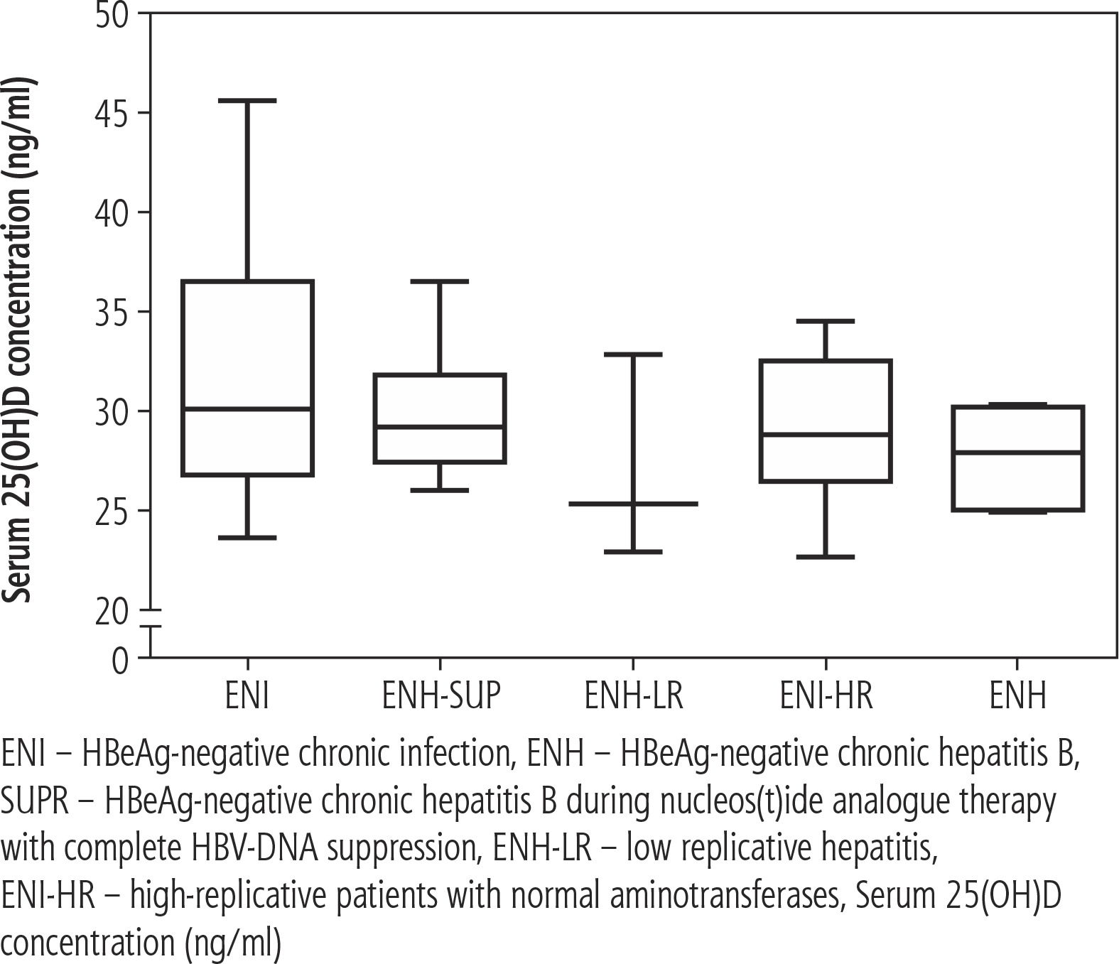
Fifty-eight patients with chronic HBeAg-negative HBV infection (CHB), 6 with spontaneously resolved HBV infection with seroconversion HBsAg/anti-HBs ≥ 20 years earlier (RES) and 4 with treatment-associated seroconversion HBsAg/anti-HBs ≥ 2 years earlier (S-CONV) were enrolled in the study. The long-term follow-up (> 36 months) in CHB patients allowed three patterns of chronic infection to be distinguished according to current European Association for the Study of the Liver (EASL) recommendations:
HBeAg-negative chronic infection (ENI, n = 17) – HBV-DNA < 2000 U/l, normal aminotransferases,
HBeAg-negative chronic hepatitis B naïve-to-treatment (ENH, n = 7) – HBV-DNA > 2000 U/l and elevated aminotransferases,
ENH during nucleos(t)ide analogue therapy with complete HBV-DNA suppression > 24 months (ENH-SUP, n = 17).
Two additional groups were distinguished based on the HBV-DNA load and aminotransferase level:
low replicative hepatitis (ENH-LR) (n = 3) – HBV-DNA < 2000 U/l, elevated aminotransferases,
high-replicative patients with HBV-DNA > 2000 U/l, normal aminotransferases (ENI-HR) (n = 14).
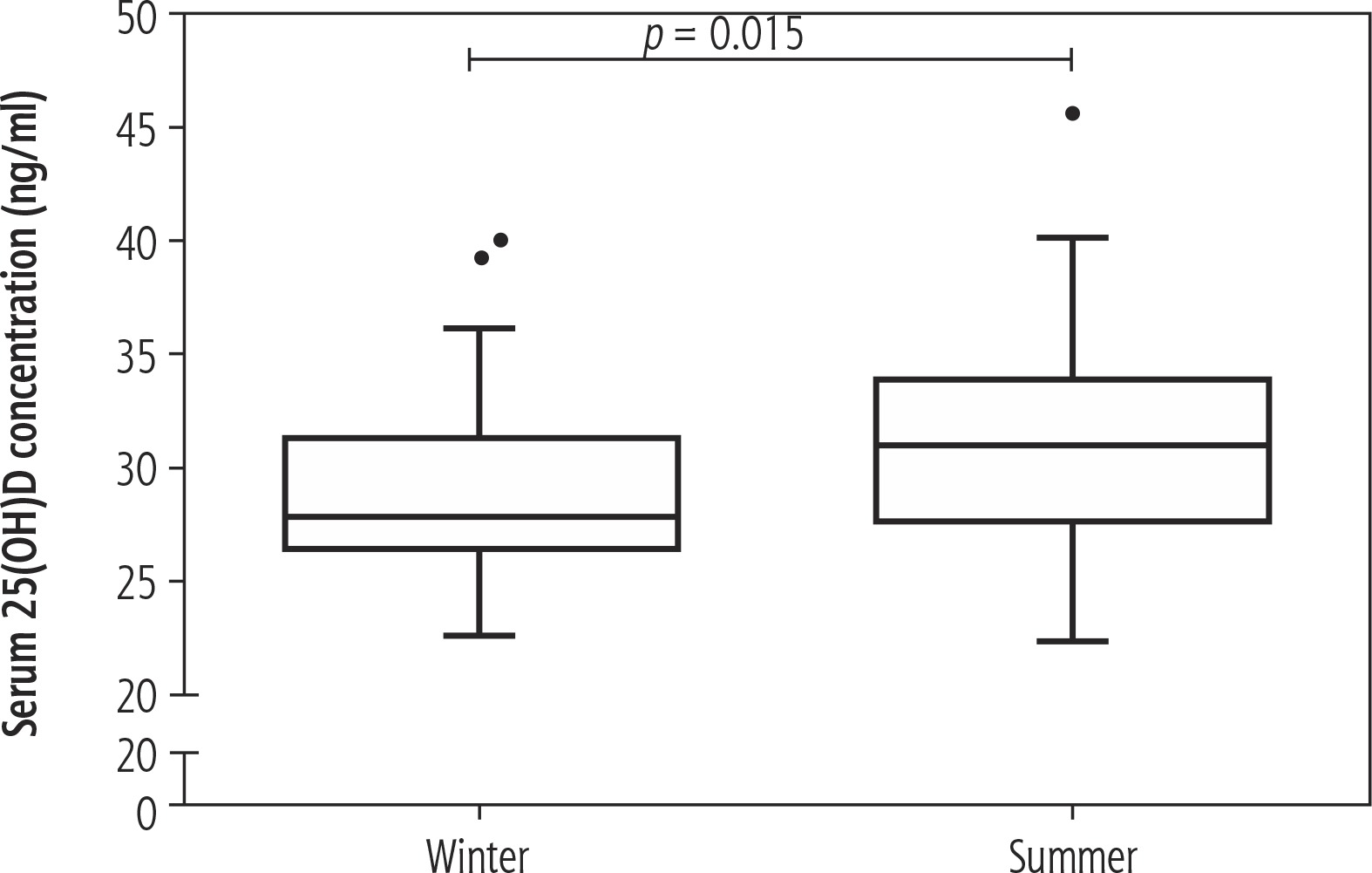
The characteristics of the studied groups are shown in Table 1. The control group (HC) consisted of 9 healthy volunteers. Blood samples were collected from fasting individuals without clinically evident acute inflammatory infection. One sample at a certain time was obtained from each patient, without further follow-up. To assess the bias of seasonal fluctuations of 25(OH)D level, all groups were additionally analysed with reference to the time of sample collection, i.e. winter (for samples collected in the November-April period) and summer (for samples collected in the May-October period) (Tables 1 and 2). Informed consent was obtained from all individuals included in the study.
Table 1
Demographic, clinical and laboratory characteristics of studied groups
Table 2
Period of sample collection in studied CHB groups
| Group | Winter | Summer |
|---|---|---|
| ENI (n, %) | 8, 47.1 | 9, 52.9 |
| ENI-HR (n, %) | 3, 21.4 | 11, 78.7 |
| ENH-LR (n, %) | 3, 100 | 0, 0 |
| ENH (n, %) | 3, 42.9 | 4, 57.1 |
| ENH-SUP (n, %) | 6, 35.3 | 11, 64.7 |
[i] ENI – HBeAg-negative chronic infection, ENI-HR – high-replicative patients with normal aminotransferases, ENH-LR – low-replicative hepatitis, ENH – HBeAg-negative chronic hepatitis B, ENH-SUP – HBeAg-negative chronic hepatitis B during nucleos(t)ide analogue therapy with complete HBV-DNA suppression
The study was approved by the institutional bioethical committee (no. R-I-002/497/2014). All procedures performed in the study were in accordance with the ethical standards of the institutional and national research committee and with the 1964 Declaration of Helsinki and its later amendments or comparable ethical standards.
Immunoassays
Serum 25-hydroxyvitamin D (25-OH vitamin D) levels were quantified by means of a commercially available 25-OH-Vitamin-D ELISA kit (BioVendor, Czech Republic), according to the manufacturer’s instructions (range: 4-120 ng/ml). The ELISA test chosen for the purpose of this study uses a monoclonal antibody which is equally specific for both forms of the vitamin, D3 and D2.
Statistical analysis
The results were expressed as a medians and range, unless indicated. The following non-parametric tests were applied: Mann-Whitney U test and Kruskal-Wallis ANOVA test for univariate comparisons, and Spearman rank test for correlation analysis. Statistically significant p values were < 0.05. Statistica 12 for Windows was used to perform the analysis (StatSoft Inc., Tulsa, USA), and graphical presentation of the results was performed using GraphPad Prism software (GraphPad Prism Software Inc., San Diego, CA, USA).
Results
Serum 25(OH)D concentration in chronic HBeAg-negative HBV patients
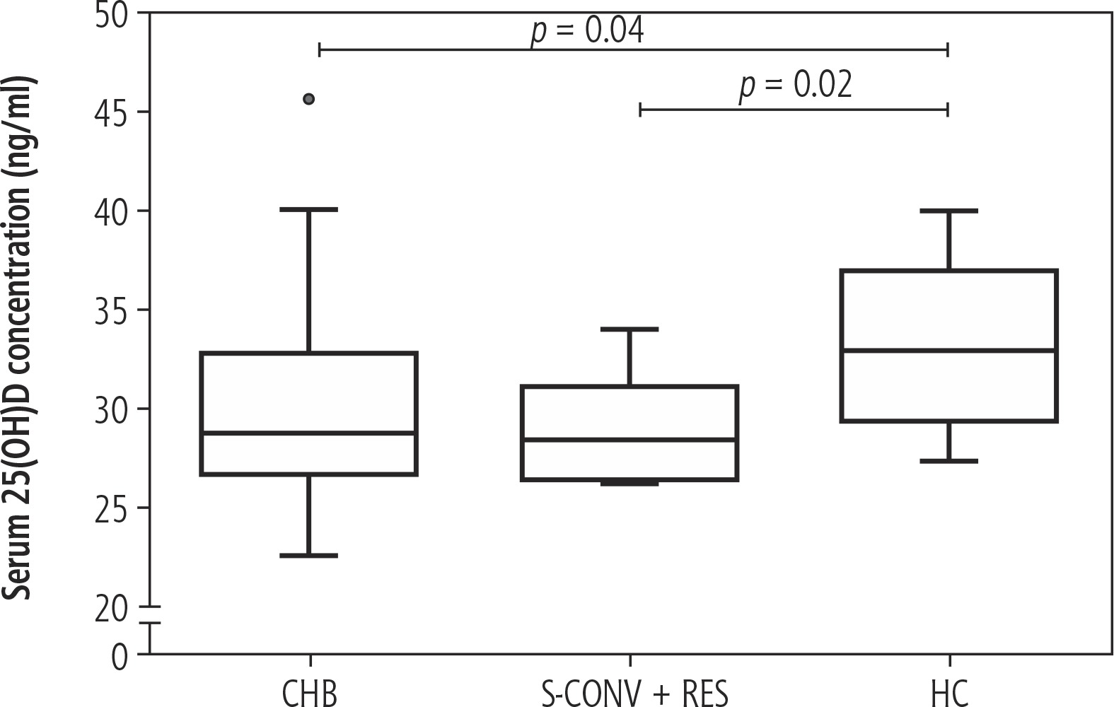
To examine whether HBV infection may have an effect on serum concentration of 25(OH)D we performed a comparative analysis of enrolled groups. We found significantly decreased serum concentration of 25(OH)D in CHB patients (28.81 ng/ml, 22.64-45.67 ng/ml) and in the S-CONV+RES group (28.42 ng/ml, 26.38-34.03 ng/ml) in comparison to the HC groups (32.97 ng/ml, 27.43-40.02 ng/ml) (p = 0.04 and p = 0.02, respectively) (Fig. 1). We did not find differences in 25(OH)D concentration across CHB groups (Fig. 2). To eliminate the bias of the period of sample collection we performed corresponding analysis with division of the groups with reference to the time of sample collection. Similarly, we did not observe significant differences among analysed groups.
Correlation of viral (VL, qHBsAg) and biochemical parameters (ALT, APRI) with serum 25(OH)D
We did not find any significant associations between serum 25(OH)D concentration and viral parameters such as viral load or HBsAg concentration. We observed a significant negative correlation between serum 25(OH)D and serum alanine aminotransferase concentration (p = 0.046). Moreover, serum 25(OH)D significantly correlated negatively with the frequency of peripheral blood monocytes (p = 0.031). There were no significant associations between serum 25(OH)D and the APRI (aspartate aminotransferase to platelet ratio index) score or liver histology (Table 3). The results of correlations were similar with regard to the time of sample collection.
Table 3
Correlations between serum 25(OH)D level and viral, biochemical and demographic parameters
Demographic and seasonal factors and serum 25(OH)D concentration
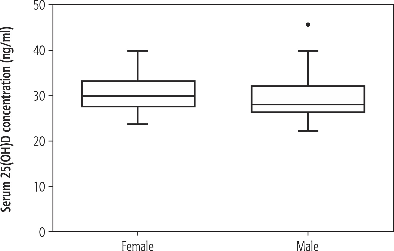
There was no significant association between age and serum 25(OH)D concentration. We did not observe a difference in serum 25(OH)D level between female and male populations enrolled in our study (Fig. 3). There was a significantly higher serum 25(OH)D concentration in the samples collected in the summer in comparison to the pool of samples collected during the winter period (p = 0.015) (Fig. 4).
Discussion
Vitamin D deficiency is a common problem affecting the general population. It is estimated that in Poland the burden of vitamin D deficiency of varying severity may affect even 90% of adults, children and adolescents. According to a large cross-sectional study in the adult population of Poland carried out in the winter and early spring period, in the region of north-eastern Poland, where our study was conducted, the mean value for the general population was 25.8 ±14.0 ng/ml, and appeared to be the highest in Poland [4]. The recommendations underline the seasonal fluctuations of vitamin D serum concentrations and emphasise the need for additional supplementation from October to March, when the endogenous skin synthesis of this prohormone is insufficient in the regions above 35° latitude (including Poland; 49°N-54°N). As the first step of cholecalciferol activation occurs in the liver, patients with liver diseases belong to the risk group for functional deficiency of vitamin D, tested by measuring the serum 25(OH)D level [8]. The available data from the Polish population of patients with liver diseases demonstrated vitamin D insufficiency in chronic HBV or HCV infections, with mean values 17.6 ng/ml and 19.3 ng/ml, respectively [9]. Taking into account immunomodulatory effects exerted by vitamin D, we expect that insufficient supply of vitamin D may drive unfavourable outcomes of disease. On the other hand, the severity of liver disease may have an impact on the bioavailability of the intermediate metabolite 25(OH)D, thus contributing to the occurrence of vitamin D deficiency [7].
Data presented in our study demonstrated a significantly decreased serum 25(OH)D level in CHB patients in comparison to HC, which correlated negatively with serum ALT activity and frequency of peripheral blood monocytes. We also observed seasonal fluctuations of vitamin D level with significantly decreased values during the winter period across all the groups.
There are numerous studies showing an association between serum vitamin D deficiency and liver diseases of various aetiology, including HBV and HCV infections, non-alcoholic liver disease, HCC and autoimmune liver diseases [7, 8, 10-12]. In our study we found a significantly decreased serum vitamin D level in comparison to the HC group. These observations are in line with the available data from HBV-infected populations reported by other authors [13-16]. The median value of serum 25(OH)D level in the CHB group was 28 ng/ml, which corresponds to the suboptimal vitamin D level. This observation is in contrast with some reports showing vitamin D deficiency in HBV infection. For instance, Chan et al. reported mean serum 25(OH)D3 levels as low as 7.83 ng/ml, which corresponds to severe vitamin D deficiency. This discrepancy between our study and other reports might be explained by the absence of patients with advanced liver disease in our group, as serum 25(OH)D has been reported to be significantly reduced in cirrhotic patients in several studies [13, 14].
There is evidence that links serum vitamin D level with the severity of the clinical outcomes of chronic hepatitis B, such as viral load control, inflammation and progression of the fibrosis, as well as treatment outcomes related to the immune responsiveness [13, 16-18]. Therefore we performed comparison across the CHB groups divided by the phases of HBV infection. We did not find any association between serum vitamin D status and phase of HBV infection, even when the groups were stratified by the period of sample collection. In our analysis serum 25(OH)D was significantly decreased in all the samples collected during the winter period in comparison to the summer, which is an expected observation, as sunlight exposure increases the serum level of vitamin D and its metabolites.
There is available evidence for the association between serum 25(OH)D level and the intensity of viral replication. The study of Farnik et al. demonstrated a strong predictive value of low serum 25(OH)D3 level for high HBV viral load [18]. Similarly, an inverse correlation between serum 25(OH)D level and HBV viral load was observed by Hoan et al. [13]. However, the data are conflicting in this matter. We did not find a significant association between serum 25(OH)D and serum HBV DNA concentrations, which is in line with the reports of Zhao et al. and Chan et al. [14, 15]. Serum 25(OH)D concentration in our enrolled CHB group correlated significantly with serum ALT level, but not with APRI or liver histology. The disproportion of cases with successive histological stages of liver disease is the major limitation of our study. However, serum ALT is considered an established marker that reflects the extent of present necroinflammatory activity. Significant correlations of 25(OH)D and serum ALT are reported by other authors. In the research of Chan et al. high baseline pre-treatment serum 25(OH)D was significantly associated only with ALT, after adjusting for age, gender, HBeAg and HBV genotype [15].
Vitamin D has immunomodulatory properties and vitamin D deficiency may influence the composition of the peripheral blood leucocyte pool [19]. We found a significant negative correlation between serum 25(OH)D concentration and peripheral blood monocyte frequency, which may reflect the immunomodulatory effect of vitamin D.
In conclusion, our results show that serum 25(OH)D level is significantly decreased in our population of CHB patients; however, the levels are at the border of suboptimal and optimal. We did not find associations between 25(OH)D and phases of HBV infection or HBV viral load, which suggests that serum 25(OH)D does not have an impact on the immune control of HBV infection. The inverse correlation between serum 25(OH)D and ALT level may support the observations indicating that the decreased 25(OH)D level might be secondary to the liver disease.


