
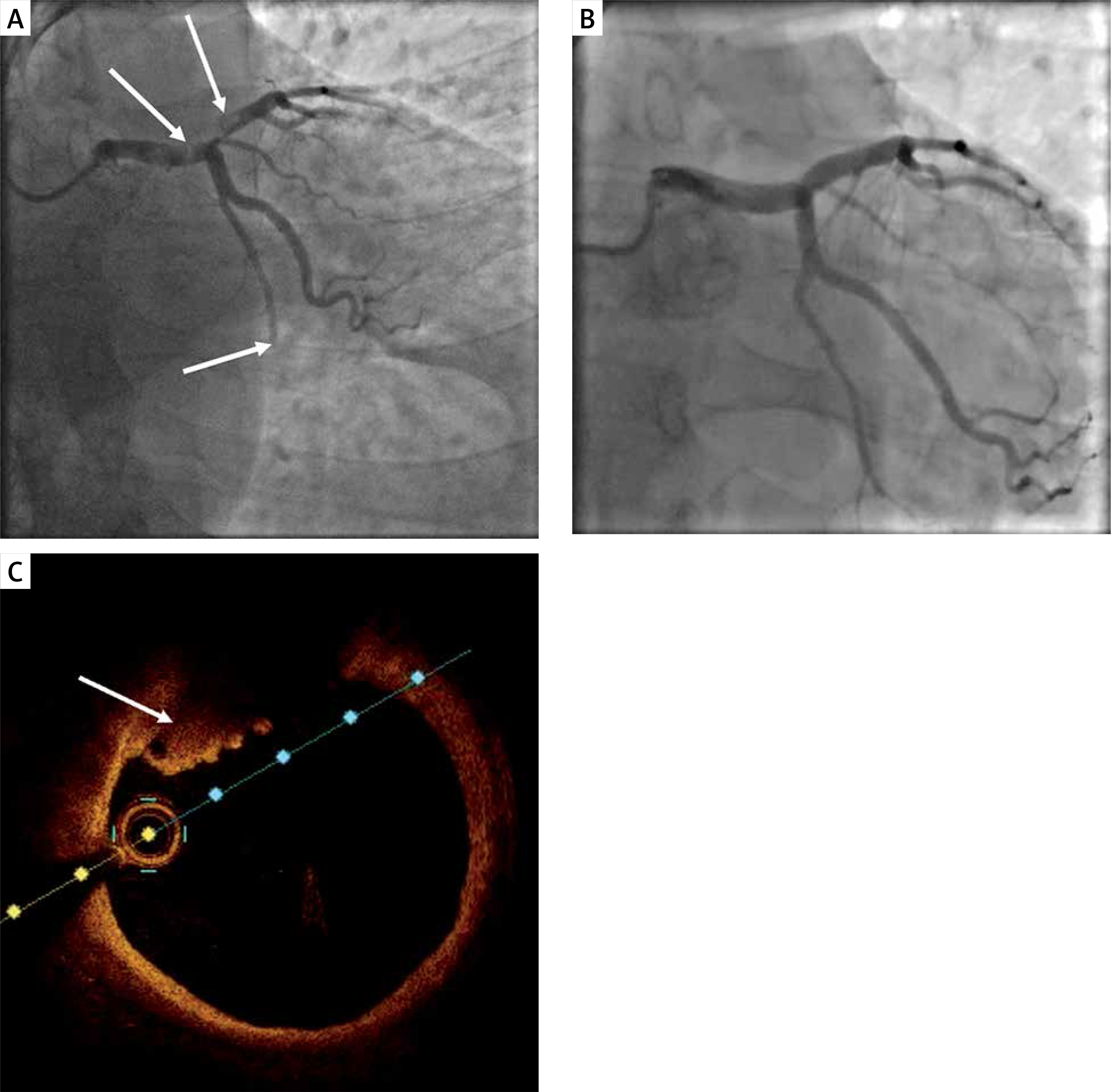
We present a case of a 45-year-old man, who was admitted due to anterior wall ST-segment elevation myocardial infarction of 3 h duration. Coronary angiography showed a normal RCA, thrombotic occlusion of the distal circumflex artery (Cx) and a massive thrombus in the distal left main artery (LM) protruding to the proximal left anterior descending artery (LAD) with preserved distal flow (Figure 1 A). Cautious, but unsuccessful thrombus aspiration (Hunter 6F) was performed. As the patient was stable he was qualified for further conservative treatment with administration of 72-hour infusion of eptifibatide, enoxaparin, acetylsalicylic acid and prasugrel. Echocardiography revealed extensive regional contractility abnormalities of the left ventricle with reduced ejection fraction (38%). Laboratory results showed a maximal troponin I (TnI) level of 83 355 pg/ml.
Figure 1
A – coronary angiography showing a massive thrombus in the distal left main coronary artery trunk, proximal left anterior descending and chronic total occlusion of distal left circumflex; B – coronary angiography eleven days after admission, no thrombus visible; C – optical coherence tomography showing a wall-mounted thrombus in the left main coronary artery trunk
After 4 days repeated coronary angiography showed only slight reduction of the thrombus. Based on this result the patient was discussed by the local Heart Team and qualified for pharmacotherapy continuation and another coronary angiography again in a few days.
Eleven days after admission, the second follow-up was performed, in which no LM thrombus was visible (Figure 1 B). OCT showed presence of a non-flow limiting fibrotic plaque (< 30% stenosis) in the LM and LAD with wall-mounted red thrombus and small elements of white thrombus (Figure 1 C). We did not observe plaque rupture, dissections or any other features of plaque instability. Taking the result into consideration the patient was qualified for conservative treatment without stent implantation.
At the follow-up 3 months after hospitalization the patient reported only mild dyspnea during physical activity without recurrence of chest pain or other major adverse cardiovascular events (MACE). Follow-up echocardiography showed a non-enlarged left ventricle with slightly reduced ejection fraction (44%).
Angiographic evidence of thrombus can be seen in 91.6% of patients who present with ST-elevation myocardial infarction (STEMI) [1]. The use of OCT is essential to reveal the underlying pathophysiology, and optimize therapeutic options [2]. Massive intracoronary thrombus has been reported in 16.4% of patients with acute coronary syndrome [3]. The recommended treatment for patients presenting with STEMI is timely primary percutaneous coronary intervention (PCI), including stent implantation, but in some patients the coronary flow may be restored and stabilized without stenting [4]. Coronary lesions causing STEMI, in general, have a luminal stenosis of less than 75% [5]. Thus, the risk of a plaque rupture with formation of thrombus and subsequent myocardial infarction is not determined by the degree of luminal stenosis, but by the plaque content [6].
In patients with a large thrombus burden the deferred strategy may potentially limit the risk of distal coronary embolization. This can allow 24–48 h of intense antithrombotic therapy, including prolonged intravenous GPIIb/IIIa inhibition and heparin. In some cases with a short ischemia time thrombolysis is a valid therapeutic option. Subsequent angiography frequently shows reduced thrombus burden and allows one to perform intracoronary imaging and PCI with a significantly lower risk of distal embolization. As shown in the recent analysis of the DANAMI-3-DEFER study deferred stenting was not associated with an increased risk of all-cause mortality, target vessel revascularization, or recurrent myocardial infarction in patients with preserved flow and residual stenosis < 30% [7].
In conclusion, there is no ideal management strategy for the present case. In general, potent dual antiplatelet agents must be used and effective anticoagulation should be maintained. Manual thrombectomy is usually the first line-treatment. If it fails and a large thrombus burden remains with preserved distal flow and without ongoing ischemia, GPIIb/IIIa inhibitors and heparin infusion should be maintained for 24–48 h. This strategy allows one to “buy some time” for the patient and finally choose the best treatment option, as in the present case.


