
INTRODUCTION
Breast cancer is the most common malignant neoplasm in women and remains the leading cause of cancer-related mortality in this population [1]. Treatment depends on the disease stage and prognostic factors and may include surgery, chemotherapy, hormone therapy, radiotherapy, and anti-human epidermal growth factor receptor 2 (HER2) therapy.
In patients with advanced breast cancer, various skin lesions may be observed. The differential diagnosis includes cutaneous manifestations associated with cancer progression [2], exacerbations of preexisting dermatological conditions, adverse cutaneous drug reactions to systemic oncologic therapies, and inflammatory dermatoses not directly associated with the coexistent malignancy [3].
Because an accurate diagnosis has a substantial impact on further therapeutic management and prognosis, rapid and precise identification of the nature of cutaneous lesions is essential.
Cutaneous metastases of breast cancer are an adverse prognostic factor, indicating disease recurrence or resistance to ongoing therapy [4]. Breast cancer is the most common source of cutaneous metastases in women. These lesions are characterized by considerable morphological heterogeneity and require careful correlation of the clinical and histopathological findings to establish the correct diagnosis [5]. In patients with breast cancer, cutaneous metastases tend to occur primarily on the chest and upper torso, which likely reflects the pattern of regional lymphatic drainage from the breast [6]. It is important to distinguish local spread, defined as direct tumor extension into the skin, from cutaneous metastases, which occur at sites distant from the primary tumor through lymphatic or hematogenous dissemination and indicate systemic disease.
OBJECTIVE
This case underscores the complexity of evaluating newly developed skin lesions in patients with advanced breast cancer, particularly those receiving anti-HER2 therapy. It highlights the potential for false-negative histopathological findings and the overlap in clinical features between cutaneous metastases and drug-induced eruptions.
CASE REPORT
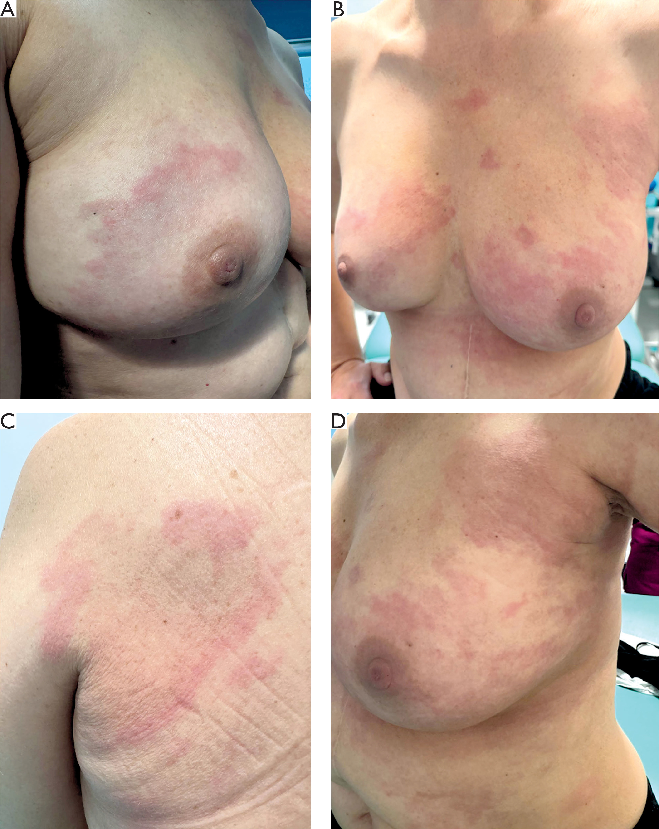
A 52-year-old woman diagnosed with advanced non-luminal HER2-positive cancer of the left breast (pT4pN1cM1) was admitted to the dermatology department because of erythematous, sharply demarcated, festoon-shaped patches with elevated borders (fig. 1). The lesions were located on both breasts, left axilla, and the left side of the back, being accompanied by localized pruritus. Their morphology initially raised suspicion of a drug-induced reaction to anti-HER2 targeted therapy.
Figure 1
Erythematous, sharply demarcated, festoon-shaped patches with elevated borders involving both breasts, left axilla, and left upper side of the back
The diagnosis of breast cancer had previously been confirmed by core needle biopsy, which revealed invasive carcinoma of no special type (NOS), histological grade 2, non-luminal, HER2-positive, with a Ki-67 proliferation index of 20.5%. Fine-needle aspiration of the left axillary lymph nodes demonstrated malignant cells. Computed tomography revealed focal hepatic lesions consistent with metastases. No BRCA1/2 mutations were detected.
Given the advanced stage of the disease, the patient was initially treated with a combination of docetaxel, trastuzumab, and pertuzumab. During treatment, the previously described erythematous lesions first appeared on the left breast. Histopathological examination of a skin biopsy obtained from that site revealed intravascular tumor emboli consistent with invasive breast carcinoma (NOS type).
After 4 months, treatment was continued with trastuzumab and pertuzumab in combination. In addition, radiotherapy was administered to the left breast, including the area affected by the skin lesions, at a total dose of 20 Gy in five fractions. Following radiotherapy, the skin lesions resolved.
One month later, the skin lesions recurred, with additional involvement of the right breast. A biopsy of the right breast skin was performed but showed no evidence of malignant cells.
Mammography performed at that time revealed microcalcifications in the left breast, classified as Breast Imaging Reporting and Data System (BI-RADS) category 6, indicating histologically confirmed malignancy. The right breast was classified as BI-RADS category 0, indicating an inconclusive examination requiring further evaluation.
The skin lesions did not resolve despite treatment with topical corticosteroid and systemic clemastine, an H1 receptor antagonist.
During hospitalization at the dermatology department, laboratory investigations were performed, including antinuclear antibody testing with immunoblot analysis, complement components C3c and C4, and routine laboratory tests. All results were within the reference range.
Given the previous resolution of the skin lesions following radiotherapy and the prolonged use of HER2-targeted therapy, further diagnostic evaluation was performed to assess possible hypersensitivity reactions to anti-HER2 agents. The results were negative.
In consultation with the treating oncologist, anti-HER2 therapy was temporarily discontinued to exclude a drug-induced etiology of the eruptions. A short course of intravenous methylprednisolone was administered at doses of 40 mg, 30 mg, and 20 mg on three consecutive days, and topical treatment with 0.1% mometasone furoate ointment was initiated. This treatment resulted in partial resolution of the erythematous lesions, particularly at their periphery.
Subsequently, the skin biopsy specimens were obtained from persistent lesions on the right breast and in the left scapular region. The specimens were collected from the elevated margins of the lesions.
The patient was discharged in good general condition while awaiting the biopsy results.
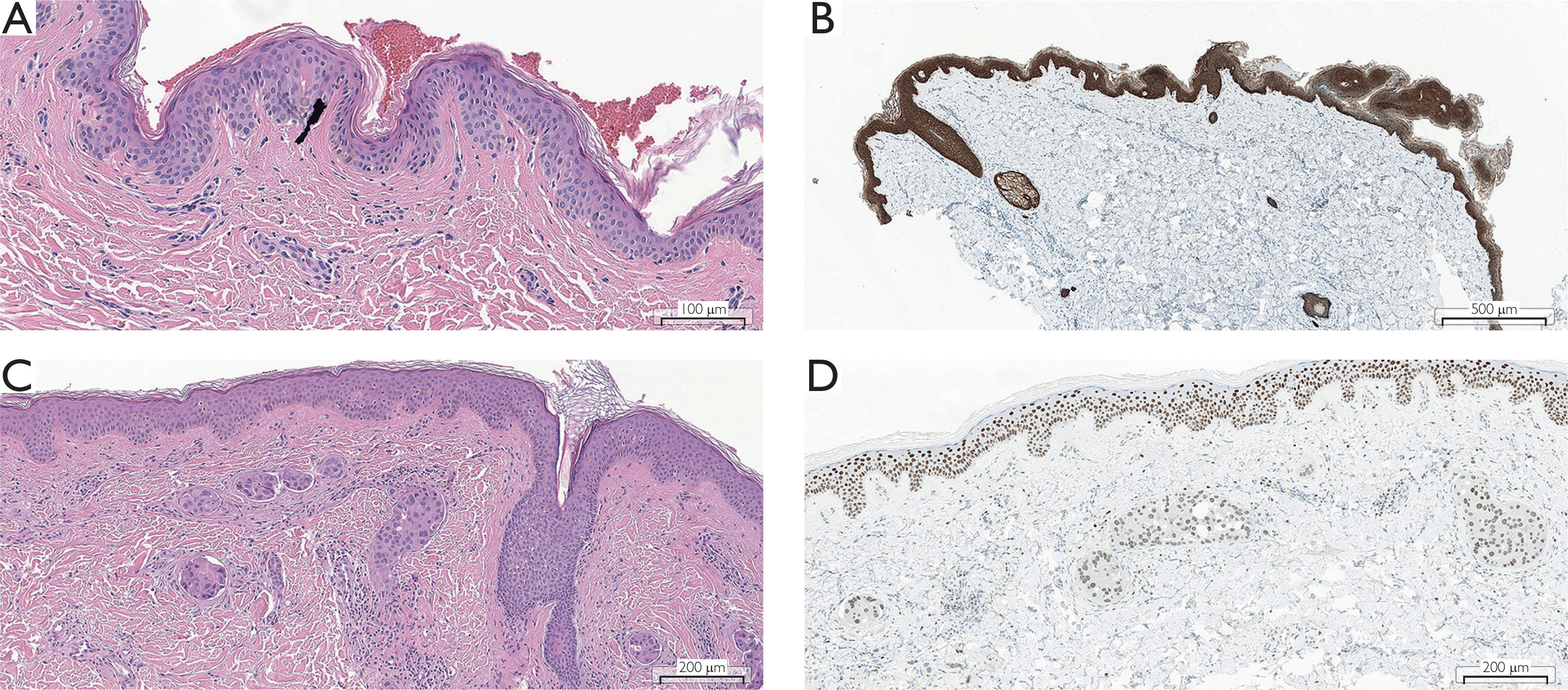
Histopathological examination of both biopsy specimens revealed intravascular tumor emboli composed of carcinoma cells that were estrogen receptor-negative, progesterone receptor-negative, HER2-positive, CK7-positive, and GATA3-positive, consistent with non-luminal HER2-positive breast cancer (fig. 2). The Ki-67 proliferation index was approximately 30%.
Figure 2
Histological scans of a skin biopsy specimen obtained from the margin of erythematous lesions on the right breast. Slides were scanned using an Aperio GT 450 DX scanner (Leica Biosystems). A – Section without evidence of carcinoma cells (H&E; scale bar: 100 μm). B – Immunohistochemical staining for pancytokeratin showing no carcinoma cells (scale bar: 500 μm). C – Section presenting with tumor emboli within dermal vessels, accompanied by a chronic inflammatory infiltrate (H&E; scale bar: 200 μm). D – Immunohistochemical staining showing GATA3 expression in carcinoma cells (scale bar: 200 μm)
Because cutaneous recurrence of the malignancy was confirmed, restaging computed tomography of the chest and abdomen was performed and demonstrated further disease progression. Consequently, the patient was started on trastuzumab emtansine at a dose of 3.6 mg/kg every 3 weeks and received treatment for 3 months. Due to lack of radiological response, treatment was subsequently switched to trastuzumab deruxtecan.
At the time of manuscript preparation, the patient remained under regular dermatologic and oncologic follow-up, with follow-up computed tomography of the chest and abdomen scheduled for the following month.
DISCUSSION
Cutaneous metastases occur in approximately 25% of patients with metastatic breast cancer and might manifest early in HER2-positive and triple-negative subtypes [5, 7]. They typically involve the chest wall and upper extremities. These metastases most commonly result from hematogenous or lymphatic dissemination of malignant cells, although direct tumor extension into the skin may also occur, leading to neoplastic infiltration and proliferation within the dermis [8].
The clinical presentation is diverse and may include erythematous-edematous or infiltrated papules and plaques, as well as nodular lesions [9]. Erosions and ulcerations may also develop. In some cases, distinct morphological patterns emerge, such as carcinoma erysipeloides, carcinoma telangiectoides, or carcinoma en cuirasse [10]. A drug-induced reaction to anti-HER2 therapy was included in the differential diagnosis because cutaneous adverse effects, including maculopapular eruptions and erythematous lesions, sometimes accompanied by pruritus, have been reported [11, 12]. Excluding this possibility was essential for the appropriate planning of further treatment.
The diagnosis is based on histopathological examination, which may reveal intravascular tumor cell emboli within dermal vessels, sometimes accompanied by a lymphocytic infiltrate. In diagnostically uncertain cases, dermoscopic evaluation may facilitate the selection of an appropriate biopsy site. Dermoscopic findings suggestive of cutaneous metastases from breast cancer include various vascular patterns, such as linear, branching, irregular, and polymorphic vessels, homogenous structureless areas that may be white, pink, yellow, or orange, with the latter sometimes containing linear fissure-like structures; peripheral white lines; and multiple brown dots and globules [13, 14].
However, the associated inflammatory response may extend beyond the area of metastatic involvement, potentially leading to false-negative biopsy results. This may explain the initial negative histopathological findings despite clinical suspicion of disease recurrence.
This case demonstrates that obtaining skin biopsies from multiple sites may be necessary to establish a definitive diagnosis. Notably, systemic anti-inflammatory treatment administered before biopsy may have reduced nonspecific inflammation surrounding the metastatic foci, thereby facilitating histological assessment. Nevertheless, the temporal relationship between anti-inflammatory treatment and biopsy timing does not allow definitive conclusions to be drawn regarding strategies to optimize diagnostic accuracy.
The skin, along with the central nervous system, has been proposed as a potential sanctuary site for breast cancer cells during anti-HER2 therapy with trastuzumab and pertuzumab. This phenomenon may be associated with immunological mechanisms, including reduced major histocompatibility complex class II expression on antigen-presenting cells, increased transforming growth factor-β production, and increased programmed death-ligand 1 expression in the dermal papillae [15]. Importantly, cutaneous involvement is frequently associated with visceral disease progression in patients with breast cancer [16]. Therefore, the management of such skin lesions remains particularly challenging, and their recurrence during therapy may represent an unfavorable prognostic indicator.
Treatment of cutaneous metastases from breast cancer primarily involves systemic anticancer therapy and palliative local measures, including chemotherapy, anti-HER2 therapy, radiotherapy, and photodynamic therapy. Local radiotherapy may be particularly useful in patients with symptomatic, ulcerated, bleeding, or painful lesions. Additional local and supportive treatment may include analgesics, topical corticosteroids, imiquimod 5% cream, 5-fluorouracil 5% cream, and appropriate wound care [17, 18].
In selected patients with isolated or limited cutaneous metastases, surgical excision may be beneficial, particularly when the lesions are small, draining, or bleeding. However, surgical treatment of cutaneous breast cancer metastases is uncommon, primarily because of the high risk of rapid local recurrence following excision [19].
Electrochemotherapy is another local treatment modality with demonstrated efficacy in the management of cutaneous and subcutaneous metastases, including those originating from breast cancer. It combines the administration of chemotherapy, most commonly bleomycin, with the local application of electric pulses, thereby increasing drug uptake by tumor cells while limiting systemic toxicity. Electrochemotherapy may be considered in patients who are not candidates for surgery or radiotherapy. Although generally well tolerated, adverse effects may include transient pain, inflammatory skin reactions, pigmentary changes, and occasional ulceration or skin necrosis requiring additional wound care [19, 20].
CONCLUSIONS
This case highlights the diagnostic challenges associated with evaluating cutaneous lesions in patients with breast cancer receiving anti-HER2 therapy. It underscores the importance of correlating clinical findings with histopathologic results and, when clinical suspicion persists despite an initially negative biopsy, of repeating the biopsy and obtaining specimens from multiple representative sites. Although systemic anti-inflammatory therapy may have reduced nonspecific inflammation in this case, its effect on diagnostic accuracy remains uncertain.












