
INTRODUCTION
Genital involvement is encountered in 33–63% of patients with psoriasis vulgaris (PV). Although quality of life is more profoundly impaired in patients with genital lesions, such lesions are overlooked by dermatologists in up to 60% of cases. In 2–5% of patients with psoriasis, genital lesions may be the sole manifestation of the disease [1]. This necessitates differentiation of psoriatic balanoposthitis (PBP) from other conditions affecting the glans penis and prepuce, including plasma cell balanitis, candidiasis, lichen planus and erythroplasia of Queyrat. Thin erythematous plaques are a common clinical presentation of PBP. Some patients may also experience itch, burning sensation, and dyspareunia. In uncertain cases, biopsy can be performed to clarify the diagnosis [2, 3]. Dermatoscopy may serve as a non-invasive diagnostic tool and, in selected cases, may reduce the need for biopsy. Reportedly, dermatoscopic features of PBP are limited to uniformly distributed dotted vessels on a light red background, while treatment of genital psoriasis is often challenging [3]. Our report focuses on pustules as a less common dermatoscopic features of PBP. Topical treatment of genital psoriasis is challenging and often requires long-term maintenance. Dermatoscopy may also be used to monitor and predict treatment response in PBP [4].
OBJECTIVE
The aim of this report was to describe potentially underreported dermatoscopic features of PBP and to evaluate treatment response using dermatoscopy.
MATERIAL AND METHODS
Patients (n = 16) with histopathologically confirmed PBP and no extragenital manifestations were enrolled in this study. Exclusion criteria were topical or systemic treatment of psoriasis for 4 weeks prior, sexually transmitted and fungal infections, circumcision and extragenital psoriasis. The mean patient age was 36.10 (range: 27–52) years. The study design was approved by an Ethics Committee.
Digital dermatoscopic images were acquired before biopsy and at the 4-week follow-up using 20× magnification (Medicam 1000s, FotoFinder Systems GmbH, Germany). The area with the most pronounced dermatoscopic features was marked, and a 3-mm punch biopsy was performed to confirm the diagnosis. Histopathological features of PBP were correlated with dermatoscopic features observed in the corresponding dermatoscopic images.
Treatment with tacrolimus 0.1% ointment was initiated 1 week after biopsy, allowing the wound to heal, and was continued twice daily for 3 weeks. The severity of PBP was graded dermatoscopically on a 4-point scale: 0 – clear, 1 – mild, 2 – moderate, and 3 – severe, before treatment and at the follow-up visit. The Wilcoxon signed-rank test was used to compare the severity of PBP before and after treatment. A p-value < 0.05 was considered statistically significant.
RESULTS
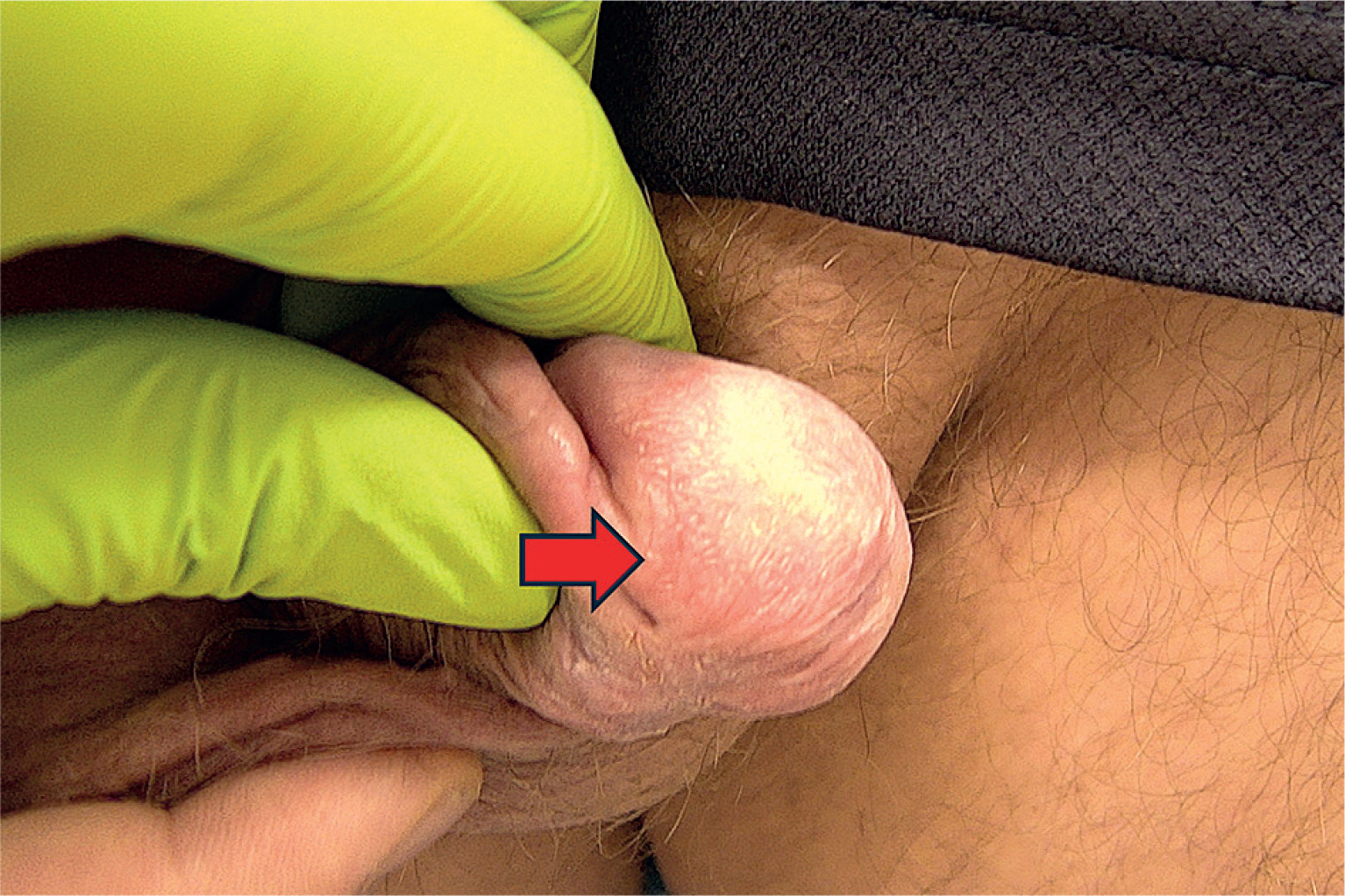
Uniformly distributed red dots were observed in all cases during dermatoscopy. In 5/16 patients (31.25%) dermatoscopy revealed pustules (fig. 1). Pustules were clinically evident in only 3 cases (fig. 2). Fine scaling was present dermatoscopically in 6/16 cases (37.50%).
Figure 1
Dermatoscopic image of psoriatic balanoposthitis showing uniformly distributed red dots (red arrow), fine scaling (white arrow), and a pustule (yellow arrow). Original magnification 20×; Medicam 1000s, FotoFinder Systems GmbH, Germany
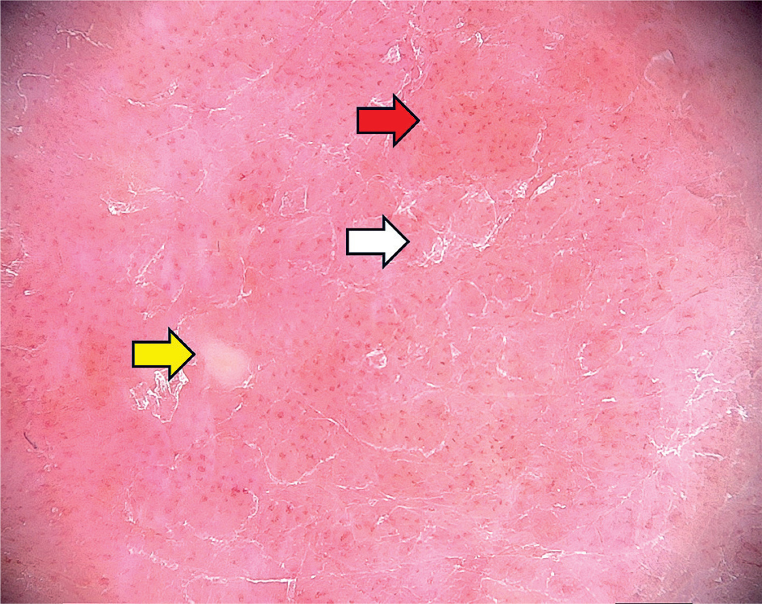
Figure 2
Clinical image of the case presented in figure 1 of psoriatic balanoposthitis, showing slight erythema and scaling involving the glans penis and prepuce. No pustule is clinically visible
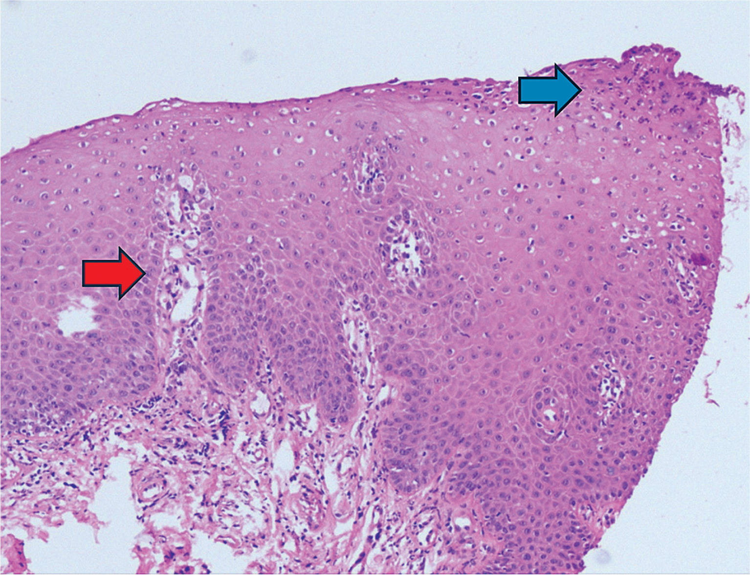
Histopathological correlation demonstrated that the red dots corresponded to tortuous, dilated capillaries located within elongated dermal papillae, while the dermatoscopically visualized pustules corresponded to spongiform pustules of Kogoj (fig. 3).
Figure 3
Histopathological image of psoriatic balanoposthitis showing dilated, tortuous vessels within elongated dermal papillae (red arrow) and a spongiform pustule of Kogoj (blue arrow). Hematoxylin and eosin staining, original magnification 200×
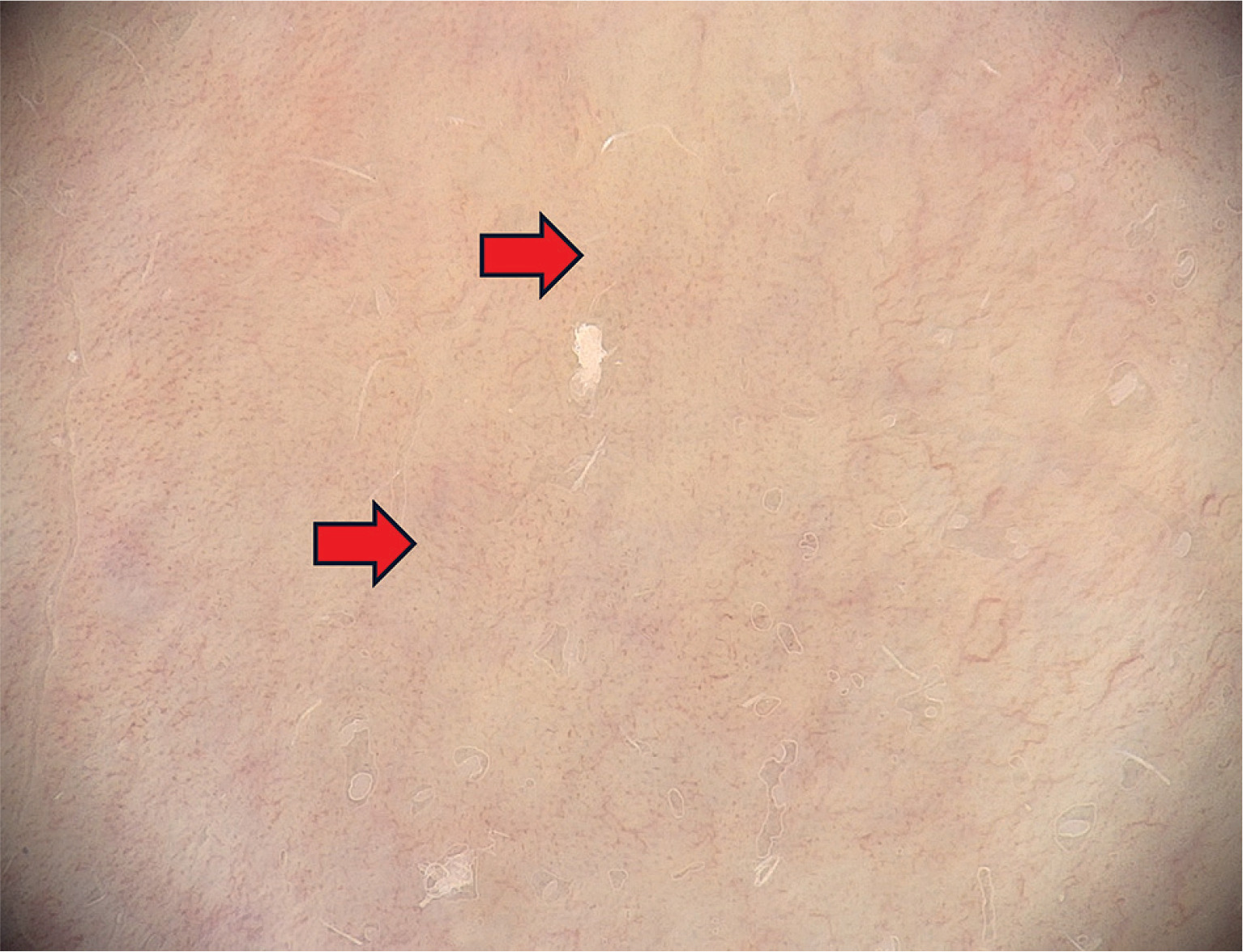
The severity of PBP was significantly lower after treatment than before treatment (Mdn = 0 vs. Mdn = 2, respectively; W = 136, p < 0.001). Complete resolution of PBP was observed in 12/16 patients, including 3/5 cases of pustular PBP. In the remaining 4/16 patients, the disease was graded as mild after treatment. In these cases, erythema resolved and the diameter of dotted vessels decreased substantially, although the vessels remained visible on dermatoscopy (fig. 4).
Figure 4
Dermatoscopic image of persistent dotted vessels (red arrow) in a patient with psoriatic balanoposthitis after a 3-week course of topical tacrolimus 0.1% ointment. Original magnification 20×; Medicam 1000s, FotoFinder Systems GmbH, Germany
A mild burning sensation was reported by 4/16 patients at the initiation of treatment with tacrolimus ointment and resolved within a few days.
DISCUSSION
To our knowledge, only two case reports of localized PBP with pustules have been published to date, and neither included dermatoscopic evaluation [5]. Spongiform pustules of Kogoj are a characteristic histopathological feature of psoriasis, describing epidermal accumulations of neutrophils that may occasionally give rise to macroscopically visible pustules within a psoriatic plaque. This condition is referred to as “psoriasis cum pustulatione” and should not be confused with pustular psoriasis (PP), a group of diseases that are genetically and phenotypically distinct from psoriasis vulgaris [6].
Syphilis and circinate balanitis secondary to chlamydia infection are possible differential diagnoses of pustular balanoposthitis, however, sexually transmitted infections, including syphilis, chlamydiosis, and gonorrhea, were excluded in all patients. Fungal elements were also absent on PAS staining, and there were no clinical signs or history of reactive arthritis in patients with pustular lesions [7]. The trigger of pustular lesions in PBP remains unclear, however, stress, the Koebner phenomenon, humidity, infections such as streptococcal tonsillitis, and smoking have been linked to exacerbations of localized pustular psoriasis [8]. Contrary to previous reports suggesting that scaling is an unusual feature of genital psoriatic lesions due to increased humidity and maceration, particularly underneath the prepuce, fine scaling was observed dermatoscopically in 6/16 patients with PBP (37.50%), although all patients were uncircumcised [3].
Genital psoriasis is considered a difficult-to-treat form of psoriasis [3]. There is an increased risk of adverse effects, such as mucocutaneous atrophy, due to enhanced percutaneous absorption of glucocorticoids. In addition, circumcision, although curative in some cases of irritant balanoposthitis, may lead to exacerbation of PBP through the Koebner phenomenon [2, 9, 10]. In our study, topical application of the calcineurin inhibitor tacrolimus 0.1% ointment appeared to be an effective and well-tolerated off-label treatment option for PBP, including cases with pustular lesions. Topical tacrolimus does not pose a risk of developing skin atrophy, although transient local irritation may occur. The mechanism of action of calcineurin inhibitors involves inhibition of T-lymphocyte activation and subsequent production of pro-inflammatory cytokines [9].
Nevertheless, some patients did not respond completely to topical treatment with tacrolimus ointment. Dermatoscopic monitoring of treatment response allowed us to observe persistent dotted vessels of decreased diameter in cases with incomplete response. It may be speculated that this finding indicates an increased risk of recurrence and that these patients may require a longer treatment course or some form of maintenance therapy. This is consistent with observations from psoriatic lesions treated in extragenital areas, where persistence of vascular structures on dermatoscopy has been associated with an increased risk of relapse [4]. Biological therapy with IL-17 and IL-23 antagonists has been reported as an effective systemic treatment modality for genital psoriasis unresponsive to topical therapy [11, 12].
The limitations of this study include the relatively small sample size and the lack of a control group consisting of patients with other types of balanoposthitis. Longer follow-up is required to draw conclusions regarding the prognostic significance of persistent dotted vessels observed during the 4-week follow-up.
CONCLUSIONS
We conclude that PBP may manifest with pustules, and that dermatoscopy can facilitate visualization of pustules when they are not clinically apparent. Topical tacrolimus is an effective and well-tolerated off-label treatment option for pustular PBP. Dermatoscopy may also be used to assess the response to treatment in PBP. A decreased diameter of dotted vessels may be observed in patients with incomplete response to topical treatment. We would like to emphasize that this is a preliminary clinical report. A longer follow-up period of several months and a larger sample size are required to draw definitive conclusions.












