
We read with great interest the review by Bętkowska et al. on clinical manifestations of mpox [1]. Following our earlier report on the first clinical cases of mpox in Poland, we wish to draw attention to the expanding differential diagnosis of genital lesions in patients evaluated for sexually transmitted infections (STIs) in Poland [2].
In May 2022 a multicountry mpox outbreak began in non-endemic regions, predominantly affecting men who have sex with men (MSM) and spreading mainly through close physical contact during sexual activity [3]. The global outbreak in 2022–2023 was caused mostly by monkeypox virus (MXV) clade II. During 2023, another outbreak caused by clade I MPXV emerged in the Democratic Republic of the Congo [4]. Although the number of newly reported cases has declined since 2022, the risk of infection persists, and mpox should remain in the differential diagnosis of genital ulcers. As demonstrated during previous overlapping epidemics, the concurrent circulation of disease, even those with different routes of transmission, may complicate the diagnostic process [5].
Genital and skin lesions may be caused by as many as 30 different pathogens. Genital ulcers are commonly observed in the course of HSV-2 and T. pallidum infections. The HSV-1 is responsible for 19–25% of genital herpes cases, whereas VZV accounts for less than 3% of genital ulcer cases [6, 7]. Less common infectious causes of genital ulcers include lymphogranuloma venereum (LGV; Chlamydia trachomatis serovars L1–L3), chancroid, granuloma inguinale (donovanosis), and mpox [8]. Some authors also report CMV and EBV infections, as well as noninfectious causes, such as bullous diseases, neoplastic, and vascular diseases [8]. Modern lifestyle, including changing patterns of sexual contact, as well as frequent intercontinental travel, broaden the spectrum of possible diagnoses in the context of the changing landscape of STI transmission worldwide.
In December 2024, we faced a diagnostic challenge in a 50-year-old MSM patient using HIV pre-exposure prophylaxis (PrEP), who presented with genital ulcers and a history of unprotected sexual encounters 2 weeks before the ulcers developed [9] (fig. 1). In the context of mpox cases being reported in Europe, mpox was considered the presumptive diagnosis; however, repeated polymerase chain reaction (PCR) testing did not confirm it. Surprisingly, PCR testing confirmed VZV infection. The overall clinical picture illustrated the difficulties associated with STI diagnosis during overlapping epidemics. Given the epidemiological situation, the symptoms were initially interpreted as possible mpox, although HSV-2 remains the most common cause of genital ulcers [10]. The incubation period of 5–12 days, the presence of black crusts on the penis, behavioral factors such as condomless sex with multiple casual partners, and the absence of previous vaccination made this diagnosis highly likely. Mpox is known for its distinctive dermatologic manifestations, particularly in the genital and perianal areas, in patients with sexual behaviors associated with an increased risk of exposure [2, 3]. However, the absence of travel to endemic areas and limited information about the patient’s sexual partner introduced uncertainty. This highlights the importance of obtaining a detailed sexual history, which may provide critical information about potential exposures and guide the diagnostic process.
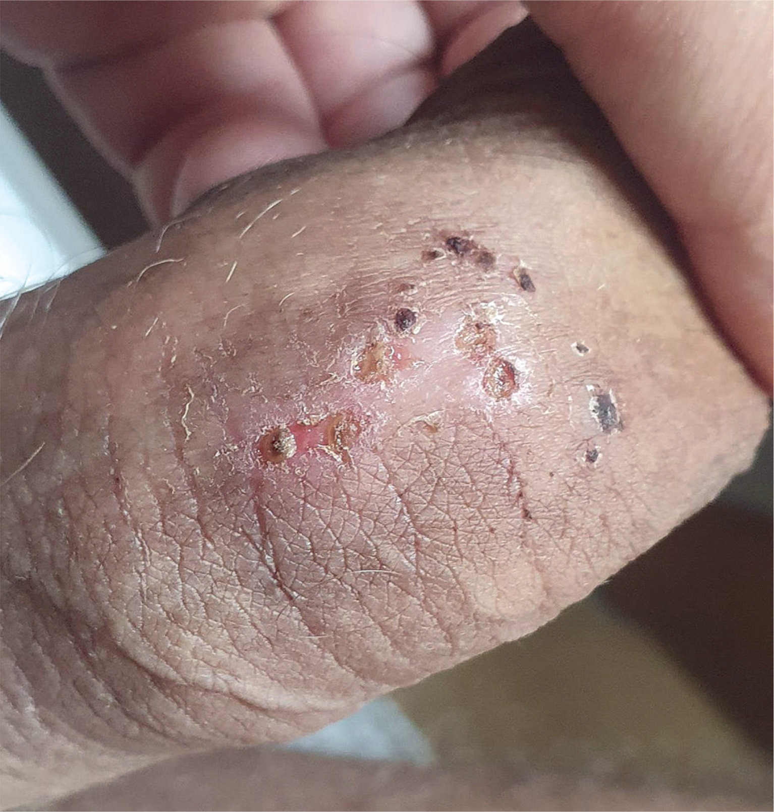
The presence of small vesicles on the glans penis, accompanied by pruritus and pain, also necessitated consideration of HSV-2 infection [10, 11]. Herpes zoster was considered the least likely diagnosis because VZV reactivation involving the sacral ganglia is uncommon. Involvement of the sacral nerves may cause ulcers on the penis and scrotum within the S2 and S3 dermatomes [6]. VZV accounts for less than 3% of genital ulcers cases, which makes the condition difficult to recognize [6, 12]. Although the findings made another STI less likely, screening remained essential given the reported sexual exposures and the possibility of coinfection. Repeat testing should be performed according to the diagnostic window period for each pathogen.
Another significant aspect of the case was the patient’s elevated liver enzyme levels, including an alanine aminotransferase level of 60 U/l (reference range: 4–50 U/l) and a γ-glutamyltransferase level of 169 U/l (reference range: 15–73 U), which prompted further investigation of possible hepatic involvement. Although the patient denied psychoactive substance use, Rosińska et al. showed that 30% of MSM report drug use during sexual encounters [13]. Abdominal ultrasonography revealed hepatic steatosis, which was considered the likely cause of the abnormal liver enzyme levels [9]. Alcohol and other psychoactive substances use may be associated with higher-risk sexual behaviors; therefore, assessment of substance use and metabolic risk factors in patients with abnormal liver tests should form part of a comprehensive evaluation.
In conclusion, the diagnosis of genital lesions is now a multifaceted challenge requiring a comprehensive medical history that includes lifestyle, sexual health, chemsex practices, and PrEP use. Moreover, detailed vaccination status, including vaccination against mpox and VZV, should be verified. This case emphasizes the importance of modern diagnostic strategies in patients presenting with genital lesions, particularly those with a history of high-risk sexual behavior. Our experience also underscores the need for a multidisciplinary approach combining infectious disease expertise, dermatological evaluation, and preventive medicine to achieve accurate diagnosis and optimal patient care.












