
Introduction
Coronary artery bypass grafting (CABG) remains the surgical benchmark in the management of patients with extensive coronary artery disease [1]. The reason for the increasing shift towards off-pump coronary artery bypass (OPCAB) surgery in the last couple of decades is two-fold – to avoid the deleterious effects of cardiopulmonary bypass (CPB) and to reduce the operation cost in developing countries [2, 3].
The introduction of dedicated instruments to optimally stabilize the coronary targets on a beating heart paved the way for the successful performance of OPCAB surgery [4]. In 1994, Borst et al. [5] developed the Octopus Tissue Stabilizer, which utilizes suction to immobilize the epicardium. In 1995, after several animal experiments, Jansen et al. [6] successfully used the Octopus Tissue Stabilizer (Medtronic Inc., Minneapolis, MN) to perform OPCAB surgery in humans. The Octopus Tissue Stabilizer consists of two suction paddles that are placed in parallel on either side of the coronary artery and it utilizes suction pressure of 300–400 mm Hg to effectively immobilize the target site. The Octopus Tissue Stabilizer in use today has evolved with several design improvements, including a lower profile, flexible heads and disposable, transparent paddles that are incorporated into a one-piece system that can be mounted directly on to the sternal retractor.
However, despite the advances in instrumentation, serious hemodynamic changes and subsequent myocardial ischemia and a decrease in left ventricular (LV) performance can be induced while displacing the beating heart and restricting the cardiac motion to expose the planned anastomosis site during off-pump coronary bypass grafting (OPCAB). The mechanism of hemodynamic instability is a temporary decrease of right ventricular (RV) output and, as a result, a decrease of LV output. If this unstable situation persists too long without proper correction, circulation will not normalize and grafting will be impossible [7]. Such hemodynamic derangements may necessitate conversion to on-pump CABG.
Aim
We therefore proposed to evaluate the hemodynamic alterations during the course of anastomosis in OPCAB surgery using the Octopus tissue stabilizer.
Material and methods
After receiving approval from the Institutional Review Board and obtaining patients’ consent, a prospective study was undertaken. A hundred consecutive patients undergoing OPCAB surgery were studied. Patients receiving at least 3 grafts were included in the study. Patients undergoing emergency CABG, concomitant aneurysmectomy and those with low left ventricular ejection fraction (LVEF) < 25%, pre-existing hepatic and renal dysfunction were excluded from the study.
OPCAB technique
The left internal mammary artery (LIMA) and veins were used in all the cases. Intravenous heparin 1 mg/kg was administered after the dissection of the LIMA to maintain the activated clotting time (ACT) > 250 s during anastomosis. A pericardial suture was placed in the posterior aspect of pericardial reflection between the left and right pulmonary veins. To expose the lateral side of the heart and elevate the apex, two deep pericardial stay sutures were placed between the atrioventricular groove and left inferior pulmonary vein. The tissue stabilizer (Octopus Tissue Stabilization System, Medtronic, USA) was applied to immobilize the target site of coronary anastomosis. Coronary anastomosis was performed in order of the left anterior descending artery (LAD), obtuse marginal branch (OM), and posterior descending artery (PDA)/right coronary artery (RCA). During displacement of the beating heart, the patients were placed in a 20–30° head-down position. Norepinephrine 0.03–0.05 mg/kg per min was infused intermittently if mean systemic arterial pressure (MAP) decreased below 60 mm Hg and epinephrine 0.03–0.05 mg/kg per min was used intermittently in case of bradycardia. Intracoronary shunts to maintain the patency of the coronary arteries during the course of anastomosis were used in all cases.
Hemodynamic monitoring
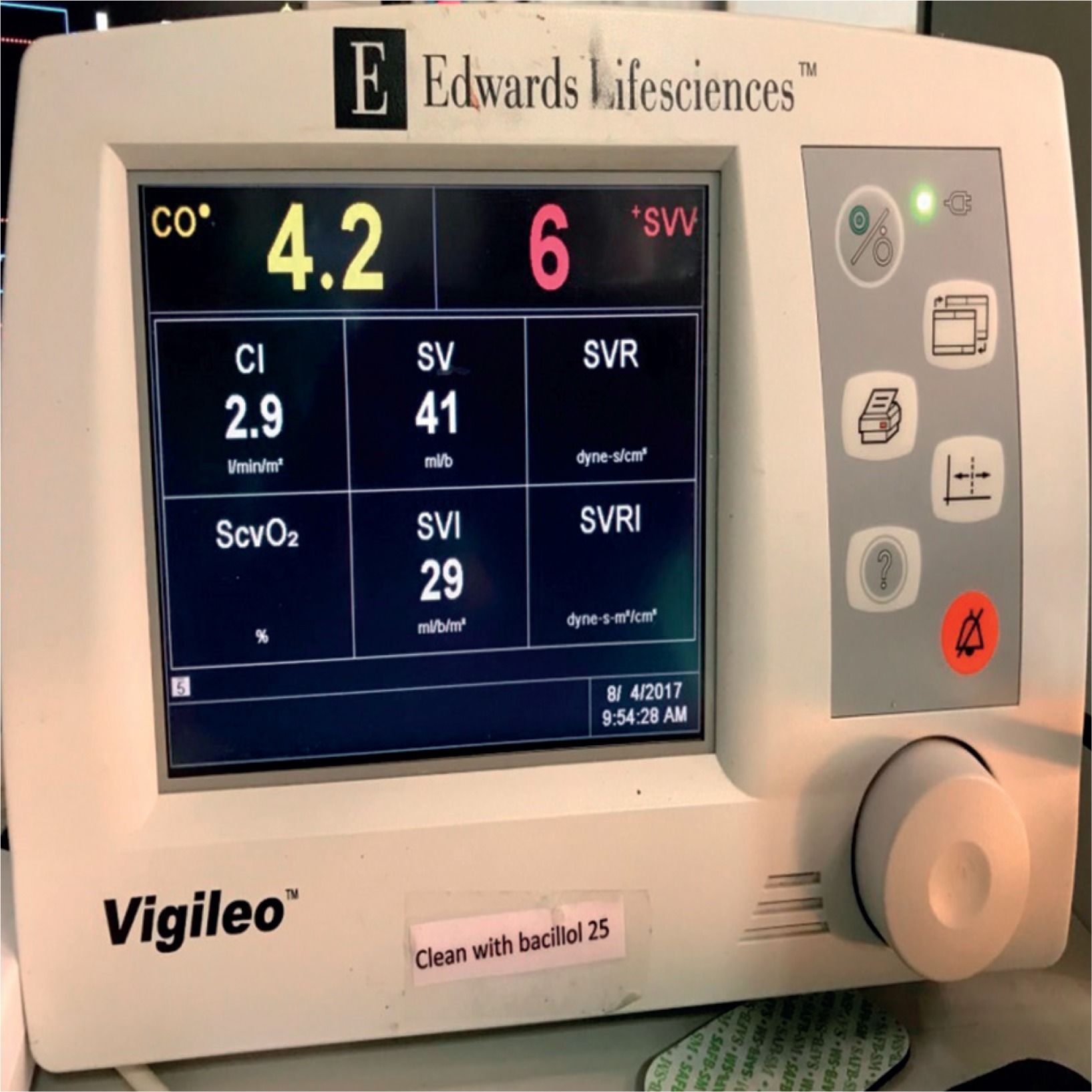
The hemodynamic variables including cardiac output (CO), heart rate (HR), mean arterial pressure (MAP) and central venous pressure (CVP) were recorded upon pericardiotomy, during each coronary artery anastomosis at intervals of 2 min, 10 min and after release of the Octopus tissue stabilizer. The non-invasive FloTrac (Vigileo system, Edwards Lifesciences Inc) was used to measure CO (Fig. 1).
Statistical analysis
All hemodynamic variables were expressed as the mean value ± standard deviation (SD). Hemodynamic changes for each coronary anastomosis were compared to baseline measures using repeated measures of ANOVA. The comparisons of variables between coronary arteries were analyzed with one-way ANOVA. A p-value of < 0.05 was considered as statistically significant. Data analysis was performed with SPSS 19 statistical software for windows (version 19; IBM Corp., NY, USA).
Results
The demographic characteristics of the entire cohort are presented in Table I. All patients received grafts to the LAD, OM and PDA or RCA. The mean number of grafts per patient was 3.21 ±0.48. There were a total of four conversions (4/100) to on-pump CABG in our study. Seven patients (7/100) required intra-aortic balloon pump (IABP) support, which permitted the completion of OPCAB surgery without the need for conversion to on-pump surgery.
Table I
Patient characteristics
| Variable | No. of patients(n = 100) | Percentage |
|---|---|---|
| Age [years] | 61.8 ±5.9* | |
| Sex (M/F) | 58/42 | 58/42 |
| Coronary artery involvement: | ||
| 3-vessel disease | 81 | 81 |
| Left main and 2-vessel disease | 4 | 4 |
| Left main and 3-vessel disease | 15 | 15 |
| Segmental wall motion abnormality: | ||
| Hypokinesia | 38 | 38 |
| Akinesia | 14 | 14 |
| Pre-operative medications: | ||
| β-Blockers | 93 | 93 |
| Calcium channel blockers | 66 | 66 |
| Angiotensin converting enzyme inhibitors | 39 | 39 |
| Diuretics | 28 | 28 |
| Comorbid conditions: | ||
| Hypertension | 43 | 43 |
| Diabetes mellitus | 24 | 24 |
| Previous myocardial infarction | 19 | 19 |
| Stroke/transient ischemic attack | 12 | 12 |
| Previous PTCA | 16 | 16 |
| Intra-operative variables: | ||
| No. of grafts received per patient | 3.21 ±0.48* | |
| Patients requiring intra-aortic balloon pump support | 7 | 7 |
| Conversion to on pump CABG | 4 | 4 |
The hemodynamic data pertaining to the intra-operative variability of cardiac output during the course of coronary anastomoses are presented in Table II. The baseline CO at pericardiotomy was 5.42 ±1.1 l/min. The mean drop in the cardiac output from the baseline noted in all territories after target stabilization (4.26 ±1.02 l/min at 2 min and 3.92 ±0.98 l/min at 10 min) was 21.4% and 27.8% at 2 and 10 min respectively and was statistically significant (p < 0.001). This decrease was the greatest during the anastomosis of the OM branch of the left circumflex artery (3.67 ±0.86 l/min at 2 min and 3.38 ±0.78 l/min at 10 min), which was a drop of 32.3% and 37.8% at 2 and 10 min respectively. The fall in CO was lowest during LAD anastomosis (4.38 ±0.77 l/min at 2 min and 4.06 ±0.76 l/min at 10 min) and intermediate during the anastomosis of RCA or PDA (3.92 ±0.81 l/min at 2 min and 3.81 ±0.79 l/min at 10 min). The recovery of cardiac output back towards the baseline was noted consistently in all territories, upon the release of the tissue stabilizer, following the completion of the anastomosis (5.07 ±0.80 l/min).
Table II
Intra-operative cardiac output
| Coronary territory | Cardiac output (baseline) | Cardiac output(2 min) | Cardiac output(10 min) | Cardiac output(release) |
|---|---|---|---|---|
| LAD | 5.42 ±1.1 | 4.38 ±0.77* | 4.06 ±0.76* | 5.18 ±0.78 |
| OM | 5.42 ±1.1 | 3.67 ±0.86* | 3.38 ±0.78* | 4.89 ±0.80 |
| PDA/RCA | 5.42 ±1.1 | 3.92 ±0.81* | 3.81 ±0.79* | 4.96 ±0.81 |
Inotropic drugs were required to maintain mean arterial pressure (MAP) > 60 mm Hg in 43 patients. Almost three-fourths (74.4%) of these were accounted for during OM anastomoses (p < 0.001), as presented in Table III. The table also shows the incidence of bradycardia requiring inotropes, which was noted to be the highest (11/16 patients) during LAD anastomosis (p = 0.002). The central venous pressure was maintained without any statistically significant intra-operative changes during all the coronary anastomoses.
Discussion
Previous studies investigating the hemodynamic effects of octopus tissue stabilizer in OPCAB surgery have used transesophageal echocardiography (TEE) as done by Mathison et al. [8] or a thermodilution pulmonary artery (PA) catheter as performed by Kwak et al. [9] for the estimation of ventricular function and cardiac output. However, TEE use has limitations during the positioning of the heart for anastomosis, and measuring right ventricular (RV) contractility is difficult because of the complex shape of the RV. Likewise, thermodilution technique is a non-specific tool to assess RV function and it does not provide real-time information regarding changes in RV volume and cardiac output.
The present study, therefore, is the first study to evaluate the effect of cardiac stabilization on cardiac output in OPCAB surgery using a non-invasive monitor – the FloTrac system. Jo et al. [10] in 2011, and more recently Joosten et al. [11] in (2017), have reported excellent reliability of FloTrac/Vigileo system in the measurement of CO, even in patients with low LVEF and showed reliability at all the time points of measurement. Hence, we believe that the FloTrac monitor, being the least invasive, would be the choice of care in the real-time monitoring of CO in OPCAB surgery.
In line with the separate studies done previously by Mathison et al. [8], Kwak et al. [9] and Shinn et al. [12], which reported higher incidence of hemodynamic instability during lateral wall (OM) anastomosis, our study confirmed the same. Shinn et al. [12] suggested that the drop in CO during revascularization of the OM artery seemed to be associated with mechanical interruption caused by manipulation of the heart rather than the progression of myocardial ischemia. Mathison et al. [8] reasoned that RV dysfunction induced by direct ventricular compression is the main cause of hemodynamic instability as observed with TEE and reported that displacement and compression of the RV to a greater extent than the LV is responsible for hemodynamic alterations when using suction type stabilizers. Also, the degree of increase in RV wall tension is greater than that in the LV and since the RV is more affected by compression compared to LV, due to the thinner RV wall and lower pressure, it is easy to be sandwiched between the thicker LV wall and pericardium when the heart is displaced. Coronary occlusion during the anastomosis can have additional effects on the LV function, depending on the status of collateral flow. These hemodynamic changes either secondary to the stabilization technique or due to transient ischemia represent an important diagnostic challenge during OPCAB surgery.
A unique finding of our study that has not been reported in previous studies is the higher incidence of bradycardia during LAD anastomosis. This can be explained on the basis of our operative strategy wherein LAD is the first in the order of anastomosis, apart from our routine practice of placing a mop beneath the LV to lift it anteriorly. The incidence of bradycardia drops with subsequent anastomoses, following the revascularization of the anterior and the anterolateral wall.
The results of our study draw attention to the risk of detrimental effects of sustained hemodynamic alterations on the heart as well as other major organ function, especially if the total time required for anastomosis is prolonged and hemodynamic stability gets worse. In this study, CO decreased well below 70% of the baseline (67.7% at 2 min and 62.2 % at 10 min) during the period of OM anastomosis. MAP requiring inotropes, so as to continue with the surgical revascularization, was encountered in 43/100 patients. However, intravenous fluid loading, Trendelenburg position, appropriate inotropes and finally the use of intra-aortic balloon pump (IABP) support enabled the successful completion of OPCAB surgery in 96% of the patients.
Our study has the limitation in being a single-centre study with a small population of 100 patients. The diagonal artery was clubbed with the LAD, while the distal right coronary artery (RCA) was clubbed with the PDA, and no separate recordings were made. Patients with severely impaired LV function (LVEF < 25%) were excluded from the study. Therefore, larger multi-centre trials are necessary to further validate our findings.
Conclusions
We found that OPCAB surgery is feasible in the vast majority of patients even with hemodynamic instability with appropriate inotropic management or additional IABP support. In this regard, the role of effective communication between the surgeon and the anesthesiologist in guiding the stabilization cannot be overstated. Therefore, careful and vigilant monitoring and management of hemodynamic variables during OPCAB surgery are of paramount importance to avoid conversion to on-pump surgery.


