
Introduction
Ovarian cancer is the fifth in terms of morbidity (incidence of 5%) and the fourth in terms of mortality (6% of cancer deaths) in cancer among women in Poland [1], the latter being over 15% higher than the average for EU countries [1]. This high rate is associated with late diagnosis. About 75% of patients are diagnosed at clinical stages III or IV because clinical symptoms are non-specific and appear when the tumour achieves significant progress [2-5]. There are some risk factors known for ovarian cancer such as infertility, low number of pregnancies, early menarche, late age of menopause, high BMI, long-term use of hormone replacement therapy, and genetic mutations [6]. Recently, the detection rate of ovarian lesions has increased, as imaging studies have become increasingly popular with different indications. Gynaecological ultrasonography is the most common ovarian cancer examination method [7].
In order to evaluate pathological changes of the ovary, it is recommended to apply the ultrasound International Ovarian Tumour Analysis (IOTA) simple rules [8]. Since 2009, the IOTA collaboration has standardised the adnexal pathology ultrasound description approach improving it in the last few years [9]. The rules are divided into two groups of features: B-rules and M-rules. B-rules for predicting a benign tumour consist of the following characteristics: unilocular, presence of solid components with the largest diameter < 7 mm, presence of acoustic shadows, smooth multilocular tumour with the largest diameter < 100 mm, and no blood flow. M-rules for predicting a malignant lesion comprise the following: irregular solid tumour, presence of ascites, at least four papillary structures, irregular multilocular solid tumour with the largest diameter ≥ 100 mm, and very strong blood flow [10-13]. If one or more M-rules apply in the absence of a B-rule, a mass is predicted as malignant (rule 1). If one or more B-rules apply in the absence of an M-rule, a mass is predicted as benign (rule 2), whereas if both M-rules and B-rules apply or no rule applies, the mass is classified as inconclusive (rule 3).The aim of the study was to evaluate how IOTA simple rules used in ultrasound examination estimate the probability of malignant and benign tumour occurrence in the studied population.
Material and methods
The study was performed on a group of 425 patients with ovarian tumours operated in the Department of Surgical and Oncological Gynaecology at the Medical University of Lodz in the years 2014-2015. Patients without complete data available were excluded, and thus the final study group consisted of 401 patients.
Based on subjective examination, we included the following in our database: IOTA ultrasound criteria (B-rules and M-rules) as originally published elsewhere [10-13]; postoperative histopathological results of adnexal tumours; and established risk factors of ovarian cancer such as: age, BMI, age of menarche, age of menopause, number of pregnancies, cancer antigen 125 (CA 125) concentration, nicotine addiction and comorbidities. Adnexal tumour appearance on ultrasonography (US) was rated according to IOTA simple rules classifying tumours as malignant, benign, or unclassified lesions. The results of the study were compared with final histopathological results.
We performed a statistical analysis using STATISTICA 13 PL with Medical Pack. The utilised methods comprised the following: the analysis of variance with the post-hoc Tukey test, the Kruskal-Wallis test with post-hoc multiple comparisons test, receiver operating characteristics curves, Bayesian analysis of diagnostic parameters, and logistic regression.
Results
Three groups were identified based on histopathological results:
In the statistical analysis, borderline tumours were grouped with malignant mases. The characteristics of the groups are presented in Table 1. There are several factors gathered in the table. Women with malignant tumours were significantly older (mean age 60.95 years), had higher BMI (mean BMI 27.28), more pregnancies, higher CA 125 concentrations (mean concentration 251.5 IU/ml), and more often suffered from diabetes and hypertension compared to women identified with benign tumours. Differences in menarche and menopause age and the proportion of smokers were statistically non-significant (Table 1). The association of age of the patients and malignancy status was also found in other studies of ovarian tumours [14].
Table 1
Comparison of examined variables for groups determined by histopath
Based on the three rules (presented in the introduction), it was determined how the IOTA system could be used to modify the probability of a malignant or benign ovarian change in the studied population.
The purpose of the rule 1 is to detect a malignant change.
Patients classified as IOTA 1 were almost five times more likely to have a malignant lesion than patients not classified as IOTA 1 (positive predictive value [PPV] increased from 11% to 51%). Among patients not classified as IOTA 1, the probability of a non-malignant lesion increased from 89% to 97% (negative predictive value – NPV) (Table 2).
Table 2
IOTA rule 1. IOTA predictive value analysis of rule 1 in malignant ovarian lesion
| Malignant | |||||
|---|---|---|---|---|---|
| Yes | No | Total | |||
| IOTA 1 | Yes | 35 | 33 | 68 | PPV 51% |
| No | 7 | 247 | 254 | NPV 97% | |
| Total | 42 | 280 | |||
| Sensitivity 83% | Specificity 88% | ||||
In patients classified as IOTA 1 or 3, the probability of a malignant lesion increased from 11% to 35%. Being excluded from category 1 or 3 increased the probability of a non-malignant lesion from 89% to 99% (NPV). The method increased the sensitivity and NPV at the expense of PPV and specificity. The calculations indicate that the IOTA rule is useful in evaluating ovarian tumour malignancy (Table 3).
Table 3
IOTA rule 1 + 3. IOTA predictive value analysis of the 1 + 3 rule for malignant ovarian lesion
| Malignant | |||||
|---|---|---|---|---|---|
| Yes | No | Total | |||
| IOTA 1 or 3 | Yes | 40 | 77 | 117 | PPV 34% |
| No | 2 | 203 | 205 | NPV 99% | |
| Total | 42 | 280 | |||
| Sensitivity 95% | Specificity 73% | ||||
Determination of the predictive value of CA 125 for comparison with IOTA
CA 125 concentration was statistically significantly higher in women with malignant lesions. Comparing two women – one with a malignant lesion, the other with a benign one – there was 87% probability that the woman with a malignant lesion had a higher CA 125 concentration than the woman with a benign lesion (Table 4 and Fig. 1).
Table 4
Parameters of ROC curve for Ca 125 in the prediction of ovarian cancer change in the examined group
| Variable: CA125 | |||||
|---|---|---|---|---|---|
| AUC | SE | AUC lower 95% | AUC upper 95% | z = (v1-0.5) / v2 | p |
| 0.867 | 0.034 | 0.8 | 0.934 | 10.75 | 0.0000 |
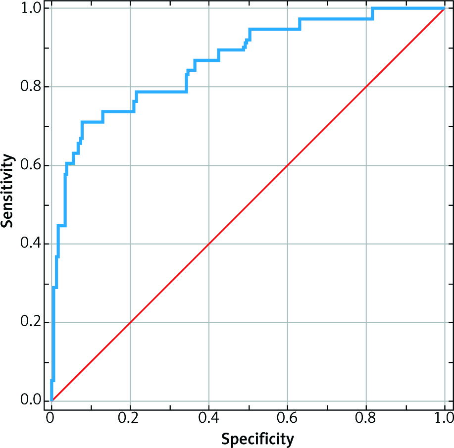
Based on the Youden index (a parameter calculated based on the highest specificity and sensitivity sum), the optimum cut-off for CA 125 was 138.5. Using that point, an analysis of the predictive value for the parameter was performed. In patients with CA 125 concentrations ≥ 138.5, the probability of a malignant change increased from 11% to 56% (PPV). In patients with CA 125 concentrations < 138.5, the probability of a non-malignant lesion increased from 89% to 96% (NPV).
A hierarchical logistic regression analysis indicated that the probability of a malignant lesion increased 36-fold when the IOTA 1 criterion was met. If the CA 125 concentration was taken into account along with IOTA rule 1, the probability of malignancy increased 51 fold. The third model included risk factors for ovarian cancer other than IOTA and CA 125. In such a case, meeting IOTA rule 1 indicated a 46-fold increase in malignant tumour occurrence regardless of age, BMI, number of pregnancies, hypertension, and CA 125 level.
The comparison of areas under the curve (AUC) for each model provided additional information. Each model was applicable for predicting malignant lesions in the studied population (AUC > 0.7). Each model had a higher AUC, which implied that taking into consideration additional risk factors increased the predictive value. However, a statistically significant difference occurred between models 1 and 3 (based on the 95% confidence interval) (Table 5).
Table 5
Hierarchical logistic regression analysis – malignant lesion prediction in the tested group
Discussion
There is a broad discussion nowadays on how to diagnose ovarian masses and by whom the diagnosis should be established. Different strategies are known worldwide with general surgeons, sonographers, and physicians carrying out the first ultrasound examination. In Poland, for many years, gynaecologists or radiologists have diagnosed ovarian masses in the first step of the diagnostic schema [15].
Transvaginal ultrasonography is the most common examination for the assessment of adnexa. Computed tomography (CT) and magnetic resonance (MR) are not more specific or sensitive compared to transvaginal ultrasonography in the assessment of adnexa, while being less accessible, more expensive, and exposing the patient to radiation [16-18]. For that reason, ultrasonography is considered to be the most appropriate approach for diagnosing adnexal masses, especially with IOTA standards. In addition to US, it is suggested that the CA 125 concentration be determined in ovarian tumour diagnosis. Significantly increased marker levels facilitate diagnosis, but normal levels do not exclude it because of the test being non-specific [19]. Another common laboratory test is CA 125 combined with HE 4 (human epididymis protein 4) [20-22].
The CA 125 concentration may increase in many conditions, such as endometriosis, uterine myomas, and pelvic inflammatory diseases. In our study, the cut-off value of 138.5 IU/ml for CA 125 was discriminating for our population. One should keep in mind that this is only a statistical cut-off point, and this value could be influenced by other factors, such as the number of patients with advanced ovarian cancer or advanced endometriosis in the studied population. Therefore, its low specificity does not allow it to be used as a single preoperational diagnostic method. On the other hand, the HE4 level does not increase in endometriosis and more correctly identifies benign lesions, which might indicate its usefulness in differential diagnosis and help limit disease overtreatment [23, 24].
Other markers are also being tested in trials, such as carcinoembryonic antigen (CEA), caudal type homeobox 2 (CDX2), cancer antigen 72-4 (CA 72-4), cancer antigen 19-9 (CA 19-9), alpha-fetoprotein (AFP), lactate dehydrogenase (LDH), and β-chorionic gonadotropin. It is not recommended that their levels be measured in the initial diagnostic process. Their sensitivity is relatively low, and the specificity varies depending on the combination of each marker with CA 125 concentration [25]. To fuse laboratory and imaging data, the ROMA test was implemented, but its usefulness strongly depends on the menopausal status of examined women.
The assessment of adnexa according to the IOTA system has been proven to show high sensitivity and specificity [26-28]. The aim of our study was to evaluate whether the IOTA system in combination with CA 125 concentration and clinical data is useful in diagnosing ovarian tumours.
The results suggest that the IOTA rule was superior to CA 125 in detecting malignant lesions, although the CA 125 concentration proved to be superior in excluding malignant lesions. Their usefulness increased when complementary methods were considered along with the clinical presentation of the patient. The advantage of our study was that the study groups were large enough to be representative, although it must be pointed out that there was no randomisation among control groups. Patients included in our study were referred to hospital for operative treatment. However, there were no data regarding which patients presented for US evaluation with clinical symptoms and which were routinely examined. What is more, some patients were not referred to hospital for surgical treatment, but rather were treated conservatively or scheduled for US re-examination – those patients were not included in our study. A disadvantage of our study was that ultrasonography was done by clinicians with different expertise, which might have influenced results of US examination. However, Tinnangwattana et al. stated that IOTA criteria are simple enough so that the clinician’s experience does not impact the examination results [29]. On admission to a reference hospital, another evaluation of adnexa should be performed.
The use of simple IOTA rules in our centre did not modify the procedure in relation to patients qualified for surgery based on the experience of a specialist in oncological gynaecology (evaluating a tumour in US, in a clinical presentation, and risk factors for cancer individually in each patient). Therefore, it is reasonable to implement IOTA rules in pre-hospital care, which is confirmed by literature reports [30].
Conclusions
To sum up our study, IOTA rules are a better diagnostic tool to confirm a malignant tumour than CA 125 concentration alone, because of its high sensitivity. CA 125 is better for excluding a malignant tumour due to its higher specificity. Both methods can be used simultaneously because of being easily available. While assessing the malignancy of a tumour, other risk factors for ovarian cancer should also be taken into account such as: age, high BMI, number of pregnancies, hypertension, or diabetes. IOTA rules implemented in a specialised oncological centre would not have changed the preoperative preparations because there was no case with malignant disease in our material not suspected before surgery.


