eISSN: 1897-4309
ISSN: 1428-2526
Contemporary Oncology/Współczesna Onkologia
Current issue Archive Manuscripts accepted About the journal Supplements Addendum Special Issues Editorial board Reviewers Abstracting and indexing Subscription Contact Instructions for authors Ethical standards and procedures
Editorial System
Submit your Manuscript
SCImago Journal & Country Rank
1/2019
vol. 23
 
Share:
Share:
Original paper

Evaluation of the effectiveness of physiotherapy in patients after oncological breast cancer treatment based on spirometric indicators

Iwona Kulik-Parobczy
1

1.
Institute of Physiotherapy, Faculty of Physical Education and Physiotherapy, Opole University of Technology, Opole, Poland
Contemp Oncol (Pozn) 2019; 23 (1): 47-51
Online publish date: 2019/02/18
Article file
- Evaluation.pdf  [0.08 MB]
Get citation
 
PlumX metrics:
 

Introduction

Advances in early detection and improved treatment of breast cancer have resulted in increased chances of survival after diagnosis, resulting in a lot of women living with the consequences of cancer treatment [1]. The adverse side effects of chemotherapy treatment include, among others, pain, fatigue, cachexia (wasting syndrome), diminished strength and lung capacity, and reduced range of movement [25].

There is evidence that women successfully treated for breast cancer have ongoing morbidity with symptoms of dyspnoea, exercise intolerance (reduced exercise tolerance as measured by peak oxygen consumption), and reduced physical capacity [610]. Effort dyspnoea and exercise intolerance in breast cancer survivors are associated not only with increased ventilatory demand secondary to deconditioning, but also with reduced inspiratory capacity (IC) and inspiratory muscle weakness [11].

Currently, physiotherapy forms the main intervention technique applied in preventing these complications. Respiratory physiotherapy forms a core specialty within physiotherapy and plays a key role in treatment of patients with respiratory diseases [12, 13]. A variety of interventions, including pulmonary rehabilitation, early mobilisation, and airway clearance techniques have demonstrated beneficial effects on symptoms associated with respiratory diseases. These include enhanced pain management, sputum clearance and cough efficacy, reduced dyspnoea, and improved physical fitness [12]. The aim of this study involved the evaluation of the effects of physiotherapy in patients following oncological treatment, in particular including determination of whether rehabilitation in patients resulted in improvement of spirometric indicators and decreased occurrence of respiratory disorders. The assessment of the effect of the duration of the intervention program on ventilation activity of lungs was also performed.

Material and methods

The research involved patients after breast cancer treatment qualified for outpatient physiotherapy at the Rehabilitation Unit at the Cancer Centre and Institute of Oncology in Gliwice in 2012–2013. In total 91 patients underwent rehabilitation. The characteristics of this group are summarised in Table 1.

Table 1

Selected characteristics of qualified rehabilitated patients

Risk factorMean ±SD (range)Median
Age57.1 ±10.5 (26-74)59
Tumour size (cm3)2.5 ±1.6 (0.1-9)2.0
BMI (kg/m2)27.3 ±4.9 (14.5-43.3)26.7
Time RT (days)36.4 ±5.7
Risk factorFraction
Degree (T)(0) = 0%(1) = 40%(2) = 43.5%(3) = 10.6%(4) = 5.9%
Degree (N)(0) = 21.4%(1) = 78.6%
Oestrogen recept. stat.(0) = 15.8%(1) = 7.9%(2) = 19.7%(3) = 56.6%
Progestogen recept. stat.(0) = 31.6%(1) = 14.5%(2) = 17.1%(3) = 36.8%
HER recept. stat.(0) = 51.4%(1) = 21.6%(2) = 14.9%(3) = 12.2%
Menopause statusPerimenopause = 12.2%Menopause = 87.8%
Type of surgeryMastectomy = 82%Breast conserving therapy = 18%
RT (dose)50 Gy = 75%60 Gy and > 25%

It can be seen in Table 1 that following the classification of malignant tumours, the prevailing tumour types were T = 2 and N = 1. A vast majority of patients had an expression of oestrogen receptors (3) and reached menopause. All women were subjected to radiation therapy, and more than half underwent concurrent chemotherapy. Patients participated in rehabilitation were on average two and a half years after the completion of radiotherapy. On average, physiotherapy took approximately three weeks and it was performed following the physiotherapy treatment. The respiratory disorders in patients were assessed based on the spirometric tests including the following air flow indicators: forced expiratory volume in one second (FEV1), vital capacity (VC), forced vital capacity (FVC), peak expiratory flow (PEF), ratio of forced expiratory volume in one second to forced vital capacity (FEV1/FVC), and forced expiratory flow (FEF). The resulting material was used for statistical analysis.

Statistical analysis

Student’s t-test for dependent observations for time intervals (preceding and following rehabilitation) was applied in the study. The assessment of the examined clinical factors on ventilation indicators relied on a linear model, which determines the relation between the dependent variable Y and the set of predicting factors X in the following form: Y = b0 + b1X1 + b2X2 + … + bkXk.

In this equation b0 is an intercept, and bi represents the regression coefficients (slopes) for explanatory variables (from 1 to k). In the statistical analysis, only univariate and bivariate regressions were considered.

Results

The study involved the use of Student’s t-test to compare the results before and after rehabilitation with regard to patients subjected to breast-conserving treatment and those after mastectomy.

Based on the results reported in Table 2, statistically significant differences in means of spirometric indicators before and after rehabilitation were estimated, especially in patients who underwent mastectomy. Linear univariate models of statistically significant effects of spirometric indicators and selected clinical factors on ventilation of lungs are reported in Table 3.

Table 2

Results of Student's t-test for subgroups of patients

Type of surgery
Breast conserving therapy
Mastectomy
Rehabilitation statusBeforeAfterBeforeAfter
Mean ±SDMean ±SDt statisticp-valueMean ±SDMean ±SDt statisticp-value
FEV1 (1)2.32 ±0.662.51 ±0.51-4.170.00242.24 ±0.422.57 ±0.48-7.10< 0.0001
FEV1(%)96.63 ±23.10105.60 ±23.58-4.010.003197.74 ±21.48112.19 ±24.29-5.91< 0.0001
FVC (1)3.05 ±0.903.03 ±0.51-1.210.25842.99 ±0.523.09 ±0.51-2.620.0107
FVC (%)106.75 ±28.13107.50 ±22.67-1.310.2223108.74 ±20.92112.16 ±22.82-1.530.1309
PEF (l/min)341.81 ±101.96336.10 ±87.23-0.490.6381321.60 ±69.17348.96 ±62.06-3.570.0007
PEF (%)90.56 ±23.8790.20 ±22.68-0.440.669587.88 ±20.8695.48 ±18.44-3.440.0010
FEV1/FVC (%)77.63 ±11.2482.30 ±8.96-1.160.274575.12 ±8.6781.65 ±12.93-3.430.0010
FEF25 (l/s)4.65 ±1.984.81 ±1.25-0.790.44794.44 ±1.184.75 ±1.05-2.260.0269
FEF25 (%)82.63 ±32.5587.40 ±21.30-0.790.448781.82 ±23.0287.67 ±19.97-2.100.0395
FEF50 (l/s)2.67 ±0.932.95 ±0.93-2.350.04342.35 ±0.933.07 ±0.98-5.92< 0.0001
FEF50 (%)71.38 ±21.9879.70 ±24.81-2.300.046763.52 ±27.2482.65 ±24.82-5.57< 0.0001
FEF75 (l/s)0.90 ±0.461.26 ±0.68-1.750.11381.76 ±8.581.38 ±0.780.420.6736
FEF75 (%)66.63 ±30.9093.90 ±50.44-1.750.114758.81 ±35.73105.52 ±61.83-5.97< 0.0001

[i] FEV1 – forced expiratory volume in one second, FVC – forced vital capacity, PEF – peak expiratory flow, FEV1/FVC – ratio of forced expiratory volume in one second to forced vital capacity, FEF – forced expiratory flow

Table 3

Linear model of changes in lung ventilation indicators in patients without combined chemotherapy

Response variableRisk factorRegression coefficientMeanStandard errorp-value
FEV1 (%) – after rehabilitationTime of rehabilitation (days)Intercept
Slope
-23.22
1.80
10.78
0.50
0.0345
0.0006
FVC (l) – after rehabilitationNumber of birthsIntercept
Slope
0.26
-0.08
0.08
0.04
0.0009
0.0340
PEF (l/min) – after rehabilitationStatus of lymph nodesIntercept
Slope
83.14
-63.78
23.31
24.41
0.0006
0.0108
PEF (l/min) – after rehabilitationTumour size (cm3)Intercept
Slope
57.05
-12.48
13.53
4.68
0.0001
0.0094
PEF (%) – after rehabilitationStatus of lymph nodesIntercept
Slope
19.14
-13.71
6.55
6.86
0.0046
0.0492
PEF (%) – after rehabilitationTumour size (cm3)Intercept
Slope
15.88
-3.59
3.73
1.29
0.0001
0.006
FEV1/FVC (%) – after rehabilitationTime of rehabilitation (days)Intercept
Slope
-21.90
1.48
8.86
0.41
0.0158
0.0006
FEF25 (l/s) – after rehabilitationT degreeIntercept
Slope
0.96
-0.36
0.32
0.16
0.0036
0.0295
FEF25 (I/s) – after rehabilitationTumour size (cm3)Intercept
Slope
0.91
-0.23
0.25
0.09
0.0004
0.0078

[i] FEV1 – forced expiratory volume in one second, FVC – forced vital capacity, PEF – peak expiratory flow, FEV1/FVC – ratio of forced expiratory volume in one second to forced vital capacity, FEF – forced expiratory flow

The data in Table 3 show that the time of physiotherapy significantly contributed to an increase in FEV1% – approximately 1.8 units per day of rehabilitation. In contrast, lymph node status radically reduced the level of PEF by as much as 64 units. The statistically significant linear models of a possible effect of chemotherapy on lung ventilation indicators are given in Table 4.

Table 4

Linear univariate models of spirometric indicators in patients with concurrent chemotherapy

Response variablesRegression coefficientMeanStandard errorp-value
FEV1 (l) before rehabilitationIntercept
Slope
2.134
0.234
0.073
0.097
< 0.0001
0.0184
FVC (l) before rehabilitationIntercept
Slope
2.856
0.263
0.092
0.123
< 0.0001
0.0353
PEF (l/min) before rehabilitationIntercept
Slope
306
37.31
11.66
15.58
< 0.0001
0.0187
FEF25 (l/s) before rehabilitationIntercept
Slope
4.137
0.647
0.207
0.277
< 0.0001
0.0217
FEV1 (l) after rehabilitationIntercept
Slope
2.402
0.282
0.082
0.107
< 0.0001
0.0099
FVC (l) after rehabilitationIntercept
Slope
2.905
0.333
0.087
0.114
< 0.0001
0.0044
Age of lungs after rehabilitationIntercept
Slope
55.29
-8.461
2.941
3.789
< 0.0001
0.0285

[i] FEV1 – forced expiratory volume in one second, FVC – forced vital capacity, PEF – peak expiratory flow, FEF – forced expiratory flow

The results in Table 4 demonstrate the positive influence of chemotherapy on the analysed spirometric indicators before and after rehabilitation. The effect of rehabilitation on FEV1 was also estimated in a linear bivariate regression approach (Table 5).

Table 5

Linear bivariate regression of forced expiratory volume in one second (FEV1) on interval since oncological treatment and time of rehabilitation

Response variableRisk factorRegression coefficientMean95% CIp-value
FEV1 (l) before – after rehabilitationIntercept–0.198(–0.497, 0.091)0.0831
Start rehabilitation – end RT (months)Slope0.003(0.001, 0.005)0.0044
Time of rehabilitation (days)Slope0.021(0.008, 0.036)0.0038

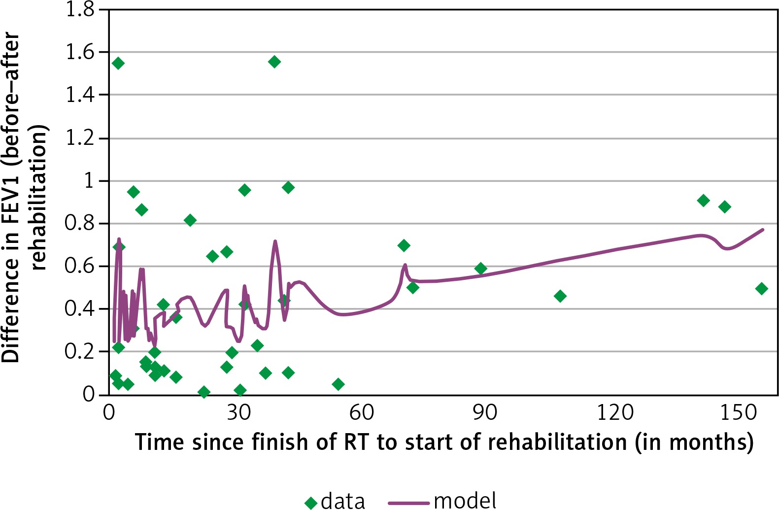
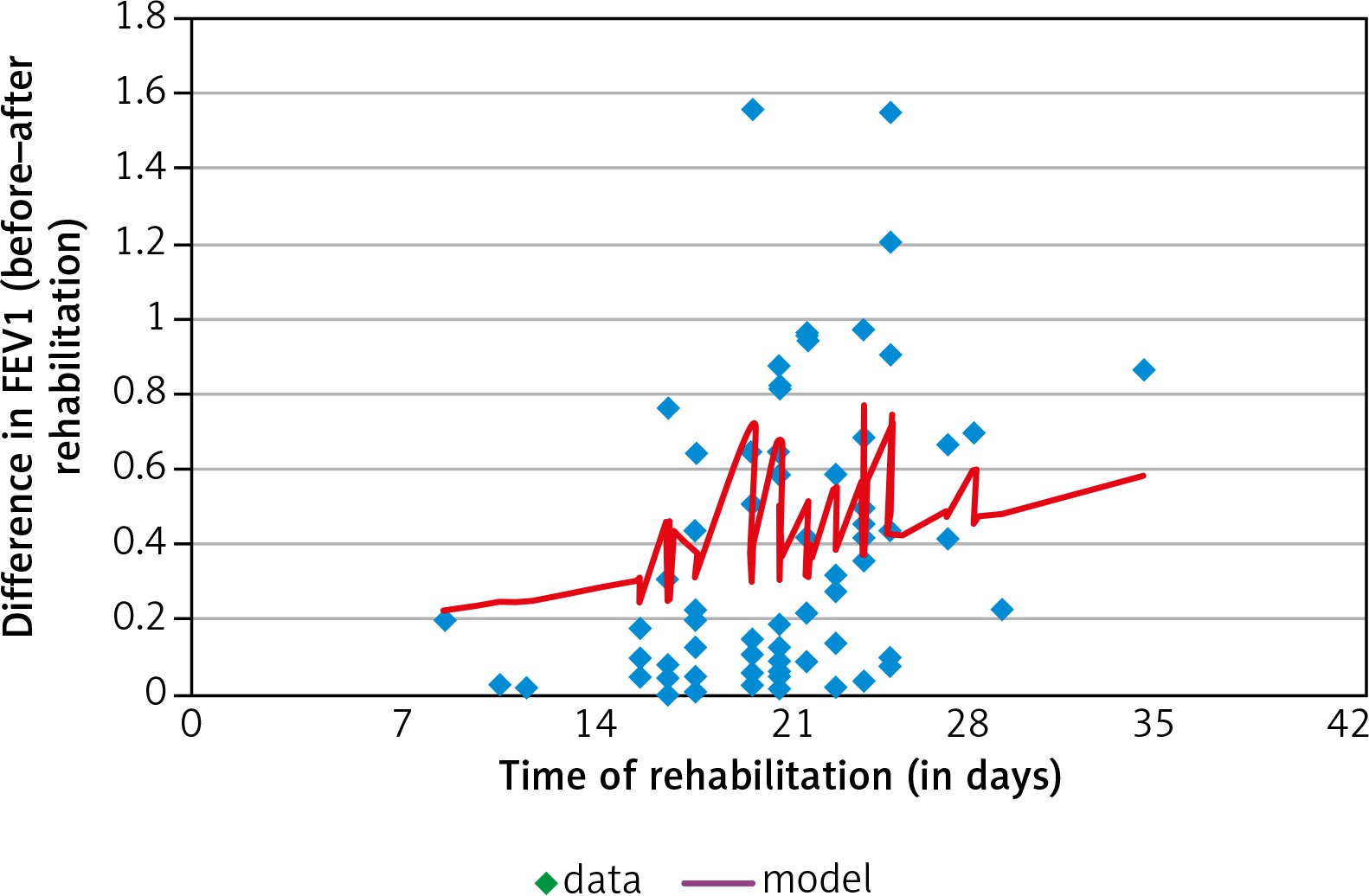
The results reported in Table 5 provide evidence of a statistically significant increase in FEV1 in patients since the end of oncological treatment (approximately 0.04 l per year of the interval). What is more, a week of rehabilitation is equivalent to half a year of the non-intervention recovery of lungs in women. These results demonstrate a significant impact of the rehabilitation on FEV1. The described model is presented graphically in Figure 1 and 2.

Fig. 1

Forced expiratory volume in one second (FEV1) increase vs. interval since the end of oncological treatment

/f/fulltexts/WO/35836/WO-23-82929-g001_min.jpg
Fig. 2

Forced expiratory volume in one second (FEV1) increase vs. time of rehabilitation

/f/fulltexts/WO/35836/WO-23-82929-g002_min.jpg

Discussion

Based on the obtained results, a possible impact of the physiotherapeutic program on the progress in spirometric functions in breast cancer patients can be established. However, overall health development was considerably stronger in radically treated women than in women who underwent breast-conserving therapy. It seems that the better rehabilitation effect is observed in radical treatment leading to a more intensive regeneration of lungs.

The estimated correlations between differences in spirometric indicators before and after rehabilitation were mainly attributable to clinical factors, i.e. tumour size and lymph node status (Table 1). This could be explained by the intensity of the conducted radiotherapy, which in turn is related to radiation injury of lungs and their stronger recovery. Similar conclusions were reached in a study [14] on FVC, FEV1, carbon monoxide in patients suffering from irradiation after breast cancer therapy. In the cited study, the spirometric tests deteriorated relatively over the period of six months following radiotherapy and returned to a level similar to the one recorded before irradiation one year after the end of observation [14].

Contrasting observations to the present study of the effect of chemotherapy with concurrent radiotherapy on the spirometric measures were found in [15]; however, they are in accordance with [16]. The latter can be explained by stronger cell regeneration and reconstruction of lung functions in patients with the more intensive medical intervention [17].

Statistical correlations were also estimated as regards spirometric efficiency with an interval since the end of oncological treatment and time of rehabilitation. These findings can be interpreted by the natural regeneration of lungs since the end of oncological treatment. However, noteworthy is the fact that a week of rehabilitation is an equivalent to half a year of the non-intervention recovery of lungs in women. This result shows significant power of the rehabilitation on lung functions after breast cancer radiotherapy.

Based on the collected material, applied methodology, and obtained statistical results, positive physiotherapeutic effects on breast cancer patients can be established. The presented findings are not alone. Similar results were previously reported in the scientific literature worldwide [1824]. They confirm the statement regarding the positive impact of rehabilitation on the condition of oncologically treated patients.

Conclusions

Physiotherapy leads to the improvement of lung function in patients with breast cancer following oncological treatment; progress is particularly evident in patients in more advanced cancer stages.

Clinical factors such as age of patients, body mass index, tumour size, nodal status, or menopause had no statistical effect on spirometry of lungs.

Time of physiotherapy played an important role in basic lung functions in breast cancer patients.

Notes

[4] Conflicts of interest The author declares no conflict of interest.

References

1 

Cancer Research UK , author. Breast Cancer Survival Statistics cited 2014. Available from: urihttp://www.cancerresearchuk.org/http://www.cancerresearchuk.org/ .

2 

Cantarero-Villanueva I, Fernández-Lao C, Fernández-de-Las-Peñas C, López-Barajas IB, Del-Moral-Ávila R, de la-Llave-Rincón AI, Arroyo-Morales M , authors. Effectiveness of water physical therapy on pain, pressure pain sensitivity and myofascial trigger points in breast Cancer survivors: a randomized, controlled clinical trial. Pain Med. 2012. 13:p. 1509–1519

3 

Hayes SC, Rye S, Disipio T, et al. , authors. Exercise for health: a randomized, controlled trial evaluating the impact of a pragmatic, translational exercise intervention on the quality of life, function and treatment-related side effects following breast cancer. Breast Cancer Res Treat. 2013. 137:p. 175–186

4 

Lee SA, Kang J, Kim YD, An AR, Kim SW, Kim YS, Lim JY , authors. Effects of a scapula-oriented shoulder exercise programme on upper limb dysfunction in breast cancer survivors: a randomized controlled pilot trial. Clin Rehabil. 2010. 24:p. 600–613

5 

Santos ALB, Marinho RC, Lima PNM, Fortes RC , authors. Subjective nutritional assessment proposed by the patient versus other methods of evaluating nutritional status in cancer patients. Rev Bras Nutr Clin. 2012. 27:p. 243–249

6 

Yee J, Davis GM, Beith JM, et al. , authors. Physical activity and fitness in women with metastatic breast cancer. J Cancer Surviv. 2014. 8:p. 647–656

7 

Burnett D, Kluding P, Porter C, Fabian C, Klemp J , authors. Cardiorespiratory fitness in breast cancer survivors. Springerplus. 2013. 2:p. 68

8 

Smoot B, Johnson M, Duda J, Krasnoff J, Dodd M , authors. Cardiorespiratory fitness in women with and without lymphedema following breast cancer treatment. Cancer Clin Oncol. 2012. 1:p. 21–31

9 

Jones W, Courneya S, Mackey R, et al. , authors. Cardiopulmonary function and age-related decline across the breast cancer survivorship continuum. J Clin Oncol. 2012. 30:p. 2530–2537

10 

Travers J, Dudgeon DJ, Amjadi K, et al. , authors. Mechanisms of exertional dyspnea in patients with cancer. J Appl Physiol. 2008. 104:p. 57–66

11 

O’Donnell DE, Webb KA, Langer D, Elbehairy AF, Neder JA, Dudgeon DJ , authors. Respiratory Factors Contributing to Exercise Intolerance in Breast Cancer Survivors: A Case-Control Study. J Pain Symptom Manag. 2016. 52:p. 54–63

12 

Bott J, Blumenthal S, Buxton M, et al. , authors. Guidelines for the physiotherapy management of the adult, medical, spontaneously breathing patient. Thorax. 2009. 64 Suppl 1:p. i1–51

13 

Stiller K , author. Physiotherapy in intensive care: An updated systematic review. Chest. 2013. 144:p. 825–847

14 

Jaen J, Vazquez G, Alonso E, León A, Guerrero R, Almansa JF , authors. Changes in pulmonary function after incidental lung irradiation for breast cancer: A prospective study. Int J Radiat Oncol Biol Phys. 2006. 65:p. 1381–1388

15 

Spyropoulou D, Leotsinidis M, Tsiamita M, Spiropoulos K, Kardamakis D , authors. Pulmonary function testing in women breast cancer treated with radiotherapy and chemotherapy. In Vivo. 2009. 23:p. 867–871

16 

Miglani A, Arora R , authors. The effect of chemotherapy on the pulmonary function tests in cancer patients with healthy lungs. J Clin Diagn Res. 2011. 5:p. 798–800

17 

Courneya K, Mackey J, Bell GJ, Jones LW, Field CJ, Fairey AS , authors. Randomized controlled trial of exercise training in postmenopausal breast cancer survivors: cardiopulmonary and quality of life outcomes. J Clin Oncol. 2003. 21:p. 1660–1668

18 

Untch M, Ditsch N, Hermelink K , authors. Immunotherapy: new options in breast cancer treatment. Expert Rev Anticancer Ther. 2003. 3:p. 403–408

19 

Woźniewski M , author; Kwolek A , author. Rehabilitacja w onkologii. Rehabilitacja medyczna. 2003. Wrocław: Urban & Partner; p. 592–619

20 

Basen-Engquist K, Taylor C, Rosenblum C, et al. , authors. Randomized pilot test of a lifestyle physical activity intervention for breast cancer survivors. Patient Educ Couns. 2006. 64:p. 225–234

21 

Irwin M, Smith A, Mc Tiernan A, et al. , authors. Influence of pre-and postdiagnosis physical activity on mortality in breast cancer survivors: the health, eating, activity, and lifestyle study. J Clin Oncol. 2008. 26:p. 3958–3964

22 

Matthews C, Wilcox S, Hanby CL, Der Ananian C, Heiney SP, Gebretsadik T, Shintani A , authors. Evaluation of a 12-week home-based walking intervention for breast cancer survivors. Support Care Cancer. 2007. 15:p. 203–211

23 

Mefferd K, Nichols J, Pakiz B , authors. A cognitive behavioral therapy intervention to promote weight loss improves body composition and blood lipid profiles among overweight breast cancer survivors. Breast Cancer Res Treat. 2007. 104:p. 145–152

24 

Pinto B, Frierson G, Rabin C, Trunzo JJ, Marcus BH , authors. Homebased physical activity intervention for breast cancer patients. J Clin Oncol. 2005. 23:p. 3577–3587

Copyright: © 2019 Termedia Sp. z o. o. This is an Open Access article distributed under the terms of the Creative Commons Attribution-NonCommercial-ShareAlike 4.0 International (CC BY-NC-SA 4.0) License (http://creativecommons.org/licenses/by-nc-sa/4.0/), allowing third parties to copy and redistribute the material in any medium or format and to remix, transform, and build upon the material, provided the original work is properly cited and states its license.
 
Quick links
© 2024 Termedia Sp. z o.o.
Developed by Bentus.