
Diaphragmatic hernia is an incidental imaging finding that may be due to congenital, traumatic, or iatrogenic causes [1]. The condition is usually benign and presents with chronic symptoms including epigastric pain, postprandial fullness, nausea, and even respiratory symptoms such as dyspnea. However, as unusual rare cases, acute respiratory distress may occur due to massive diaphragmatic hernia [2].
Post-operative diaphragmatic hernia after esophageal cancer surgery is a condition with notable morbidity and mortality. The rate of diaphragmatic hernia is especially higher in those who undergo minimally invasive esophagectomy. The condition is usually diagnosed through oncologic follow-ups for esophageal cancer [3].
The herniation of the colon in this patient may lead to strangulation of the herniated loop. In this situation, perforation of the necrotic loop and infiltration of the fecal materials are predicted. With this regard, a fecopneumothorax may happen [4, 5]. Here, we present a case of fecopneumothorax in an old man that was caused by gangrene and perforation of a herniated loop of the transverse colon.
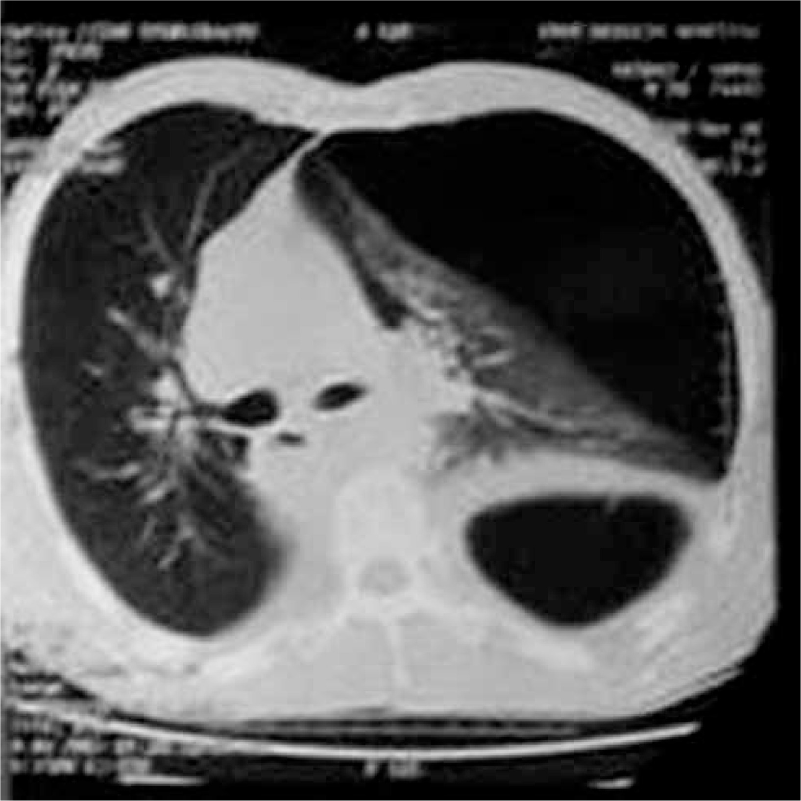
A 73-year-old man presented at the emergency room (ER) with dyspnea and chest pain that radiated to the left shoulder. He also reported a periumbilical and also hypogastric pain during the last week. However, the defecation and gas passing were normal as his routine. The assessment of past medical history revealed that the patient had undergone trans-hiatal esophagectomy along with gastric pull up surgery because of evident esophagus cancer 2 years ago. He also had a history of 30 courses of radiotherapy and 6 courses of chemotherapy. Physical examination showed that the patient had tachypnea, tachycardia, and hypotension, with no fever. The patient was previously referred to an outpatient clinic during the day of his admission to the ER and he had a high resolution computed tomography (HRCT) scan, which provided clues of a massive pneumothorax (Figure 1). Considering the imaging findings with clinical signs, a tension pneumothorax was detected. An emergency chest tube was inserted for the patient in the operating room (OR). As the patient had a diaphragmatic hernia, the tube was inserted at a higher level than usual in the fourth intercostal region and anterior axillary line. Upon the use of the chest tube, the patient’s respiratory symptoms resolved. There was no evident secretion in the chest bottle during this procedure. The patient was then admitted to the thoracic surgery ward and scheduled to undergo diaphragmatic repair for the hernia.
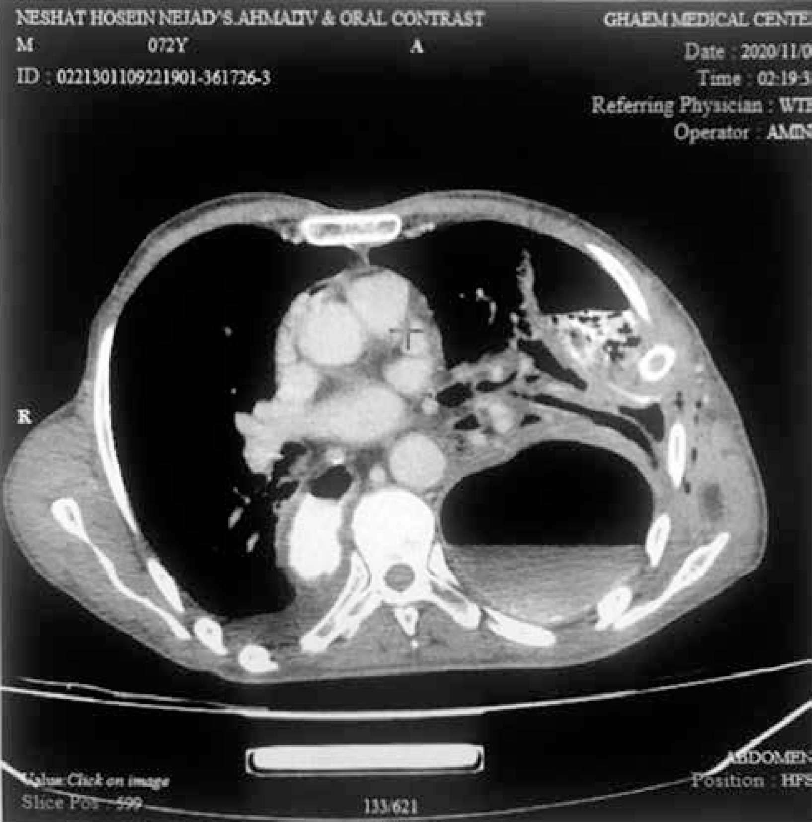
During the admission, the chest bottle secretions turned into bloody fluid, and then within 2 days the secretions turned into brownish materials. In this regard, around 24 hours after chest tube insertion, with a possible diagnosis of sepsis, antibiotic therapy with meropenem and metronidazole was initiated. Moreover, sonography was performed for the patient; it showed herniation of the transverse colon loops through the diaphragm and a mild to moderate pleural effusion with collapse consolidation of the left lung. For further complementary assessment the patient underwent abdominal and chest CT scan. The CT scan also showed a left hydropneumothorax along with collapse consolidation of the lower lobe of the left lung (Figure 2). As the contrast medium was infiltrated into the left pleural area, perforation of the left transverse colon in the pleural cavity was suspected and the patient was transferred to the OR after fluid reanimation. Then, the general condition of the patient deteriorated and thus an emergency thoracotomy was conducted. A loop of the transverse colon was herniated to the left hemithorax and was strangulated, necrotic, and perforated. There was fecaloid material in the left hemithorax. The colon was returned to the abdominal cavity, the pleural cavity was washed with normal saline, and the diaphragm was repaired; however, during the midline laparotomy for the abdominal cavity assessment and resection of the necrotic colon, the patient’s condition deteriorated and he received cardiopulmonary resuscitation that was unfortunately unsuccessful.
Post-esophagectomy diaphragmatic hernia is a rare and mortal complication that affects up to 0.2–6% of patients who undergo open surgery and 2.2–26% of those with minimally invasive approaches [6]. It is proposed that the enlargement of the hiatus during esophagectomy and partial resection of this opening are predisposing factors for the hernia [3]. In fact, this finding is incidentally diagnosed through routine imaging findings and as radiologists usually are curious about findings of the tumor recurrence, it can even be missed [7].
It is unclear whether the patients may benefit from treating the asymptomatic diaphragmatic hernia. Even surgical treatment of the diaphragmatic hernia in symptomatic cases is controversial and should be chosen taking into consideration the heavy morbidity and mortality of the surgery. However, the high mortality rate in hiatal hernia cases may be related to the high number of emergency procedures in these patients, which are usually conducted through open approaches [6]. Our study patient was first planned to undergo elective surgery; however, after development of fecopneumothorax and developing a poor condition, emergency surgery was performed.
The evident finding in this condition is a fecopneumothorax that can even be presented in clinical signs as tension pneumothorax. In these cases, fecal fluid may be present in the chest bottle after chest tube insertion, which is highly suggestive of colon necrosis and fistula formation in the pleural cavity. Therefore, plain radiography may be the most sensitive method for confirming diagnosis of a diaphragmatic hernia. However, complementary imaging including CT scan and magnetic resonance imaging (MRI) is needed for better assessment of fluid collection or abscess formation [6]. In our case, the necrosis and fistulation of the transverse colon resulted in infiltration of the intestinal gas and fecal material into the pleural cavity.
Immediate transmission of the patient to the OR is necessary, when dangerous complications such as fecopneumothorax happen. The patients usually undergo both thoracotomy and laparotomy to achieve complete thoracic and abdominal lavage. Furthermore, the herniated loop should be transposed to the abdominal cavity and the necrotic part should be resected for subsequent anastomosis [7, 8]. Our patient underwent thoracotomy, thoracic lavage, loop disposal to the abdomen, and diaphragmatic repair. However, during laparotomy and the resection of the necrotic part of the colon, the patient had a cardiopulmonary resuscitation and did not survive the operation.
In conclusion, diaphragmatic hernia may occur after esophagectomy and physicians should be aware and curious about its complications. One of the very rare complications of this type of hiatal hernia is pneumothorax along with fecal leakage in the pleural space. This complication is very dangerous, and even in the presence of emergency surgery, high mortality is expected.


