
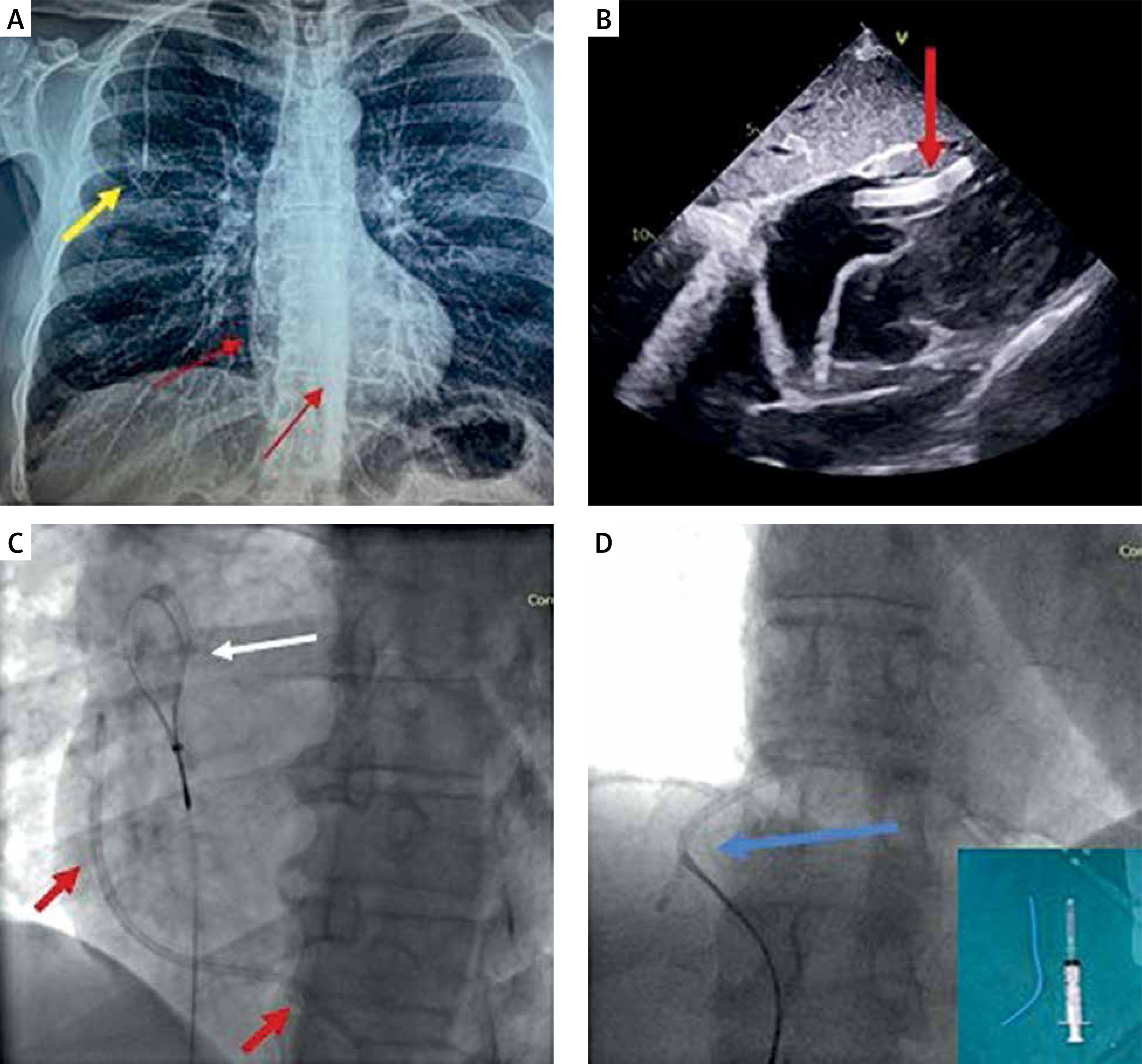
We present the case of a 52-year-old man with pancreatic cancer who had a port-a-cath implanted for chemotherapy 4 years ago and was referred to our hospital for extraction. This was an incidental finding after a routine chest X-ray. The X-ray showed detachment of the distal port-a-cath fragment which had slipped in the right ventricle (Figure 1 A). A transthoracic echocardiogram demonstrated the detached fragment floating in the right ventricle and protruding in the right atrium through the tricuspid valve (Figure 1 B).
Figure 1
A – Chest X-ray demonstrating the port under the skin (yellow arrow) and the detached fragment of the port-a-cath (red arrows). B – Transthoracic echocardiogram, subcostal view demonstrating the detached fragment lying in the right ventricle and protruding through the tricuspid valve in the right atrium (red arrows). C – Fluoroscopy image demonstrating the vascular snare (white arrow) and the detached fragment (red arrows). D – Fluoroscopy, the detached fragment is captured by the snare (blue arrow) + the detached fragment (around 6 cm in length) on the cath lab table
In the cardiac catheterisation laboratory, we decided to follow the loop snare proximal grab technique [1]. Access was obtained via the right femoral vein, and given that the size of most port-a-cath catheters used in our geographical area is 6 Fr, we decided to use a 12 Fr sheath that would be able to accommodate the folding of the port-a-cath fragment. The 7F Atrieve Vascular Snare Kit (ARGON Medical Devices, Dallas, TX, USA) was advanced through a 10F slittable outer guide catheter (CPS Direct Universal, St Jude Medical, CA, USA) in order to approach the detached port-a-cath fragment (Figure 1 C). Despite several attempts we were not able to capture the port-a-cath fragment because the guide catheter was quite soft and it did not have a favourable angle to help the snare to reach the detached fragment. We therefore decided to use a different guide catheter, specifically an 8F Multipurpose catheter (Launcher, Medtronic, Santa Rosa, CA, USA), which offered better support and torquability and helped us to capture the detached fragment with the snare. The guide catheter and the captured, folded port-a-cath fragment were removed through the 12 Fr femoral vein sheath without injury of the access site (Figure 1 D). The puncture site was closed by manual compression.
Embolization of a port-a-cath or other central venous catheter fragment in the right heart chambers or in the pulmonary artery and its branches is an uncommon complication [2]. Most often it is an incidental diagnosis after a long asymptomatic period. Percutaneous extraction using femoral venous access is the gold standard for removal of the embolised catheter fragments [2]. However, in patients with inferior vena cava syndrome or an inferior vena cava filter, where access via the femoral vein would be futile, alternative vein access such as subclavian veins should be used.


