Advances in Interventional Cardiology
eISSN: 1897-4295
ISSN: 1734-9338
Advances in Interventional Cardiology/Postępy w Kardiologii Interwencyjnej
Current Issue Archive Manuscripts accepted About the journal Editorial board Abstracting and indexing Subscription Contact Instructions for authors Publication charge Ethical standards and procedures
Editorial System
Submit your Manuscript
SCImago Journal & Country Rank
3/2025
vol. 21
 
Share:
Share:
Review paper

From bench to bedside and back: translational cardiovascular interventions in veterinary medicine

Sebastian Popiel-Dziewierz
1
,
Agnieszka Noszczyk-Nowak
2
,
Artur Dziewierz
3, 4

  1. Interdepartmental Student Scientific Society of Physiologists, Section of Regulatory Processes and Organ Physiology at the Department of Animal Physiology, Faculty of Veterinary Medicine, University of Life Sciences, Lublin, Poland
  2. Division of Translational Medicine, Department of Internal Medicine and Clinic of Diseases of Horses, Dogs and Cats, Faculty of Veterinary Medicine, Wroclaw University of Environmental and Life Sciences, Wroclaw, Poland
  3. Clinical Department of Cardiology and Cardiovascular Interventions, University Hospital, Krakow, Poland
  4. 2nd Department of Cardiology, Institute of Cardiology, Jagiellonian University Medical College, Krakow, Poland
Adv Interv Cardiol 2025; 21, 3 (81): 295–304
Online publish date: 2025/09/15
Article file
- From bench.pdf  [0.91 MB]
Get citation


Introduction

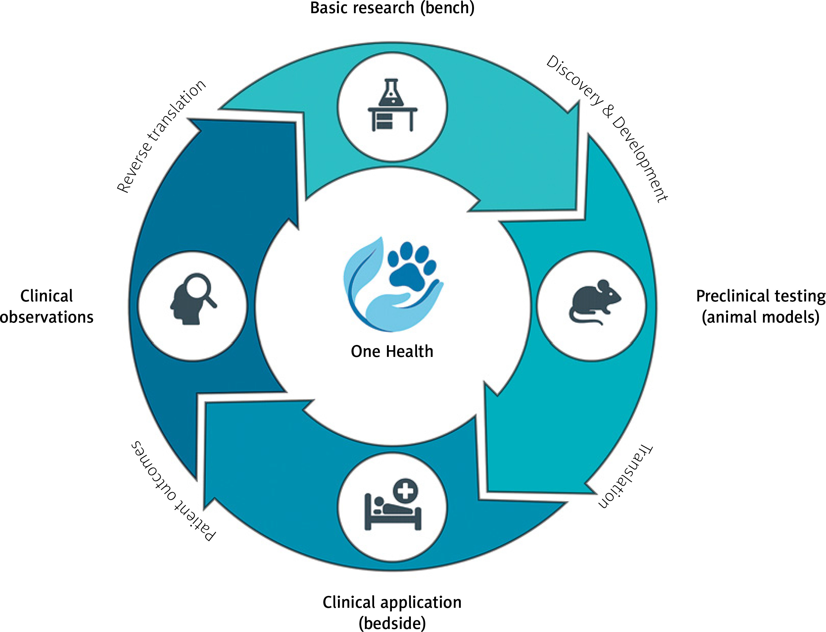
Translational research in cardiology faces a fundamental crisis [1, 2]. Despite decades of investment and thousands of promising discoveries, only 5% of therapies tested in animals ultimately receive regulatory approval for human use [3]. This “valley of death” between preclinical promise and clinical reality stems largely from the limitations of traditional animal models – young, genetically homogeneous laboratory animals with artificially induced diseases that poorly replicate the complexity of spontaneous cardiovascular disease in aging, genetically diverse human populations [1, 2]. The emerging paradigm of reverse translation offers a solution [47]. Rather than following a unidirectional path from bench to bedside, this approach creates a continuous cycle where clinical observations drive basic research, which informs better clinical trials, generating new observations (Figure 1) [5]. The One Health initiative provides the operational framework for this bidirectional exchange, formally integrating human and veterinary medicine to leverage naturally occurring diseases in companion animals as high-fidelity models for human conditions [4]. Defined as a collaborative, multisectoral, and transdisciplinary approach, One Health recognizes the inextricable links among the health of people, animals, and their shared environment [8]. While its conceptual roots trace to the 19th century, the modern movement gained momentum in the 21st century through collaborations between major medical bodies like the American Medical Association (AMA) and American Veterinary Medical Association (AVMA), largely in response to growing zoonotic disease threats [4].

Figure 1

The bidirectional translational cycle in cardiovascular medicine. The traditional bench-to-bedside pathway is complemented by reverse translation, creating a continuous cycle of discovery. The One Health approach integrates all stages, recognizing the interconnection between human and animal health

/f/fulltexts/PWKI/56684/PWKI-21-3-56684-g001_min.jpg

Veterinary interventional cardiology demonstrates this principle in action [47]. Procedures like cardiac pacing and balloon valvuloplasty, initially developed through animal experimentation, refined in human medicine, and then adapted for veterinary patients, now generate valuable clinical data that inform the next generation of human devices. This review examines how this translational cycle operates, its economic implications, and the regulatory changes needed to fully realize its potential [47, 9].

The economic reality of cardiovascular innovation

Bringing a cardiovascular device to market represents a staggering financial commitment. Class II devices approved via the 510(k) pathway cost approximately $30 million to develop, while high-risk Class III devices requiring Premarket Approval average $94 million [10]. Complex therapeutic devices can exceed $500 million when accounting for failed studies. Clinical trials alone cost $1–20 million and take 4–6 years, with failure rates exceeding 30%. FDA user fees add another layer of expense – $24,335 for 510(k) submissions and over $540,000 for PMA applications in fiscal year 2025 [11, 12].

Against this backdrop, veterinary clinical trials offer compelling economic value (Figure 2). A well-designed study in dogs with naturally occurring heart disease provides crucial safety and efficacy data at a fraction of the cost of human trials [12]. More importantly, these studies de-risk development by identifying failure modes early, refining device design based on real-world performance, and selecting optimal patient populations for expensive pivotal human trials. This strategic investment increases the probability of success in later stages, reducing financial exposure for sponsors and investors [13, 14]. However, understanding the cardiovascular system has required a diverse array of animals, from lower organisms to large mammals, each offering distinct advantages and limitations as detailed in Table I [47, 9].

Figure 2

Key stages of medical device development with associated costs, challenges, and potential contributions of veterinary (“One Health”) models to enhance translatability and regulatory success
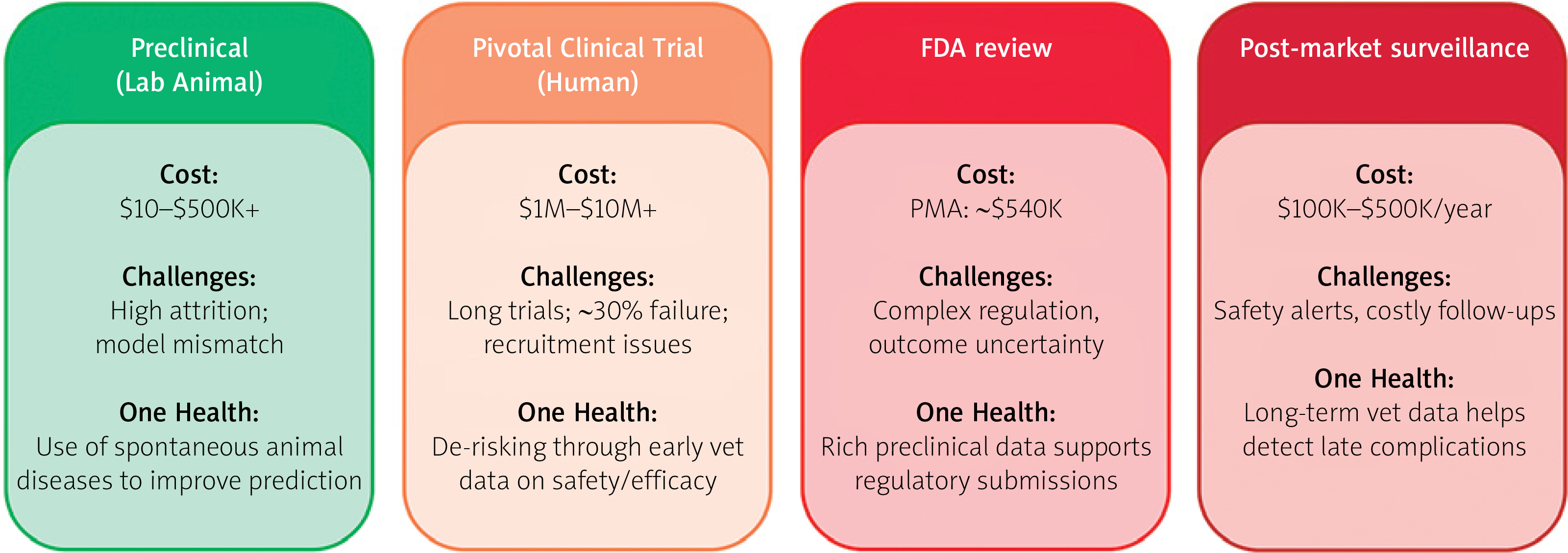
/f/fulltexts/PWKI/56684/PWKI-21-3-56684-g002_min.jpg
Table I

Comparative analysis of animal models in cardiovascular research

SpeciesCommonly modeled conditionsKey advantagesCritical limitations and translatability issues
ZebrafishHeart development, regeneration, organogenesis, basic geneticsHigh-throughput screening, low cost, transparent embryos for live imaging, strong regenerative capacityPoikilothermic (cold-blooded), simple two-chambered heart, significant physiological distance from mammals
Mouse/RatMI, DCM, AAA, atherosclerosis, hypertensionLow cost, high reproductive rate, short lifespan, extensive genetic tools (transgenic, knockout models)Very high heart rate (~600 bpm), different ion channel distribution, small size limits device testing, poor atherosclerosis model without genetic modification
RabbitAtherosclerosis, AAALarger size than rodents, develops atherosclerosis on high-cholesterol diet, historical use in AAA modelsHerbivore physiology, less genetically tractable than mice, higher cost and housing requirements
PigMI, CHF, ASD, device testing, stem cell therapyAnatomical and physiological similarity to humans (heart size, coronary circulation, cardiac output), predictable infarct sizeHigh cost, specialized housing and handling required, rapid growth can complicate long-term studies, less developed collateral circulation than dogs
SheepMI, CHF, valvular disease, ASD/device testingDocile temperament, similar heart size to humans, good model for post-MI remodeling and ischemic cardiomyopathyHigh cost, seasonal breeding, different immune response compared to humans, less commonly used for spontaneous disease studies
DogPacemaker development, PDA/valvular stenosis, arrhythmias, spontaneous disease (SAS, DCM, PH)Excellent model for spontaneous disease, similar heart:body weight ratio, well-developed electrophysiology, human-like response to many drugsHigh cost, significant ethical scrutiny, extensive collateral coronary circulation can confound MI studies, specific anatomical variations (e.g., crista terminalis)

[i] AAA – abdominal aortic aneurysm, ASD – atrial septal defect, bpm – beats per minute, CHF – congestive heart failure, DCM – dilated cardiomyopathy, MI – myocardial infarction, PDA – patent ductus arteriosus, PH – pulmonary hypertension, SAS – subaortic stenosis.

High-fidelity models: spontaneous disease in companion animals

The superiority of spontaneous disease models lies in their biological authenticity. Unlike induced laboratory models, companion animals develop cardiovascular diseases through the same complex interplay of genetics, environment, and aging that drives human disease (Table II) [57, 1517].

Table II

Summary of key interventional procedures and their translational pathway

Interventional procedureKey animal models and rolePivotal “bench” discoveryCurrent “bedside” application in veterinary medicine
Cardiac pacingDogProof-of-concept for external, transvenous cardiac pacing in a stopped heart (Bigelow/Callaghan, 1950)Standard of care for symptomatic bradycardias (e.g., AV block, sick sinus syndrome); high success rate
PDA closureDogFeasibility, safety, and efficacy testing for transcatheter occluder devices (e.g., Amplatzer)Standard of care for most PDAs; use of veterinary-specific devices like the Amplatzer Canine Duct Occluder
Balloon valvuloplastyDog (English Bulldog)First-ever successful balloon valvuloplasty performed on a patient with spontaneous, severe pulmonic stenosis (1980)Preferred treatment for moderate-to-severe pulmonic stenosis; improves clinical signs and quality of life
RF catheter ablationDogDevelopment and validation of catheter ablation techniques, first with DC shock (Scheinman, 1970s) and later with safer RF energyCurative, first-choice treatment for accessory pathway-mediated supraventricular tachycardias; high success rate (> 98%)
ASD closureDog (early); pig/sheep (later)Early device feasibility in dogs; later shift to porcine/ovine models for superior anatomical fidelity to test modern occluders and study endothelializationPerformed in select canine cases with suitable septal anatomy and significant shunting
MitraClipPigDevice feasibility confirmed in pigsModified design for canine myxomatous mitral valve disease – V-Clamp system

[i] ASD – atrial septal defect, AV – atrioventricular, DC – direct current, PDA – patent ductus arteriosus, RF – radiofrequency.

Canine dilated and arrhythmogenic right ventricular cardiomyopathies: a window into arrhythmogenesis

Canine dilated cardiomyopathy provides exceptional insight into heart failure pathophysiology [9, 15]. The tachycardia-induced canine model faithfully replicates the neurohumoral activation characteristic of human heart failure, with marked elevations in plasma norepinephrine, epinephrine, renin, and aldosterone [15, 18]. Most critically, it reproduces the specific electrophysiological remodeling underlying sudden cardiac death – prolonged action potential duration and increased spatial dispersion of repolarization that create substrate for lethal arrhythmias. This makes it invaluable for testing anti-arrhythmic therapies. Arrhythmogenic right ventricular cardiomyopathy (ARVC) occurs spontaneously in Boxer dogs, providing a naturally occurring animal model that faithfully recapitulates the clinical and pathological features of human disease [19, 20]. This canine model exhibits the hallmark characteristics of ARVC: ventricular tachycardia originating from the right ventricle, progressive structural abnormalities including right ventricular dilation, and the pathognomonic histological triad of myocyte loss, fibrofatty replacement, and inflammatory infiltrates [21]. Affected dogs demonstrate myocardial apoptosis and may experience sudden cardiac death, paralleling the human phenotype [22]. Notably, familial clustering of cases has been documented, supporting an inherited basis for canine ARVC and further strengthening its validity as a translational model for understanding the genetic mechanisms underlying this cardiomyopathy.

Canine subaortic stenosis: natural experiments in disease management

Subaortic stenosis in dogs offers unique translational opportunities through its divergent management compared to humans [23]. Certain breeds, such as Boxers, show high disease prevalence, providing genetically concentrated populations for investigating hereditary disease basis [2325]. While human patients undergo surgical resection, dogs are managed medically with β-blockers. This creates a natural experiment – decades of veterinary data on disease progression under conservative therapy that could never be ethically obtained in humans. The discovery of PICALM gene mutations in Newfoundland dogs with subaortic stenosis provides a concrete genetic target for investigating disease mechanisms [23, 26]. The shared pathophysiology – abnormal aortoseptal angle generating high shear stress and driving fibrous ridge formation – ensures direct relevance to human disease [23].

Additional models

Canine heartworm disease (Dirofilaria immitis) provides a natural model for pulmonary hypertension through chronic inflammatory endarteritis [27]. Also, idiopathic pulmonary fibrosis in West Highland white terriers serves as a natural model of this disease [28, 29]. Patent ductus arteriosus, the most common congenital defect in dogs, offers insights into volume overload pathophysiology and device closure techniques. Also, increased pressure in the left atrium resulting in left-sided congestive heart failure is a common sequela to multiple cardiac conditions in dogs, with the most common being myxomatous mitral valve disease [15]. These spontaneous models share environmental exposures, genetic diversity, and comorbidities with human patients, providing data superior to any laboratory simulation [57, 1517].

Case studies in translational success

Patent ductus arteriosus: completing the circle

Patent ductus arteriosus closure exemplifies the full translational cycle [3032]. After surgical ligation’s introduction in 1938, researchers developed transcatheter devices tested extensively in canine models [32, 33]. The Amplatzer Duct Occluder underwent rigorous evaluation in dogs before human use, ultimately leading to the Amplatzer Canine Duct Occluder – the first device designed specifically for canine anatomy. Today, transcatheter patent ductus arteriosus closure is standard care in veterinary medicine, generating continuous data on device performance that inform next-generation human pediatric occluders [33, 34].

Balloon valvuloplasty: veterinary patients leading the way

In a remarkable reversal of the typical pathway, the world’s first balloon valvuloplasty was performed on an English Bulldog with pulmonic stenosis in 1980, 2 years before the first human case [35]. This veterinary success provided crucial proof-of-concept enabling human application. Today, balloon valvuloplasty remains the treatment of choice for canine pulmonic stenosis [36, 37]. However, long-term human studies reveal significant pulmonary regurgitation in up to 29% of patients – a complication less studied in dogs, presenting a clear opportunity for veterinary research to inform human practice [36].

Cardiac pacing: a century of innovation

The development of cardiac pacing began with Bigelow and Callaghan’s 1950 demonstration of transvenous pacing in dogs [38]. This foundational work enabled all subsequent advances in both human and veterinary pacing [39]. Today, pacemaker implantation in dogs achieves excellent outcomes (86% 1-year survival), though complication rates (13–35%) mirror human experience [40]. The shared challenge of lead-related complications makes long-term veterinary data invaluable for predicting device durability and refining management protocols. The Department of Internal Medicine with Clinic of Diseases of Horses, Dogs and Cats at the Faculty of Veterinary Medicine, Wroclaw University of Environmental and Life Sciences, has performed cardiac pacemaker implantation in dogs since 1992. The program predominantly utilizes single-chamber pacemakers, particularly in small and miniature breed dogs. Current procedural volume averages 20 implantations annually (unpublished data, Figure 3 A). The primary indications for pacemaker therapy include third-degree atrioventricular block with ventricular escape rhythm and symptomatic paroxysmal second-degree atrioventricular block presenting with syncope. Unlike human procedures performed under conscious sedation, canine pacemaker implantation requires general anesthesia, representing a key procedural distinction between veterinary and human electrophysiology practice.

Figure 3

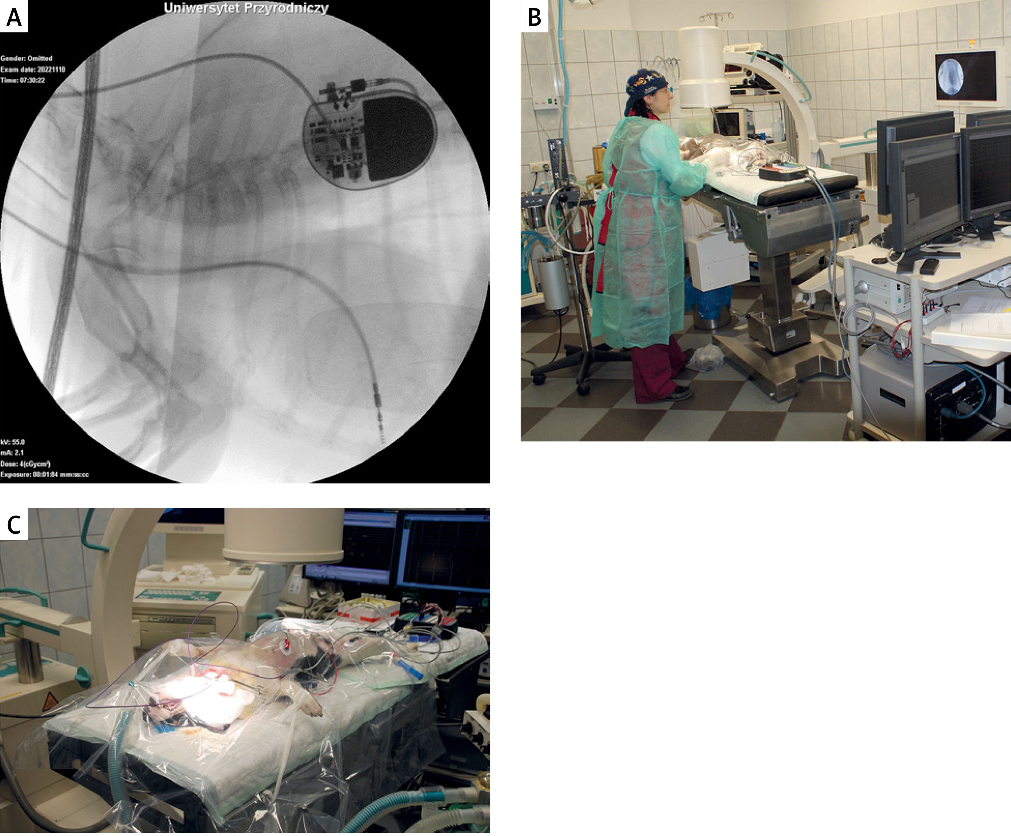
Cardiac electrophysiology procedures in a canine model. A – Fluoroscopic image demonstrating implanted cardiac pacemaker with visible lead placement in a canine subject. B, C – Procedural setup during cardiac electrophysiology study and radiofrequency catheter ablation, showing patient positioning and equipment arrangement. Images courtesy of Prof. Agnieszka Noszczyk-Nowak, Department of Internal Medicine and Clinic of Diseases of Horses, Dogs and Cats, Wroclaw University of Environmental and Life Sciences, Poland

/f/fulltexts/PWKI/56684/PWKI-21-3-56684-g003_min.jpg

Radiofrequency ablation: from crude to curative

Catheter ablation evolved from Scheinman’s initial direct current (DC) shock experiments in dogs through meticulous refinement to today’s precise radiofrequency techniques [41]. The technology now offers > 98% cure rates for accessory pathway-mediated tachycardias in dogs [42]. Veterinary experience with challenging anatomies and novel mapping systems continues to inform human electrophysiology [4345]. In Europe, catheter ablation procedures in dogs are performed at three specialized centers: the Veterinary Department of Ghent University, Clinica Veterinaria Malpensa, and the Department of Internal Medicine with Clinic of Diseases of Horses, Dogs and Cats at Wroclaw University of Environmental and Life Sciences, Poland [46]. The Polish center pioneered ablation therapy in the country in 2010, collaborating with electrophysiologists from the 4th Military Hospital in Wroclaw. Since inception, the Wroclaw veterinary team has successfully performed ablations for accessory pathway-mediated tachycardias, focal atrial tachycardia, and ventricular premature complexes (Figures 3 B, C). Notably, many patients presented with tachycardia-induced cardiomyopathy, which demonstrated complete or partial reversal following successful arrhythmia ablation, paralleling outcomes observed in human electrophysiology [18].

Septal defects: the importance of anatomical fidelity

Atrial septal defect closure device development revealed critical lessons about model selection [47, 48]. Early devices tested successfully in dogs failed to account for anatomical differences in atrial septum structure. Researchers pivoted to porcine and ovine models with more human-like anatomy, demonstrating that model fidelity trumps convenience [4951]. Despite this, modern devices like the Amplatzer Septal Occluder are now successfully used in selected canine patients.

Transcatheter edge-to-edge repair in canine mitral valve disease: the V-Clamp system

While percutaneous mitral valve repair using the MitraClip system (Abbott Vascular, Abbott Park, Chicago, IL, USA) has been successfully employed in human medicine since 2008, device dimensions preclude its use in veterinary patients [5254]. To address this gap, the V-Clamp (HongYu Medical Technology, Shanghai, China) was developed as a species-specific transcatheter edge-to-edge repair (TEER) device for canine myxomatous mitral valve disease (MMVD). The V-Clamp functions by approximating mitral valve leaflets to reduce regurgitant flow, thereby improving cardiac hemodynamics. The device demonstrates optimal efficacy when regurgitation is localized to specific leaflet segments without cleft pathology. Compared to conventional open-heart surgery, this minimally invasive approach eliminates the need for cardiopulmonary bypass, significantly reduces procedural time, and minimizes perioperative morbidity. Initial clinical studies have demonstrated promising safety and efficacy profiles for the V-Clamp in dogs with MMVD [55, 56]. However, comprehensive evaluation awaits longer-term follow-up studies with larger patient cohorts to establish definitive outcomes and refine patient selection criteria.

The return journey: clinical insights driving innovation

The true power of reverse translation emerges when veterinary clinical experiences generate new research questions [57, 1517]. When patients respond unexpectedly to therapy, these “clinical puzzles” drive investigations into novel mechanisms and applications. Veterinary hospitals functioning as clinical research sites provide real-world device performance data impossible to obtain in laboratory settings [57, 58]. Successful collaborations demonstrate this synergy: University of Florida’s joint human-veterinary team pioneered hybrid surgical-interventional techniques; Colorado State University’s advanced imaging integration provides real-world performance data to manufacturers while delivering cutting-edge care to animals; multi-institutional registries track long-term outcomes across species (Table III). This bidirectional exchange accelerates innovation while expanding access to advanced therapies [59].

Table III

Examples of One Health collaborations in cardiovascular medicine

InstitutionCollaboration typeKey innovationMutual benefits
University of FloridaHuman pediatric interventional cardiologists + veterinary cardiologistsFirst-of-its-kind hybrid surgical-interventional stent placement in a small dog with severe pulmonic stenosis
  • Saved patient’s life

  • Pioneered technique for both fields

  • Cross-training opportunity

Colorado State UniversityVeterinary cardiology + human cardiologists + medical device companiesAdvanced imaging integration (Philips EchoNavigator fusion technology)
  • Dogs: cutting-edge care access

  • Companies: real-world data

  • Clinicians: accelerated expertise

Cornell UniversityGlobal veterinary networkInternational ECG database and arrhythmia protocol development
  • Rapid pattern identification

  • Accelerated therapy refinement

  • Global knowledge sharing

Multi-institutionalCardiomyopathy in Cats RegistryComparative feline-human cardiomyopathy research
  • Novel disease insights

  • Bidirectional translation

  • Expanded patient database

Department of Internal Medicine with Clinic of Diseases of Horses, Dogs and Cats, Faculty of Veterinary Medicine, Wroclaw University of Environmental and Life SciencesVeterinary cardiology + human cardiology + medical device companiesChronic tachycardia-induced cardiomyopathy pig model; myocardial infarction pig model (old drug-new application);
novel endo-epicardial radiofrequency ablation techniques
  • Direct therapeutic applications saving companion animal lives

  • New models of diseases

  • New technologies

Regulatory evolution: bridging the gap

Current FDA guidance for animal studies makes no distinction between induced disease in laboratory animals and spontaneous disease in clinical patients. This regulatory gap creates uncertainty for sponsors considering veterinary clinical trials, forcing continued reliance on less predictive traditional models [60]. A new FDA-CVM guidance document should establish clear pathways for including veterinary clinical data in human device submissions, addressing study design standards, data collection requirements, ethical oversight, and reporting formats. This evolution would align regulatory frameworks with scientific reality, accelerating safer therapies to market [6062].

The clinical reality: technology and expertise

Modern veterinary interventional cardiology requires sophisticated infrastructure (Table IV) [6365]. Fusion imaging systems like Philips EchoNavigator combine echocardiography’s soft tissue detail with fluoroscopy’s device visualization, enabling complex structural repairs. Specialized anesthetic protocols balance adequate sedation with cardiovascular stability [66]. Advanced biomaterials – nitinol’s shape memory, anti-thrombogenic coatings, endothelialization-promoting surfaces – enable long-term device success [67].

Table IV

Imaging modalities in interventional cardiology

ModalityPrimary functionKey advantagesKey limitationsCommon veterinary application
FluoroscopyReal-time 2D visualization of radiopaque devices and cathetersExcellent device visualization, universally usedIonizing radiation, poor soft tissue detailGuiding all catheter-based procedures (pacing, device deployment, angiography)
Transesophageal echocardiography (TEE)Real-time 2D/3D visualization of soft tissue, valves, and blood flowExcellent soft tissue detail, no radiation, well-establishedRequires general anesthesia, esophageal intubation, can have poor acoustic windowsGuiding structural heart repairs (septal defects, valve repair), assessing device position and function
Intracardiac echocardiography (ICE)High-resolution, real-time 2D/3D visualization of intracardiac structures from within the heartSuperior near-field resolution, overcomes poor TEE windows, may reduce need for general anesthesiaInvasive (requires separate vascular access), high cost of single-use catheters, smaller field of viewGuiding complex procedures where TEE is inadequate (e.g., challenging PDA closure, septal puncture)

[i] PDA – patent ductus arteriosus.

Board certification in veterinary cardiology requires completion of research projects and rigorous examinations through either the American or European College of Veterinary Internal Medicine. The scarcity of residency positions globally has created a critical shortage of veterinary cardiologists, resulting in significant geographic disparities in access to specialized cardiac care. This challenge is particularly acute in countries like Poland, where no veterinary cardiology residency programs currently exist. To address these limitations, many veterinary centers have established collaborative partnerships with human cardiology departments. These interdisciplinary collaborations enhance procedural expertise, improve patient safety, and advance the standard of care through shared knowledge and technical resources.

Procedure costs ($5,700–7,000) and insurance limitations pose ethical challenges, forcing clinicians to balance technological capabilities with economic realities while maintaining patient welfare [68].

Future directions: convergent technologies

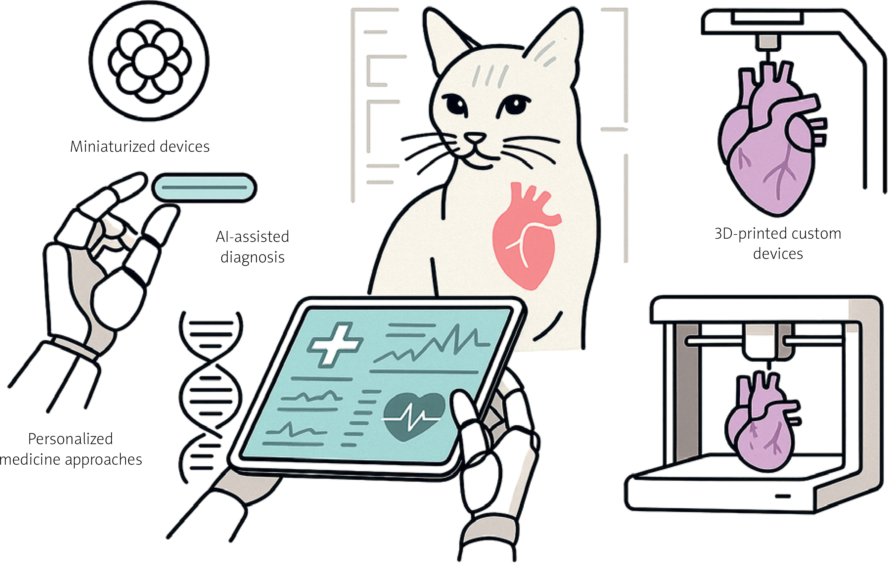
Emerging innovations promise to revolutionize cardiovascular care across species (Figure 4). Miniaturized devices like leadless pacemakers eliminate lead complications; experimental bioresorbable devices dissolve after restoring the function [69]. Artificial intelligence enables predictive medicine through automated arrhythmia detection and disease staging [7072]. 3D printing creates patient-specific implants [73, 74]. Precision medicine uses genetic profiling and biomarkers to individualize therapy [75, 76]. These technologies, developed through bidirectional translation, will create increasingly predictive, preventive, and personalized cardiovascular care. More importantly, their convergence will transform today’s untreatable conditions into manageable diseases, fundamentally reshaping the therapeutic landscape of veterinary cardiology [77].

Figure 4

Emerging technologies in veterinary cardiology. Future advances include injectable miniaturized devices, AI-assisted diagnostic tools, personalized medicine based on genetic profiles, and patient-specific 3D-printed cardiac devices

/f/fulltexts/PWKI/56684/PWKI-21-3-56684-g004_min.jpg

Conclusions

Veterinary interventional cardiology demonstrates that the future of translational medicine is cyclical, not linear. By embracing spontaneous disease models in companion animals, leveraging economic advantages of veterinary trials, and creating regulatory frameworks that recognize their value, we can overcome the valley of death that has long plagued cardiovascular innovation. The One Health approach transforms veterinary medicine from a recipient of human medical advances to an active partner in discovery. This bidirectional bridge ensures that insights flow continuously between species, accelerating progress toward safer, more effective therapies for all. As we advance toward an era of miniaturized, intelligent, and personalized cardiovascular interventions, the lessons learned from our animal companions will prove more valuable than ever.

Acknowledgments

The authors declare that AI-assisted technology (Claude, Anthropic) was used for grammar checking and language editing during the preparation of this manuscript. The AI tool was used solely to improve the clarity and readability of the text. The authors take full responsibility for the scientific content, accuracy, and integrity of the work presented.

Ethical approval

Not applicable.

Conflict of interest

The authors declare no conflict of interest.

References

1 

Ceballos G, Ortiz-Flores M, Nájera N. Medicina/investigación translacional: ¿quién recibe los beneficios? Cardiovasc Metab Sci 2021; 32: 64–5.

2 

Jia X. Translational medicine: creating the crucial bidirectional bridge between bench and bedside. Int J Mol Sci 2016; 17: 1918.

3 

Ineichen BV, Furrer E, Grüninger SL, et al. Analysis of animal-to-human translation shows that only 5% of animal-tested therapeutic interventions obtain regulatory approval for human applications. PLoS Biol 2024; 22: e3002667.

4 

Schneider B, Balbas-Martinez V, Jergens AE, et al. Model-based reverse translation between veterinary and human medicine: the One Health initiative. CPT Pharmacometrics Syst Pharmacol 2018; 7: 65–8.

5 

Shakhnovich V. It’s time to reverse our thinking: the reverse translation research paradigm. Clin Transl Sci 2018; 11: 98–9.

6 

Zaragoza C, Gomez-Guerrero C, Martin-Ventura JL, et al. Animal models of cardiovascular diseases. J Biomed Biotechnol 2011; 2011: 497841.

7 

Mukherjee P, Roy S, Ghosh D, et al. Role of animal models in biomedical research: a review. Lab Anim Res 2022; 38: 18.

8 

Danasekaran R. One health: a holistic approach to tackling global health issues. Indian J Community Med 2024; 49: 260–3.

9 

Hasenfuss G. Animal models of human cardiovascular disease, heart failure and hypertrophy. Cardiovasc Res 1998; 39: 60–76.

10 

Miller BJ, Blanks W, Yagi B. The 510(k) third party review program: promise and potential. J Med Syst 2023; 47: 93.

11 

Sertkaya A, DeVries R, Jessup A, et al. Estimated cost of developing a therapeutic complex medical device in the US. JAMA Netw Open 2022; 5: e2231609.

12 

Fogel DB. Factors associated with clinical trials that fail and opportunities for improving the likelihood of success: a review. Contemp Clin Trials Commun 2018; 11: 156–64.

13 

Farag A, Hendawy H, Emam MH, et al. Stem cell therapies in canine cardiology: comparative efficacy, emerging trends, and clinical integration. Biomolecules 2025; 15: 5371.

14 

Pyle WG. Cardiology’s best friend: using naturally occurring disease in dogs to understand heart disease in humans. J Mol Cell Cardiol Plus 2025; 13: 100474.

15 

Houser SR, Margulies KB, Murphy AM, et al. Animal models of heart failure: a scientific statement from the American Heart Association. Circ Res 2012; 111: 131–50.

16 

Cesarovic N, Lipiski M, Falk V, et al. Animals in cardiovascular research. Eur Heart J 2020; 41: 200–3.

17 

Jia T, Wang C, Han Z, et al. Experimental rodent models of cardiovascular diseases. Front Cardiovasc Med 2020; 7: 588075.

18 

Paslawska U, Gajek J, Kiczak L, et al. Development of porcine model of chronic tachycardia-induced cardiomyopathy. Int J Cardiol 2011; 153: 36–41.

19 

Vischer AS, Connolly DJ, Coats CJ, et al. Arrhythmogenic right ventricular cardiomyopathy in Boxer dogs: the diagnosis as a link to the human disease. Acta Myol 2017; 36: 135–50.

20 

Basso C, Fox PR, Meurs KM, et al. Arrhythmogenic right ventricular cardiomyopathy causing sudden cardiac death in boxer dogs: a new animal model of human disease. Circulation 2004; 109: 1180–5.

21 

Camacho P, Fan H, Liu Z, et al. Large mammalian animal models of heart disease. J Cardiovasc Dev Dis 2016; 3: 30.

22 

Akdis D, Medeiros-Domingo A, Gaertner-Rommel A, et al. Myocardial expression profiles of candidate molecules in patients with arrhythmogenic right ventricular cardiomyopathy/dysplasia compared to those with dilated cardiomyopathy and healthy controls. Heart Rhythm 2016; 13: 731–41.

23 

Crofton AE, Kovacs SL, Stern JA. Subvalvular aortic stenosis: learning from human and canine clinical research. Cardiol Res 2023; 14: 319–33.

24 

Chetboul V, Trollé JM, Nicolle A, et al. Congenital heart diseases in the boxer dog: a retrospective study of 105 cases (1998–2005). J Vet Med A Physiol Pathol Clin Med 2006; 53: 346–51.

25 

Bussadori C, Amberger C, Le Bobinnec G, et al. Guidelines for the echocardiographic studies of suspected subaortic and pulmonic stenosis. J Vet Cardiol 2000; 2: 15–22.

26 

Stern JA, White SN, Lehmkuhl LB, et al. A single codon insertion in PICALM is associated with development of familial subvalvular aortic stenosis in Newfoundland dogs. Hum Genet 2014; 133: 1139–48.

27 

Falcón-Cordón S, Falcón-Cordón Y, Caro-Vadillo A, et al. Association between thoracic radiographic changes and indicators of pulmonary hypertension in dogs with heartworm disease. Animals 2024; 14: 1900.

28 

Serres FJ, Chetboul V, Tissier R, et al. Doppler echocardiography-derived evidence of pulmonary arterial hypertension in dogs with degenerative mitral valve disease: 86 cases (2001–2005). J Am Vet Med Assoc 2006; 229: 1772–8.

29 

Clercx C, Fastrès A, Roels E. Idiopathic pulmonary fibrosis in West Highland white terriers: an update. Vet J 2018; 242: 53–8.

30 

Israël NV, Dukes-McEwan J, French AT. Long-term follow-up of dogs with patent ductus arteriosus. J Small Anim Pract 2003; 44: 480–90.

31 

Yarboro MT, Gopal SH, Su RL, et al. Mouse models of patent ductus arteriosus (PDA) and their relevance for human PDA. Dev Dyn 2022; 251: 424–43.

32 

Gruenstein DH, Bass JL. Experimental evaluation of a new articulated Amplatzer® ductal occluder device without fabric. Catheter Cardiovasc Interv 2009; 74: 482–7.

33 

Rao PS. Percutaneous closure of patent ductus arteriosus – current status. J Invasive Cardiol 2011; 23: 517–20.

34 

Szeliga J, Dryżek P, Rudziński A, et al. Combination of modified techniques for percutaneous closure of a large secundum atrial septal defect. Adv Interv Cardiol 2023; 19: 178–81.

35 

Buchanan JW, Anderson JH, White RI. The first balloon valvuloplasty: an historical note. J Vet Intern Med 2002; 16: 116–7.

36 

Winter RL, Rhinehart JD, Estrada AH, et al. Repeat balloon valvuloplasty for dogs with recurrent or persistent pulmonary stenosis. J Vet Cardiol 2021; 34: 29–36.

37 

Johnson MS, Martin M. Results of balloon valvuloplasty in 40 dogs with pulmonic stenosis. J Small Anim Pract 2004; 45: 148–53.

38 

Bakytzhanuly A. Evolution of cardiac pacemakers: a journey from galvanic experiments to leadless pacemakers. Heart Vessels Transplant 2023; 7, doi: 10.24969/hvt.2023.407.

39 

Buchanan JW. First pacemaker in a dog: a historical note. J Vet Intern Med 2003; 17: 713–4.

40 

Akshata B. Pacemaker implantation in small animal practice: indications, types of pacing and implantation technique. J Anim Res 2024; 14: 39–46.

41 

Scheinman MA, Rutherford JD. The development of cardiac arrhythmia ablation: a conversation with Melvin A. Scheinman, MD. Circulation 2017; 135: 1191–3.

42 

Wright KN, Connor CE, Irvin HM, et al. Atrioventricular accessory pathways in 89 dogs: clinical features and outcome after radiofrequency catheter ablation. J Vet Intern Med 2018; 32: 1517–29.

43 

Kulakowski P, Derejko P, Kusnierz J, et al. Fatal complication of endo-epicardial bipolar radiofrequency ablation during animal experiment: a warning for clinical electrophysiologist? JACC Clin Electrophysiol 2023; 9: 886–8.

44 

Elvan A, Pride HP, Eble JN, et al. Radiofrequency catheter ablation of the atria reduces inducibility and duration of atrial fibrillation in dogs. Circulation 1995; 91: 2235–44.

45 

Derejko P, Janus I, Kułakowski P, et al. Bipolar endo-epicardial RF ablation: animal feasibility study. Heart Rhythm 2024; 21: 790–8.

46 

Noszczyk-Nowak A, McAulay G, Fuglewicz A, et al. Radiofrequency catheter ablation of a concealed accessory pathway of the heart in a Labrador Retriever. Med Weter 2017; 73: 505–9.

47 

Węglarz P, Węgiel M, Kuszewski P, et al. Atrial septum anatomy as a predictor of ischemic neurological episodes in patients with a patent foramen ovale. Kardiol Pol 2024; 82: 303–7.

48 

Węglarz P, Węgiel M, Kuszewski P, et al. Characteristics of patients diagnosed for cardiac cause of ischemic neurological events and prescreened with a transcranial Doppler examination. Pol Arch Intern Med 2024; 134: 16832.

49 

Prochownik P, Bielecka K, Przewłocki T, et al. Cardiac troponin I release after transcatheter closure of atrial septal defects is associated with supraventricular arrhythmias on early follow-up. Adv Interv Cardiol 2024; 20: 338–44.

50 

Gordon SG, Miller MW, Roland RM, et al. Transcatheter atrial septal defect closure with the AMPLATZER® atrial septal occluder in 13 dogs: short- and mid-term outcome. J Vet Intern Med 2009; 23: 995–1002.

51 

Jalal Z, Seguela PE, Baruteau AE, et al. Role of animal models for percutaneous atrial septal defect closure. J Thorac Dis 2018; 10: S2966–74.

52 

Czober T, Darocha T, Mendrala K, et al. Routine use of procedural sedation and analgesia for transcatheter edge-to-edge mitral valve repair. Kardiol Pol 2023; 81: 778–80.

53 

St Goar FG, Fann JI, Komtebedde J, et al. Endovascular edge-to-edge mitral valve repair: short-term results in a porcine model. Circulation 2003; 108: 1990–3.

54 

Polat F, Inanç İH, Doğru M, et al. EMCAPT study: the effect of MitraClip treatment on the mitral annulus and left atrial appendage evaluation by transoesophageal echocardiography. Adv Interv Cardiol 2023; 19: 56–63.

55 

Potter BM, Orton EC, Scansen BA, et al. Clinical feasibility study of transcatheter edge-to-edge mitral valve repair in dogs with the canine V-Clamp device. Front Vet Sci 2024; 11: 1448828.

56 

Petchdee S, Pongkan W, Lei J, et al. Transcatheter edge-to-edge repair of the mitral valve in four dogs: preliminary results regarding efficacy and safety. Animals 2024; 14: 3068.

57 

Baillie S, Dilly M, Ciappesoni JL, et al. The rapid and international expansion of veterinary clinical skills laboratories: a survey to establish recent developments. J Vet Med Educ 2024; 51: 215–28.

58 

Bruno R. Use of real-world data as pivotal evidence in veterinary regulatory applications. Front Vet Sci 2025; 12: 1588068.

59 

Frydrychowski P, Michałek M, Bil-Lula I, et al. Cardioprotective effect of acetylsalicylic acid in the myocardial ischemia-reperfusion model on oxidative stress markers levels in heart muscle and serum. Antioxidants 2022; 11: 1432.

60 

Zushin PJH, Mukherjee S, Wu JC. FDA Modernization Act 2.0: transitioning beyond animal models with human cells, organoids, and AI/ML-based approaches. J Clin Invest 2023; 133: e175824.

61 

Bertout JA, Baneux PJR, Robertson-Plouch CK. Recommendations for ethical review of veterinary clinical trials. Front Vet Sci 2021; 8: 715926.

62 

Ponmurugesan K, Murthannagari VR, Devaraj H, et al. A comparative review of human and veterinary generic drug approval systems. Asian J Pharm Clin Res 2025; 18: 47–53.

63 

Divekar AA, Arar YM, Clark S, et al. Transcatheter device therapy and the integration of advanced imaging in congenital heart disease. Children 2022; 9: 497.

64 

Chetboul V, Damoiseaux C, Behr L, et al. Intracardiac echocardiography: use during transcatheter device closure of a patent ductus arteriosus in a dog. J Vet Cardiol 2017; 19: 293–8.

65 

Zhang X, Gosnell J, Nainamalai V, et al. Advances in TEE-centric intraprocedural multimodal image guidance for congenital and structural heart disease. Diagn 2023; 13: 2981.

66 

Grubb T, Sager J, Gaynor JS, et al. 2020 AAHA anesthesia and monitoring guidelines for dogs and cats. J Am Anim Hosp Assoc 2020; 56: 59–82.

67 

Strohbach A, Busch R. Predicting the in vivo performance of cardiovascular biomaterials: current approaches in vitro evaluation of blood-biomaterial interactions. Int J Mol Sci 2021; 22: 11390.

68 

Quain A, Ward MP, Mullan S. Ethical challenges posed by advanced veterinary care in companion animal veterinary practice. Animals 2021; 11: 3010.

69 

Rav Acha M, Soifer E, Hasin T. Cardiac implantable electronic miniaturized and micro devices. Micromachines 2020; 11: 902.

70 

Chu CP. ChatGPT in veterinary medicine: a practical guidance of generative artificial intelligence in clinics, education, and research. Front Vet Sci 2024; 11: 1395934.

71 

Akinsulie OC, Idris I, Aliyu VA, et al. The potential application of artificial intelligence in veterinary clinical practice and biomedical research. Front Vet Sci 2024; 11: 1347550.

72 

Engel-Manchado J, Montoya-Alonso JA, Doménech L, et al. Machine learning techniques for canine myxomatous mitral valve disease classification: integrating anamnesis, quality of life survey, and physical examination. Vet Sci 2024; 11: 118.

73 

Jędrzejek M, Peszek-Przybyła E, Jadczyk T, et al. 3D printing from transesophageal echocardiography for planning mitral paravalvular leak closure: feasibility study. Adv Interv Cardiol 2023; 19: 270–6.

74 

Daungsupawong H, Wiwanitkit V. Application of 3D printing in veterinarian science. Res Vet Sci 2025; 186: 105571.

75 

Edvardsson M, Heenkenda MK. Precision medicine: personalizing healthcare by bridging aging, genetics, and global diversity. Healthcare 2025; 13: 1529.

76 

Johnson KB, Wei WQ, Weeraratne D, et al. Precision medicine, AI, and the future of personalized health care. Clin Transl Sci 2021; 14: 86–93.

77 

Queiroga FL. Pioneering advances in veterinary medicine: from diagnosis to treatment. Animals 2025; 15: 516.

Copyright: © 2025 Termedia Sp. z o. o. This is an Open Access article distributed under the terms of the Creative Commons Attribution-NonCommercial-ShareAlike 4.0 International (CC BY-NC-SA 4.0) License (http://creativecommons.org/licenses/by-nc-sa/4.0/), allowing third parties to copy and redistribute the material in any medium or format and to remix, transform, and build upon the material, provided the original work is properly cited and states its license.
 
Quick links
© 2025 Termedia Sp. z o.o.
Developed by Bentus.