
Introduction
Allogeneic and autologous hematopoietic stem cell transplantation (HSCT) is potentially curative for several malignancies. Acute graft-versus-host disease (GVHD) is an immunologically mediated process, involving donor T cell responses to host alloantigens and the dysregulation of inflammatory cytokine cascade after allogeneic hematopoietic stem cell transplantation (allo-SCT) [1–3]. Graft-versus-host disease (GVHD) in patients who received autologous hematopoietic stem cell transplantation (HSCT) is less common compared to patients who received allogeneic stem cell transplantation. Due to the obvious lack of genetic disparity between donor and recipient, the existence of autologous GVHD (AGVHD) has been a topic of controversy [2–5]. But beyond the technical inaccuracy of the terminology, it is a well-known disease that can affect the skin, gastrointestinal tract, and liver.
We present the case of a 4-year-old girl with metastatic neuroblastoma who spontaneously developed AGVHD after autologous HSCT. An accurate diagnosis can minimize unnecessary hospitalizations and repeat diagnostic tests. Prompt treatment can improve morbidity and prevent disease progression.
Case report
A 4-year-old girl with neuroblastoma stage 4 first received 6 courses of chemotherapy and surgery according to the Turkish Pediatric Oncology Group (TPOG) Neuroblastoma 2009 protocol. Chemotherapy treatment consisted of three courses of chemotherapy containing vincristine, dacarbazine, ifosfamide, and doxorubicin, and three other courses that contained cyclophosphamide, cisplatin, and etoposide. She was randomized to a high risk stem cell transplantation group with a small remnant of neuroblastoma (partial response).
She was then treated with autologous HSCT. Melphalan (140 mg/m2) and busulfan (intravenous busulfan – IVBU) 1.1 mg/kg were used for the first dose; thereafter, the IVBU dose was modified to achieve a final area under the concentration-time curve (AUC) at steady state of 1150 μmol/l/min per dose followed by stem cell support in a cell dose of 5.7 × 10⁶ CD34+ cells/kg. Fluconazole was given from conditioning therapy until day 75 after stem cell infusion. She also received prophylactic low-dose acyclovir. She had weekly cytomegalovirus (CMV) screening with a CMV pp65 antigenemia assay. Engraftment of neutrophils and platelets took place on day +11 and +14 respectively after autologous HSCT. At post-engraftment day +20 intermittent nausea and diarrhea started. Vital signs were normal except mild tachycardia. Physical examination was normal except dry oral mucosa and hyperactive bowel sound. There were no skin lesions. The blood biochemistry tests were within the normal range. Elevation of hepatic enzymes was not detected. The diarrhea worsened, color changed to green and reached a daily volume up to 1500 ml/m2 during the next three days. Albumin and immunoglobulin G decreased to 2.3 g/dl and 322 mg/dl respectively. Intravenous immunoglobulin and albumin were replaced intravenously.
Infectious studies of stool and blood including influenza A and B, parainfluenza, adenovirus, Epstein-Barr virus, amebiasis, Cryptosporidium parvum, cytomegalovirus, Clostridium difficile, Salmonella, Campylobacter, Yersinia, and Shigella were all found negative.
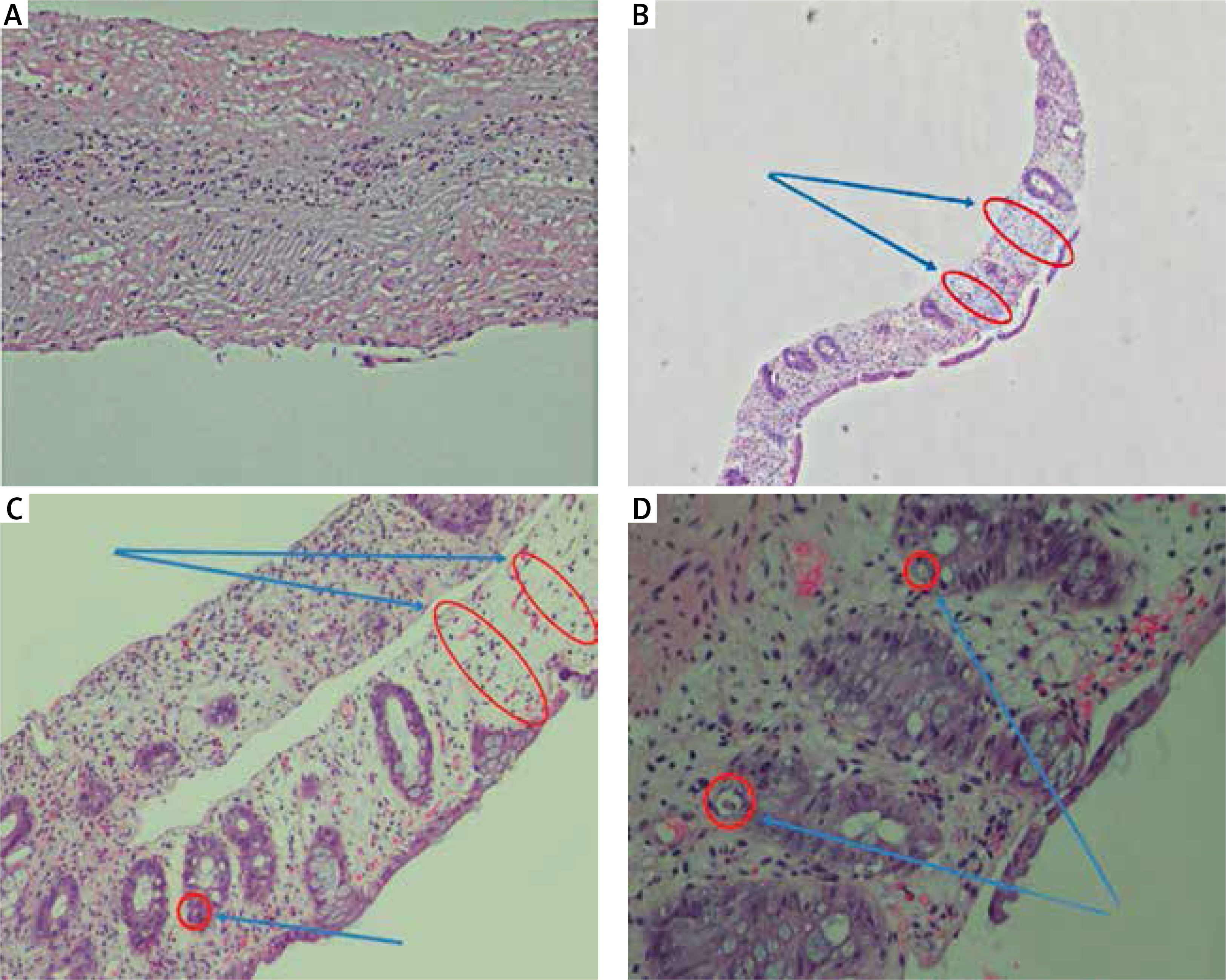
Fecal calprotectin was high at 822 mg/dl (n: 0–120). The diarrhea lasted longer and the amount increased. The contrasted computed tomography of the abdomen and pelvis revealed diffusely thickened walls of small bowel and colon suggestive of diffuse enterocolitis. Total parenteral nutrition (TPN) was started because of difficulty in tolerating enteral feeding. After radiological evidence of gastrointestinal system involvement and persistent worsening of clinical symptoms colonoscopy and rectoscopy were performed by an experienced pediatric gastroenterologist at the 34th day of autologous HSCT and endoscopic images were found suspicious for severe GVHD. Colonoscopy and rectoscopy revealed severe inflammatory changes, friability and patchy dark exudates on the mucosa of the rectum. Endoscopy revealed erosions, ulcers in the esophagus and a pale mucosal surface with reticulated submucosal vessels accompanied by erosion and erythema in the antrum of the stomach (Fig. 1). Diffuse crypt dropout and mucosal erosion on rectal mucosal biopsy that is consistent with grade III GVHD was confirmed by pathologists (Figs. 2A and 2B). Mucosal erosions, apoptosis of epithelial cells and small lymphocytic infiltration of the lamina propria that is consistent with grade II GVHD were found on duodenal biopsy (Figs. 2C and 2D). Immunostaining for cytomegalovirus was negative. There was no evidence of pathologic surface organisms, fungi or viral inclusions on microscopic examination.
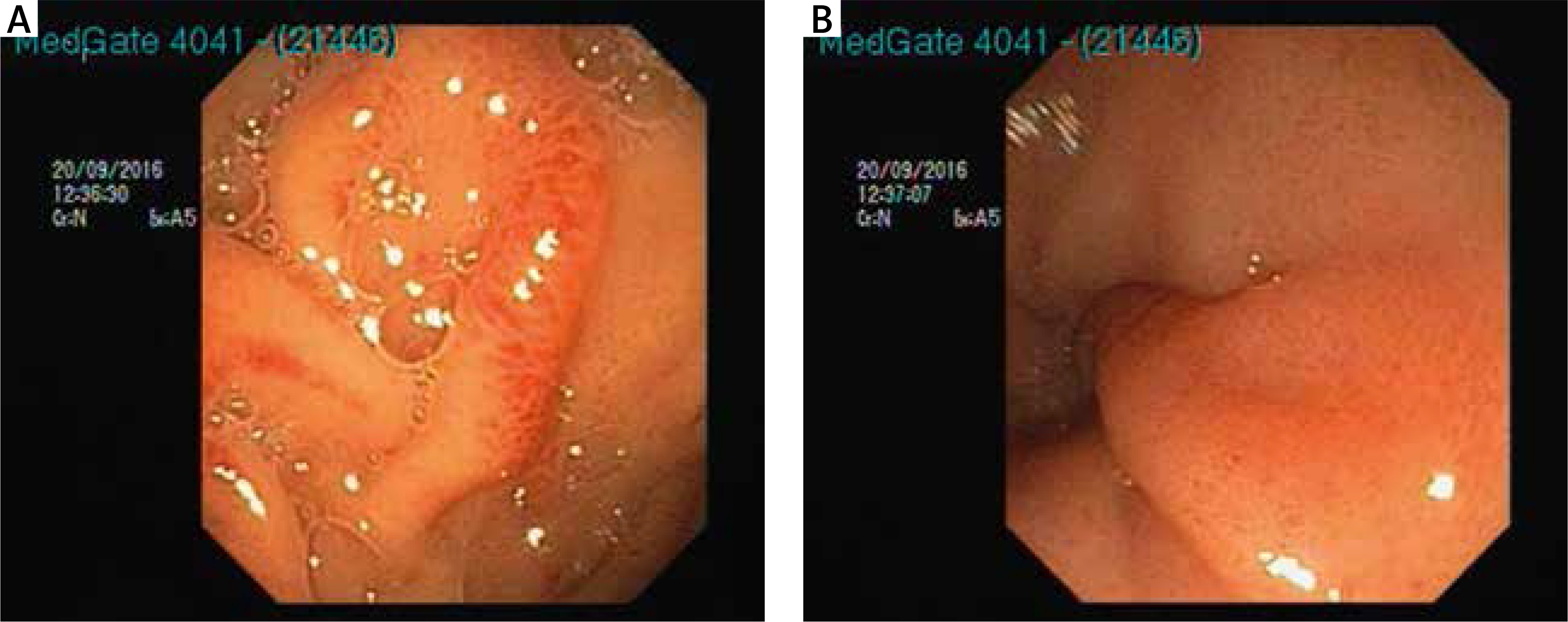
Fig. 2
A) Esophagus 10× mucosal erosions, lymphocytic infiltration of lamina propria. B) Colon 10×, loss of crypt. C) Colon 10×, loss of crypts and apoptosis. D) Colon 20×, apoptosis
After these results, we started methylprednisolone intravenously at a dosage of 2 mg/kg/day for GVHD of the gastrointestinal tract. Because of unresponsiveness to treatment on the fourth day of treatment we increased the dosage to 5 mg/kg/day and added cyclosporine and budesonide to the treatment. The patient needed prolongation of TPN. Because of an inadequate response to treatment, we decided to administer third-party mesenchymal stem cells (MSC) (1 × 10⁶ CD73+/CD105+ cells/kg). These were given intravenously at day +49 of HSCT as first infusion. The second dose was given at the same dosage on day +56. Within 5 days after first application of MSC, the frequency of diarrhea decreased to one third. At day +16 after the second dose of MSC, the patient’s stool became nearly normal. We changed methylprednisolone to oral prednisolone; prednisolone was tapered gradually and stopped over a period of 75 days. We stopped cyclosporine at day +92 after HSCT. Budesonide was stopped at day +110 HSCT. Over the following 12 months there was no recurrence of symptoms.
To our knowledge, this is the second case report of spontaneous severe autologous GVHD in a child with a solid tumor [2] and this is the first case report of severe autologous gastrointestinal GVHD in a child in the literature.
Discussion
Graft-versus-host disease is a common complication of allo-SCT which is induced by donor T cell recognition of recipient alloantigens [6]. GVHD after autologous HSCT or transplantation from a genetically identical twin may cause clinical and histopathologic findings consistent with GVHD [1]. Some investigator propose that all forms of AGVHD (i.e., cutaneous, GI, and liver) be included under the umbrella term “engraftment syndrome” [5]. Beyond the nomenclature AGVHD is an under-diagnosed complication of autologous HSCT resembling GVHD occurring after allogeneic HSCT [1]. There are several hypotheses to explain the pathogenesis of AGVHD. The development of GVHD-like syndrome after autologous HSCT is associated with baseline immune dysregulation as reported in the allogeneic GVHD model. If appropriate treatment is not initiated the prognosis may be poor [7]. The pathogenesis is attributed to the failure in self-tolerance. Alteration of T regulatory cells by previous chemotherapy may be the key point. Endogenous cells that survive conditioning and assist in post-transplant maintenance of self-tolerance may be affected [2]. Microchimerism due to maternal cells transmitted during fetal development and persisting throughout adult life has also been postulated as another cause. However, it is not very clear which factors might contribute to the pathogenesis of this rare disease.
Skin involvement is the most common involvement site of AGVHD. But all other systems can be affected. Althouh Adams et al. reported acute GVHD in 18% of recipients of genetically identical twin transplants [8], there has been estimated a 4–15% incidence of GVHD after autologous HSCT in adults. Among multiple myeloma patients, the incidence is slightly higher and can be as high as 5–20% [3–6]. Skin AGVHD is reported with an 8% incidence by Hood et al. for patients who received autologous and syngeneic transplantation. Patients’ rashes were typically self-limited, but some needed treatment with steroids. Gastrointestinal GVHD incidence was reported to be 13% among autologous stem cell transplantation patients by Holmberg et al. In their study they found AGVHD as a limited disease; a majority of their patients resolve spontaneously and often require no treatment [9]. In patients with multiple myeloma some experts report pathologically verified gastrointestinal GVHD as high as 6% [3]. Responses to steroids are generally good but can be variable. A significant proportion improve dramatically after early therapeutic intervention. So clinicians and pathologists should be aware in suspecting and recognizing GVHD in patients with diarrhea to guide therapy as soon as possible. However, Drobyski et al. reported five cases refractory to corticosteroids and treated with different medications. Four of them died because of AGVHD-related problems [3]. GVHD must be considered when making the differential diagnosis of skin lesions and diarrhea syndromes that occur even in the autologous posttransplantation period.
In treatment of AGVHD, immunosuppression using steroids remains the first line of intervention. Several novel therapeutic options are being investigated for treatment of steroid-refractory AGVHD including the use of mesenchymal stem cells, anti-thymocyte globulin and extracorporeal photophoresis. Third party MSC can support hematopoietic cells and also have immunomodulatory functions. They can directly inhibit proliferation of T cells introduced by alloantigens. It has been shown that MSC can suppress lymphocyte alloreactivity in mixed lymphocyte cultures (MLC) [10, 11]. Ex vivo expansion and infusion of MSC have been demonstrated to be safe for therapeutic use with no acute toxicity. Due to low immunogenicity and immunomodulatory effects, MSC have been used in an allogeneic hematopoietic stem cell transplantation setting [12]. MSC are safe to use. MSC therapy seems to be more efficient in the treatment of gastrointestinal and liver acute GVHD [13].
Conclusions
To our knowledge this report is the second report of spontaneous severe AGVHD and first report of autologous gastrointestinal GVHD in a child with a solid tumor malignancy. Even after an extensive search of the PubMed literature, we could not find any report about the use of MSC in AGVHD. We think that in severe cases of AGVHD, MSC may be an alternative treatment option. In conclusion, this report aims to increase awareness of this rare complication in children and to give an idea about different treatment options.


