
Introduction
Current treatment modalities for colorectal liver metastases (CRLM) have led to increased survival rates through the development of various treatment options, such as hepatectomy, chemotherapy, and localized paracentesis (e.g., radiofrequency ablation). Among these, hepatectomy is currently the most reliable; hepatectomized patients can now achieve long-term survival. However, recurrence has been reported in 50–80% of hepatectomized patients, with remnant liver recurrence being the most common and representing approximately half of all recurrences [1, 2]. An increasing number of medical institutions perform a repeat hepatectomy if a recurrent liver tumor following a hepatectomy can be resected. Although there is no strong evidence that a repeat hepatectomy is effective for management of liver metastasis, repeat hepatectomies are commonly performed in many medical institutions, regardless of the extent of metastasis or number of prior hepatectomies. A history of CRLM and hepatectomies can represent a challenge when faced with recurrent liver metastases.
There are some reports about the efficacy of a repeat hepatectomy for recurrent liver tumor after a hepatectomy. Few studies have investigated remnant liver regeneration after a repeat hepatectomy. This article focuses on treatment results, including volumetric and functional liver regeneration after repeat hepatectomy for CRLM, which has been increasingly performed in recent years.
Material and methods
Patient population and selection
We retrospectively assessed 386 consecutive patients who underwent hepatic resection for CRLM at Osaka Medical College Hospital between January, 2008 and December, 2018. Hepatic resection was performed when a liver tumor could be curatively resected. There was no limitation on the number or size of liver tumors with regard to hepatic functional reserve after resection. All patients provided written informed consent to participate and the study design was approved by the Ethics Committee on Clinical Investigation of Osaka Medical College Hospital (approval no. 2001 and 2059). Patients who underwent additional therapy, such as repeat hepatic resection or radiofrequency ablation, during the first six postoperative months were excluded.
Assessment of liver volume
The Volume analyzer SYNAPSE VINCENT image analysis system (Fujifilm Medical, Tokyo, Japan) automatically calculated an approximate total liver volume (TLV) on preoperative computed tomography (CT) scans. Remnant liver volume (RLV) was measured using multidetector CT on postoperative day 7 and at 1, 2, 5, 12, and 24 months postoperatively. We calculated the RLV at day 0 after surgery as follows: (TLV + tumor volume) – resected liver volume, and the regeneration rate as follows: (RLV at day 7 and at 1, 2, 5, 12, and 24 months / TLV) × 100.
Surgical procedure
Details of the surgical technique routinely used at our department have been described in previous reports [3, 4]. Briefly, a standard diagnostic and staging laparotomy was performed. Central venous pressure was maintained at 0–3 mm Hg during parenchymal transection. Parenchymal transection was achieved using the Sonop 5000 ultrasonic dissector (Hitachi Aloka Medical Ltd., Tokyo, Japan). Small vessels were ligated or coagulated using a soft-coagulation system. Intraparenchymal control of major vessels was accomplished using non-absorbable sutures, and biliary and vascular vessels were ligated with stapling devices or non-absorbable sutures. The hepatic pedicle was always isolated in order to use the Pringle maneuver when required. The surgical margin was carefully confirmed using intraoperative ultrasonography; a surgical margin of 2–10 mm was obtained when possible.
Definitions
The “R” classification was used to denote the absence or presence of residual tumor after surgery [5]. Morbidity was graded according to the Clavien-Dindo classification [6, 7]. Postoperative bile leakage and post-hepatectomy liver failures (PHLF) were defined based on the criteria of the International Study Group for Liver Surgery [8, 9]. We defined massive ascites as ascites that could not be mobilized or satisfactorily prevented with medical therapy [10]. The extent of hepatic fibrosis was scored as follows: stage 0, no fibrosis; stage 1, portal fibrosis without septa; stage 2, portal fibrosis with rare septa; stage 3, numerous septa without cirrhosis; and stage 4, cirrhosis [11].
Statistical analysis
To minimize the influence of potential confounders on selection bias, propensity scores were generated using binary logistic regression, which included the following variables: age, sex, body mass index, pathological diagnosis, viral hepatitis infection status, presence of diabetes mellitus, total bilirubin, albumin, prothrombin time (PT), platelet count, indocyanine green retention rate at 15 min (ICGR-15), tumor number, largest tumor size, tumor location, number of hepatic resections, and type of hepatic resections. One-to-one matching between groups was accomplished using the nearest-neighbor matching method performed without replacement, using a caliper width of 0.2 standard deviations of the logit of the estimated propensity score. After propensity score matching (PSM), the two matched groups were handled as unpaired independent groups. Continuous variables are expressed as medians ±standard deviations. Continuous variables were compared using Student’s t-test and the χ2 test. Univariate analyses of categorical variables were performed using the likelihood-ratio test, Fisher’s exact test, or the Mann-Whitney U test as appropriate. Factors that were found to be significant in the univariate analysis were subjected to a multivariate logistic regression analysis to determine the adjusted odds ratios. Overall survival (OS) rates and recurrence-free survival (RFS) rates were calculated using the Kaplan-Meier method and compared using the log-rank test (univariate analysis) or Cox proportional hazards regression (multivariate analysis). Values of p < 0.05 were considered significant. All statistical analyses were performed using JMP version 14 (SAS Institute, Inc., Cary, NC, USA).
Results
Patient demographics
Two hundred and eighty patients underwent an initial hepatectomy. Of the 105 patients who underwent a repeat hepatectomy, 26 underwent three or more hepatectomies. According to PSM, 84 of the 280 patients in the initial hepatectomy group were matched with 84 of the 44 patients in the repeat hepatectomy group (Table 1). The estimated volume of blood loss was significantly lower in the initial hepatectomy group (105 ±428 ml; range, 0–3460 ml) than in the repeat hepatectomy group (438 ±877 ml; range, 0–5040 ml) (p < 0.001). There was no significant difference between the two groups in terms of postoperative bile leakage, PHLF, massive ascites, or complication rate (Clavien-Dindo grade > IIIA).
Table 1
Baseline characteristics and surgical outcomes of study population
Resected liver volume and remnant liver regeneration after hepatectomy
The median regeneration rate of all 168 patients on postoperative day 7 and at 1, 2, 5, 12, and 24 months postoperatively was 98.7%, 94.5%, 101.4%, 100.6%, 98.0%, and 101.1%, respectively. Table 2 shows the correlations between resected liver volume and remnant liver regeneration throughout the postoperative period (p = 0.708, 0.511, 0.055, 0.053, 0.102, and 0.110, respectively). The liver regeneration rate peaked at 1 week postoperatively, and gradually decreased thereafter. The RLV plateaued around 2 months postoperatively, when liver regeneration was almost complete.
Table 2
Resected liver volume and remnant liver regeneration
Similarly, there were no differences in the rates of liver volume regeneration throughout the postoperative period among patients who had undergone a second hepatectomy compared with that among those who had undergone three or more hepatectomies (p = 0.399, 0.458, 0.513, 0.643, 0.664, and 0.905, respectively).
Postoperative changes in laboratory data
The postoperative levels of aspartate aminotransferase and alanine aminotransferase peaked on day 1 and had almost normalized on day 7. Postoperative serum albumin levels, white blood cell counts, C-reactive protein levels, PT values, and platelet counts peaked on day 2 and then gradually normalized. Postoperative serum albumin levels and platelet counts were significantly better in the initial hepatectomy group than in the repeat hepatectomy group (p = 0.002 and < 0.001, respectively), particularly on the days they reached peak values. Two months postoperatively, however, there were no significant differences in any of the laboratory results between the two groups; the data showed near-normal levels.
Prognostic factors for 1-, 2-, 3-, and 5-year overall survival and recurrence-free survival
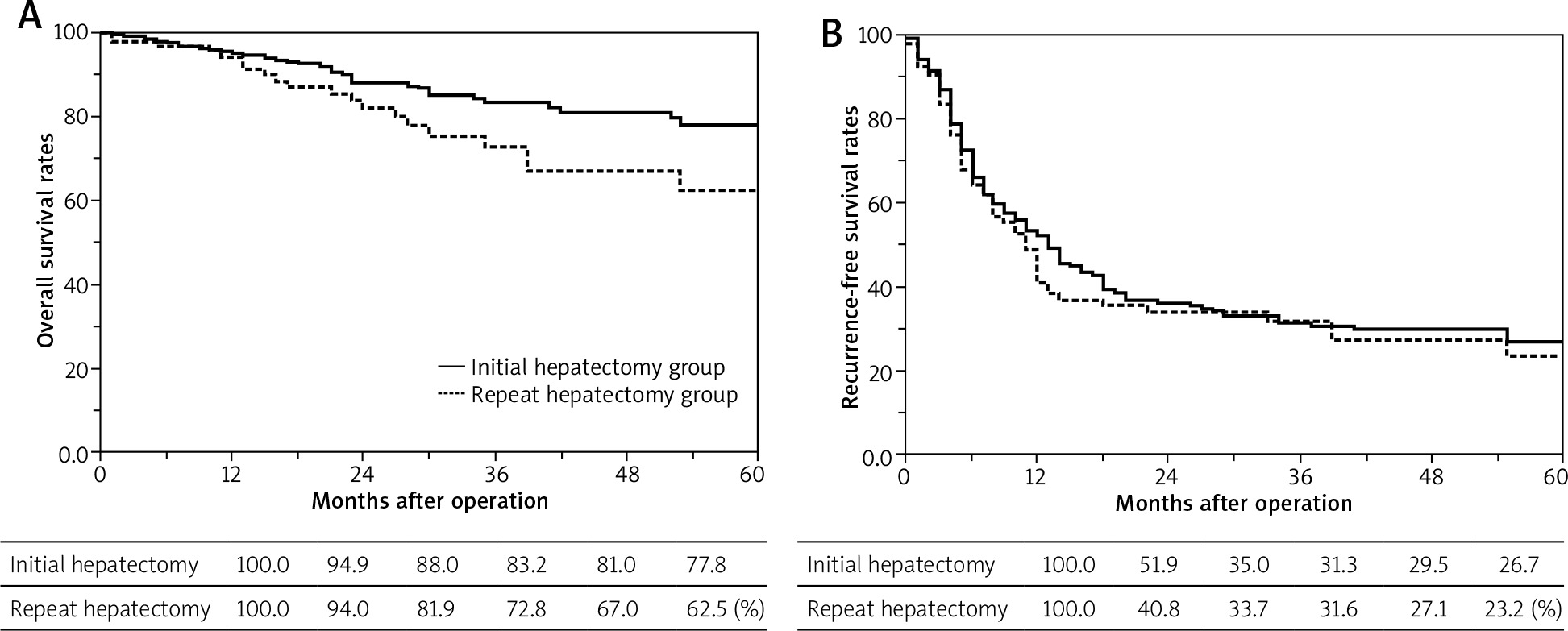
The 1-, 2-, 3-, and 5-year OS rates after initial hepatectomy and repeat hepatectomy were 94.9%, 88.0%, 83.2%, and 77.8% vs. 94.0%, 81.9%, 72.8%, and 62.5%, respectively (p = 0.071). The 1-, 2-, 3-, and 5-year RFS rates after initial hepatectomy and repeat hepatectomy were 51.9%, 35.9%, 31.3%, and 26.7% vs. 40.8%, 33.7%, 31.6%, and 23.2%, respectively (p = 0.387). There were no significant differences in OS and RFS between the initial and repeat hepatectomy groups after hepatic resection (Fig. 1). The 1-, 2-, 3-, and 5-year OS rates of patients treated for recurrent liver tumors were 100.0%, 92.2%, 82.2%, and 67.4% for resection-based treatments; 93.3%, 78.8%, 45.0%, and 22.5% for chemotherapy; and 59.9%, 9.0%, 0%, and 0% for palliative care, respectively (p < 0.001). There were no differences in the 1-, 2-, 3-, and 5-year OS rates between patients who underwent three or more hepatectomies and those who underwent a repeat hepatectomy (94.1%, 66.7%, 58.4%, and 46.7% vs. 94.0%, 81.9%, 72.8%, and 62.5%, respectively) (p = 0.288).
Fig. 1
Overall survival (OS) and recurrence-free survival (RFS) rates. The 1-, 2-, 3-, and 5-year OS rates after initial hepatectomy were 94.9%, 88.0%, 83.2%, and 77.8%, respectively. The 1-, 2-, 3-, and 5-year OS rates after repeat hepatectomy were 94.0%, 81.9%, 72.8%, and 62.5%, respectively. The 1-, 2-, 3-, and 5-year RFS rates after initial hepatectomy were 51.9%, 35.9%, 31.3%, and 26.7%, respectively. The 1-, 2-, 3-, and 5-year RFS rates after repeat hepatectomy were 40.8%, 33.7%, 31.6%, and 23.2%, respectively. There were no significant differences in (A) OS and (B) RFS between the initial and repeat hepatectomy groups after hepatic resection (p = 0.071 and 0.387, respectively)
Discussion
In order to assess liver regeneration, the recovery of liver function was evaluated in addition to measuring changes in RLV. In this study, the RLV increased markedly within 1–2 months after surgery, and a rapid increase in the RLV was particularly observed until the seventh day after surgery. Moreover, there was no difference in the change in RLV even after multiple hepatectomies. Remnant liver regeneration is caused by the enlargement of the remaining liver due to the same amount of portal venous blood inflow as before surgery. Therefore, there is no difference in the progression of liver regeneration after initial or repeat hepatectomy. The remnant liver function also recovers along with the progression of RLV regeneration. The recorded levels of test items, such as serum albumin, total bilirubin, PT, and platelet count, for evaluating functional aspects returned to normal values approximately 1–2 months after surgery.
This implies that liver volume and functional regeneration almost completely recovered to their pre-hepatectomy states within 1–2 months after surgery. Even if recurrence occurred in a shorter period of time after the initial surgery, it sufficed to perform various treatments, such as repeat hepatectomy and chemotherapy, for the volume and functional aspects [12]. In the management of patients experiencing tumor recurrence, the possibility of resection should be considered rather than worrying about remnant function, given that resection often yields the most favorable results. The approach to the treatment of post-hepatectomy recurrence should be determined based on the same criteria as for the initial treatment, regardless of when recurrence was observed or prior treatments.
On the other hand, with repeat hepatectomies, there have been reports that the technical difficulties are much greater because anatomical landmarks may be lost due to adhesions related to the initial procedure, and damage to other structures, such as the blood vessels, bile ducts, intestinal tract, and diaphragm, is more likely [13]. However, a review of 748 single and 288 repeat hepatectomy cases for CRC-related liver metastasis by Wicherts et al. showed that there were no significant differences in morbidity (27.1% vs. 34.4%, p = 0.069) and mortality (0% vs. 2.4%, p = 0.831) between initial and repeat hepatectomies [14]. The same was also true for short-term prognoses at our institution. Since approximately 96.8% of cases of CRLM result in a normal liver, hepatectomy can be performed safely because repeat hepatectomy does not increase the risk of complications, such as PHLF or massive ascites, thanks to standardized surgical techniques and improved instrument performance. In recent years, some articles have reported on the efficacy of laparoscopic repeat hepatectomies [4]. Laparoscopic hepatectomy has some disadvantages, such as lack of tactile sensation and decreased spatial recognition; however, intraoperative bleeding is reduced by effectively using pneumoperitoneum pressure, and surgery can proceed safely with a dry liver transection surface.
For long-term prognoses, initial and repeat hepatectomies showed similar long-term survival rates. The same long-term survival rates were obtained for repeat hepatectomy as for three or more hepatectomies. In patients undergoing treatments other than hepatectomy, given that only a 20% survival rate is possible, there is no definitive evidence with regard to the technical difficulty of surgery and remnant liver function; however, repeat hepatectomy seems to be beneficial. Similar to the reports of Antoniou et al., a repeat hepatectomy for a post-hepatectomy recurrence demonstrated a long-term survival rate similar to that of the initial hepatectomy in a meta-analysis, and treatment should be considered since the frequencies of complications and surgery-related deaths are similar [15].
Conclusions
Initial hepatectomy and repeat hepatectomy showed similar results of remnant liver regeneration, and short-term and long-term prognosis. However, the small sample size could lead to bias, including inconsistencies in perioperative chemotherapy, liver tumor status, and the type and quality of hepatectomy. Future randomized controlled trials and meta-analyses will be required to validate the study results.


