
Introduction
Renal cell carcinoma (RCC) constitutes about 3% of all malignant neoplasms in adults [1]. The most common histologic type of RCC, about 65% of cases, is clear cell carcinoma [1]. Renal cell carcinoma is asymptomatic in most cases. The main symptoms presenting mainly in the advanced disease are painless haematuria, flank pain, and palpable mass in the abdomen [2]. In 4–15% of cases RCC invades venous system and may form tumour thrombi, occasionally reaching the right atrium and causing inferior vena cava syndrome (IVCS) [3, 4]. Diagnosis of RCC is usually based on medical examination, computed tomography (CT), or magnetic resonance imagining (MRI) of the abdomen and pelvis, and the result of a histopathological examination [5]. Treatment is based on the stage of the disease determined according to TNM classification. In all the stages, nephrectomy, either partial, radical, or cytoreductive, is considered a standard procedure [5]. However, in the case of RCC with inferior vena cava tumour thrombus (RCC IVC-TT) surgical treatment is associated with poor prognosis and relatively high morbidity [6].
Renal cell carcinoma is relatively resistant to conventionally fractionated radiotherapy, and this method is rarely used in standard care [7]. However, some data suggest hypofractionated or stereotactic radiotherapy [8].
We present a case report of a 43-year-old female patient diagnosed with advanced RCC IVC-TT, who underwent nephrectomy and thrombectomy followed by hypofractionated high-dose radiotherapy.
Case report
At the beginning of 2018, a 43-year-old female patient was diagnosed with RCC IVC-TT (stage IV according to Mayo Clinic RCC Tumour Thrombus Level Classification System) [3]. At first a pathological mass was found accidentally in an ultrasonography examination. Later a CT scan was performed. The primary tumour size in CT was 81 × 62 mm with invasion of the inferior pole of the right kidney and right renal artery, and a 57-mm-long tumour thrombus in the IVC reaching the heart and obstructing the hepatic veins – cT3cN0M0 [9]. The patient reported periodic lower-back pain and, besides chronic hypertension, had no other comorbidities.
In 2018, the patient underwent nephrectomy with the subtotal resection of the tumour thrombus from the lumen of the IVC. After surgery the patient received anticoagulant therapy with dalteparin for 28 days. The surgery specimen included the right kidney with the 7.7 × 5.5 × 5.5 cm tumour and 8.1-cm-long tumour thrombus. The pathological examination of the tumour and thrombus confirmed the diagnosis of clear cell renal carcinoma WHO/ISUP (International Society for Urological Pathology) grade 4, corresponding to tumour stage pT3bN0M0 [9].
Three months after the initial treatment the patient noted fatigue and swelling of the lower parts of the body. She was hospitalized twice, at 5th and 6th month after the surgery due to, respectively, IVC thrombosis and massive oedema of lower limbs. Fluorodeoxyglucose-positron emission tomography-computed tomography (FDG-PET-CT) was performed during the second hospitalization to determine the cause of IVC thrombosis. The scans showed the presence of highly metabolically active tumour thrombus blocking the IVC (58 × 42 × 116 mm) and a single lesion of 12 × 10 mm in the inferior lobe of the left lung with elevated standardized uptake value (SUVmax = 11.7) of fluorodeoxyglucose (18F) and heterogeneous lesion in the liver (SUVmax = 6.6).
The patient was deemed inoperable and referred for palliative radiotherapy of the tumour thrombus. At admission she presented massive oedema of lower limbs, ascites, fatigue, severe pain, disturbed sleep, and anxiety but had good performance status (ECOG 1). The clinical presentation was typical for the IVC syndrome. Laboratory tests showed highly elevated liver function tests (LFTs) and coagulopathy with increased D-dimer level.
Conventional treatment of advanced RCC with tyrosine kinase inhibitor (TKI) pazopanib was considered. However, it was abandoned due to liver function impairment.
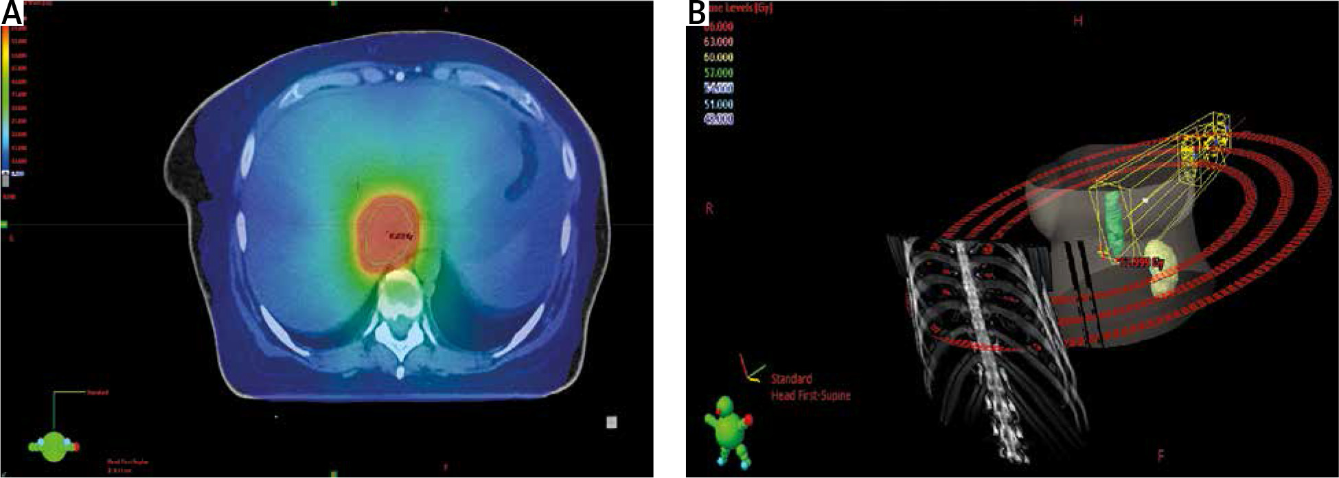
After careful analysis of all clinical data, the initially planned palliative radiotherapy was replaced by a more aggressive hypofractionated radiotherapy with the total dose of 60 Gy delivered in 12 fractions (5 Gy per fraction, 4 times a week: Tuesday, Wednesday, Thursday, Friday during the first week and Monday, Tuesday, Thursday, Friday during the latter 2 weeks). The treatment was conducted using dynamic, 4-dimensional volumetric arc therapy (4D-VMAT). Computed tomography, MRI, and PET-CT fused scans were all used for radiotherapy planning.
Gross tumour volume (GTV) of 122 cc was identified and carefully delineated according to the PET-CT and MR findings. The internal target volume (ITV) was delineated using a 4D technique and consisted of the average position of the GTV. Respiratory gating was used instead of breath hold technique because it was impossible to perform due to the aggravation of dyspnoea in the horizontal position and ascites. Additional margins to GTV consisted of superior margin: 1 mm, left lateral margin: 2 mm, right lateral margin: 2.5 mm, anterior, posterior, and inferior margin: 0 mm. However, identification of the tumour on CT gating scans and cone beam computed tomography (CBCT) performed during each fraction was extremely difficult; therefore, planning target volume (PTV) included 5–6 mm of isometric margins from ITV and was larger than initially planned (223 cc).
The planning target volume was irradiated with 100% mean dose (min 87%, max 105%) and a conformity index of 0.63. The organs at risk (OARs) were irradiated without any significant overdosing according to international guidelines [10]. Because there is no characterized OAR limit for 12 fractions, the dose limits for 10 fractions were used (Table 1).
Table 1
Organs at risk – statistics and dose limits
Because there are no guidelines and constraint values for hypofractionated or stereotactic radiotherapy of IVC tumour thrombus reaching the atrium, we implemented the therapy which, according to our best knowledge and the Tumour Board, was a compromise between possible adverse effects and local tumour control.
The position of the isocentre and irradiated PTV was controlled at each fraction with CBCT. For adequate accuracy, we assumed the borders of IVC as the matching points. Immobilization of the patient was achieved with lung base and Vac-Lok combined. The positioning of the patient was reliable and repeatable.
After the first fraction of radiotherapy the patient experienced nausea, vomiting, and abdominal pain, which were managed with thiethylperazine, drotaverine, and ranitidine. After the 6th fraction the vomiting recurred and was effectively managed with dexamethasone. Abdominal pain was completely relieved using morphine and transdermal fentanyl. Additionally, the patient was administered diuretics for severe oedema and a therapeutic dose of enoxaparin sodium as the basic anticoagulant therapy. She completed the planned radiation treatment in good general condition (ECOG 1) and with no grade 3 or higher side effects. Significant regression of IVCS was achieved according to clinical examination. The patient presented reduced peripheral oedema, and improvement of LFT values and laboratory tests.
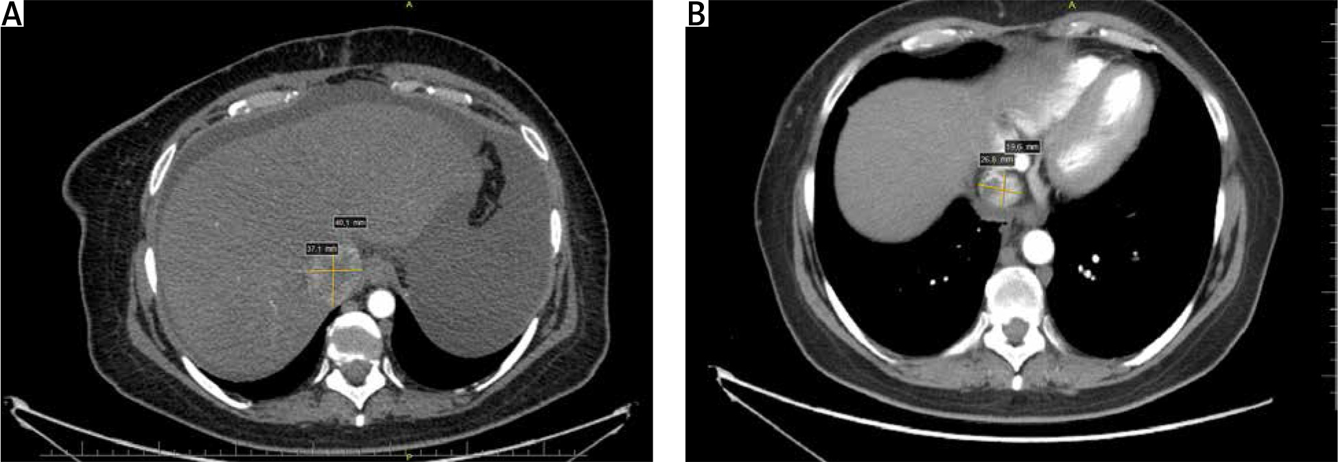
Within the first 3 months of follow-up, dysphagia and the feeling of fullness in the abdomen gradually decreased and there was no nausea, vomiting, or oedema of lower limbs. In the 2nd week after discharge, she was hospitalized due to hypokalaemia induced by the diuretic treatment. The post-treatment follow-up compliance was poor, and the first CT was performed almost 9 months after radiotherapy (Fig. 1). The initially enlarged lumen of IVC residual mass assessed at Th9-10 decreased 41.8–25.0 mm (Fig. 2). No lesions in the lungs and liver were determined. Surgical removal of persistent thrombus was considered, but the patient refused a cardiosurgical consultation and wilfully stopped enoxaparin medication. Two consecutive CT examinations performed 11 and 19 months after treatment showed no signs of local progression. Nonetheless, the last CT scan revealed atelectasis of the lower part of the lung caused by occlusion of the lower branch of the left main bronchus by a pathological mass. After 4 months the patient did not report any significant symptoms and was referred for PET-CT examination to evaluate the nature of the lung lesions. Further therapeutic plan included possible radiotherapy of present metabolically active lesions. She did not report for PET and for further follow-up visits. The systemic treatment with pazopanib (TKI) was once again considered during the follow-up period. Again, because of the poor compliance and prior refusal of the other approaches it was abandoned.
Fig. 1
Computed tomography scan of the abdomen at the Th9-10 level before radiotherapy (A), 9 months after radiotherapy (B)
The patient died in November 2021 – one year after the last follow-up visit and 3 years after radiotherapy. The direct cause of death was undefined, there was no post-mortem examination.
Discussion
The standard of care in metastatic and non-metastatic RCC is nephrectomy with thrombectomy (if indicated) [11]. The presence of tumour thrombus in vessels strongly affects staging, prognosis, and treatment. The surgical treatment of RCC-IVC-TT is mainly dependent on the thrombus size and the level of invasion (reaching hepatic vessels or right atrium) [12–15]. Potentially curative treatment includes nephrectomy and thrombectomy, invasive cardiopulmonary bypass (CPB), and deep hypothermic circulatory arrest (DHCA) [13, 16]. Although CPB with DHCA is relatively safe, the risk of platelet dysfunction, coagulopathy, hypothermia, and intensive multi-disciplinary care may be challenging [1, 14]. The radical surgery is associated with 30–35% morbidity [17]. Additionally, poor performance status of the patient, comorbidities, the presence of metastases, or the size of tumour thrombus frequently do not allow complete removal of the tumour mass [18].
Advanced RCC is also conventionally managed with molecularly targeted therapy, mainly TKIs. The VHL gene inactivation is present in most RCCs and leads to upregulation of vascular and platelet derived growth factors, making them the main targets of molecular therapy. Tyrosine kinase inhibitors such as axitinib, cabozantinib, pazopanib, sorafenib, and sunitinib are metabolized in the liver and may cause its damage. Hence, hepatic impairment is a strong contraindication for administration of TKIs [19]. In our patient, pazopanib was considered after the surgery but was abandoned due to rapid liver function impairment resulting from hepatic vein thrombosis associated with IVC-TT. Systemic treatment with pazopanib was reconsidered during the follow-up period. Due to the poor compliance and prior refusal of radicalization of the treatment systemic the treatment was abandoned.
Renal cell carcinoma has been considered a radio-resistant malignancy [18, 20–22]. Indeed, conventional radiotherapy (2 Gy per fraction) causes only a limited cytotoxic effect. However, higher fractions (6–8 Gy) may lead to exponentially higher tumour cell deaths with acceptable toxicity [7, 23]. Stereotactic ablative radiation therapy used in both primary and metastatic clear cell RCCs has been shown to achieve local control in almost 98% of cases. There are no standards for treatment of RCC with invasion of inferior vena cava. Standard stereotactic radiotherapy fractionation was not feasible due to the large volume of the PTV, close location of the fragile OARs, and potential constraint violation. Hence, the radiation oncologist team choice was a hypofractionated radiotherapy similar to the fractionation schedules preferred for centrally localised tumours in the chest [24]. Moreover, in our facility, the 60 Gy/12 fractions regimen is mostly administered in 3 weeks (4 fractions per week) – as per institutional standard.
Despite considerable risks, the effectiveness of treatment of our patient was satisfactory (nearly 3-year local control with good tolerance and quality of life). The case emphasizes the feasibility of radiotherapy as a possible salvage treatment in RCC with inferior vena cava tumour thrombus [25].
Conclusions
Our case indicates that modern hypofractionated radiotherapy may provide a relatively good palliative effect and extend survival. It is feasible option as an emergency treatment for circulation insufficiency caused by RCC-IVC-TT when other methods are contraindicated or impossible to administer.


