Imaging is a key point in the guidance of transcatheter treatment of paravalvular leak (PVL). Mostly explored by echocardiography, this imaging modality is limited by the acoustic shadowing from the valve prosthesis [1]. Cardiac computed tomography may be useful to assess anatomical characteristics (size, location, path) but is exposed to beam hardening artifacts and to cardiac movements [2]. Conventional cardiac magnetic resonance (CMR) seems to be relevant for functional evaluation of PVL after transcatheter aortic valve replacement, but is highly sensitive to metallic artefacts [3]. The question of a complete evaluation of paraprosthetic leakage therefore remains open and no conventional modality seems satisfactory. Because this question is also frequent in clinical practice, we have been led to consider an innovative imaging modality to complete the multimodal analysis; 4D flow imaging. We considered exploring new imaging techniques after percutaneous closure failure for a paravalvular mitral valve leak. The hypothesis put forward was a misguided selection of the prosthesis and therefore an unsatisfactory assessment of the leak. The initial evaluation was performed according to our usual practice, based on echocardiography (transthoracic and transesophageal) and cardiac computed tomography (CT) (Figures 1, 2). Considering that this initial evaluation was unsatisfactory, we favored an additional imaging modality rather than renewing potentially invasive or deleterious examinations. Cardiac magnetic resonance was then performed at 1.5 Tesla (Discovery MR 450 W, GEHC) in order to assess 4D flow imaging (TR 4.3, TE 2.4, VPS 3, 1.4 × 1.4 × 1.2 mm) (Figure 3). 4D flow imaging is an innovative modality of blood flow imaging with 3-dimensional time-resolved, phase-contrast cardiac magnetic resonance. This technique has already shown its feasibility and interest in many cardiovascular areas, including the study of prosthetic valves [4, 5]. Full examination was done in a free-breathing, single, acquisition (acquisition time 8 min, post-processing time 20 min) [6]. Retrospective valve tracking was used to quantify PVL after post-processing (Arterys, California, USA). Inlet and outlet dimensions were determined using planimetry on dynamic and almost isotropic acquisition after adjustment for every timeframe and direction of the regurgitant flow. Hemodynamic characteristics could be determined by direct measurement giving celerity and regurgitant volume. To our knowledge, this is the first case of PVL assessed by 4D flow imaging. This proof-of-principle case demonstrates the feasibility and the interest of this imaging modality for PVL, providing comprehensive analysis, in patients with a CMR-compatible mechanic valve. It is likely to have an appealing appraisal interest, while it offers both anatomical and hemodynamic appreciation, which the other modalities do not. Even if metal artefacts are less important than for conventional CMR, the main limit is the inability to evaluate the passway, near the ring of the prosthetic valve. This first assessment must be supported by further work. However, the fields of application of this technique seem great.
Figure 1
Paravalvular leak assessment using CT scan. A, B – 2-chambers views. The CT image at the level of the mitral annulus shows a small defect at the periphery of the prosthetic valve. The defect is continuous with the lumen of the LVOT and ascending aorta. C – Short axis view from the atrium. This view confirms the location of the mild posterolateral defect and shows surrounding structures
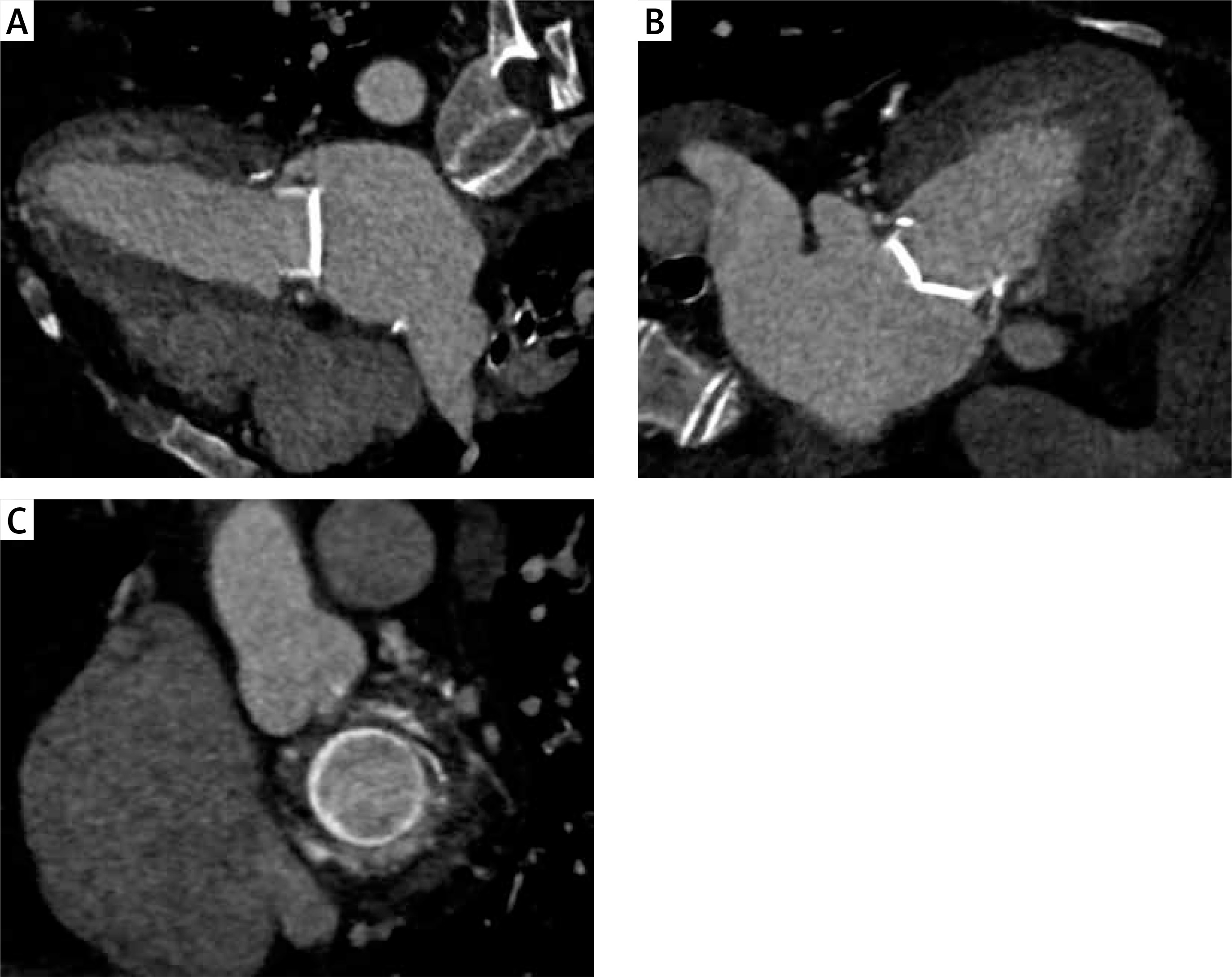
Figure 2
Paravalvular leak assessment using 3D transesophageal echography. A, B, C – “Surgeons eye view”. 3D transesophageal echography shows the paraprosthetic defect, which is located at the 5 o’clock position `
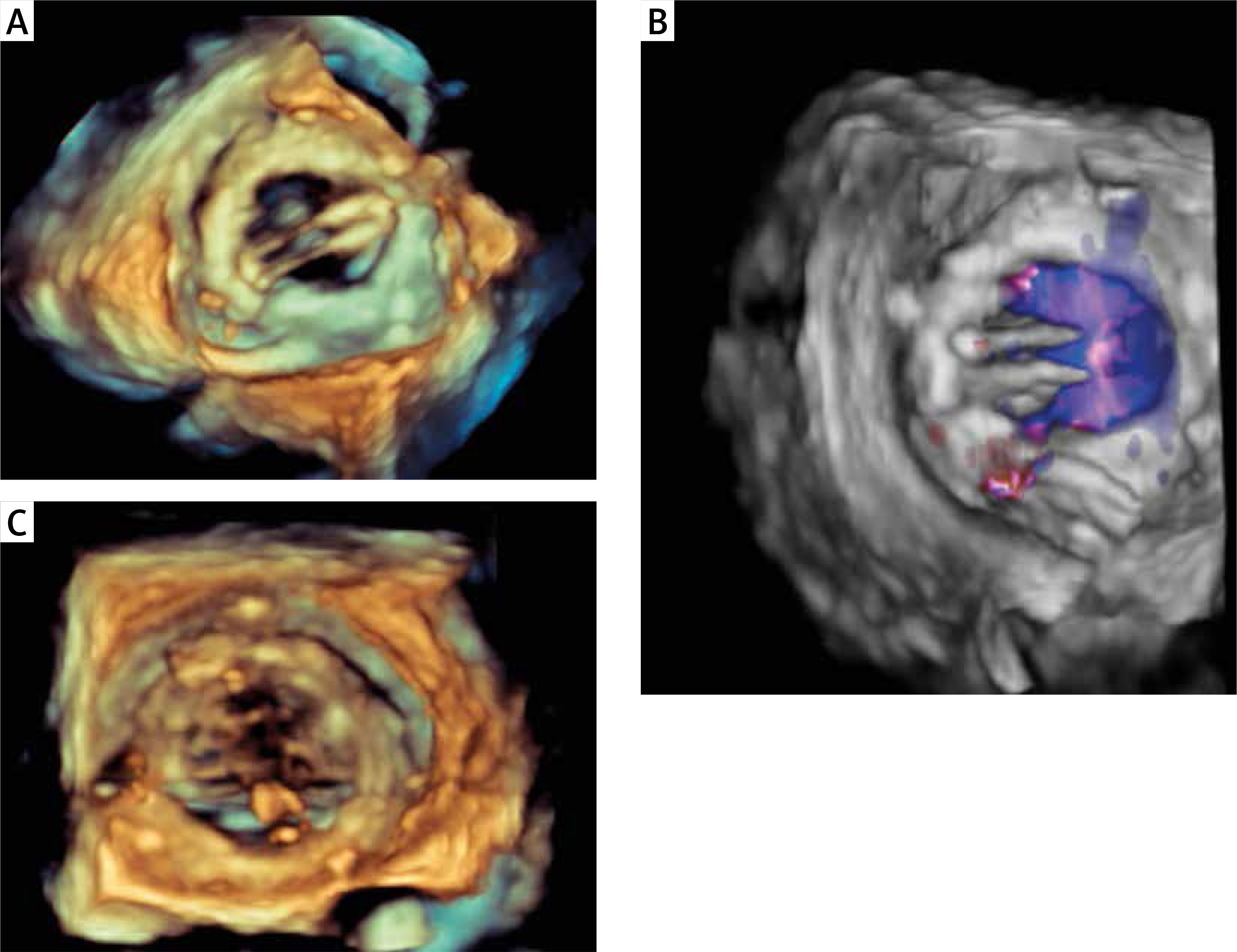
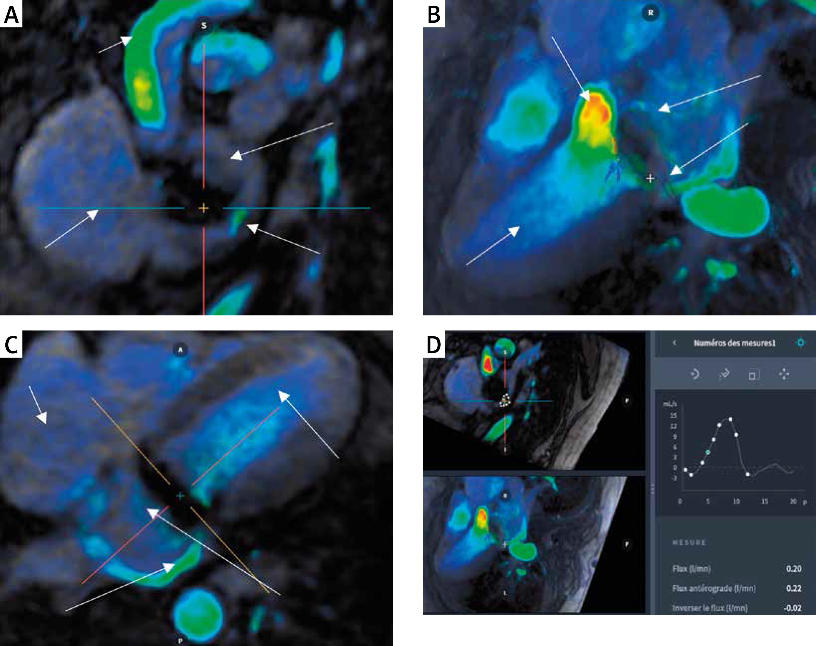
Figure 3
Paravalvular leak assessment using 4D flow imaging. A – Short axis view from the atrium. This view shows the mild posterolateral location and allows the measurement of the PVL. B – 3-chamber view. PVL jet was illustrated as a systole flow coming from left ventricle through a defect close to the mitral annulus. C – 4-chamber view. PVL’s eccentric jet is responsible for a turbulent flow in the atrium, without regurgitation in the pulmonary veins. D – Functional assessment. Flow measurement of the leak after direct contouring of the area of interest and using valve tracking. The information then collected concerns volumes as well as velocimetry