
Traumatic carotid artery perforation is a rare but serious accident [1–3]. Emergency management includes external compression (if anatomically feasible) and either surgery (reconstruction or ligation [2, 3]) or endovascular treatment. Endovascular options include carotid artery sacrifice (coiling) to stop bleeding [4, 5] or perforation sealing with the use of a covered stent (stent-graft) [4, 6–9]. In minimal-to-moderate leaks, initial strategy may involve a sealing attempt with prolonged balloon inflations [4, 6]. Prolonged balloon inflations, however, may increase the risk of symptomatic thrombosis, particularly if performed in absence of procedural anticoagulation or with premature (i.e., prior to removal of the interventional devices from the vessel lumen) heparin reversal with protamine [4].
We describe the use of a microNET-covered stent for sealing an internal carotid artery double perforation caused by a penetrating nail crossing the artery as a consequence of a nail-gun accident (Figures 1, 2).
Figure 1
Procedural images of sealing carotid artery traumatic perforation with a MicroNET-covered stent and long-term follow-up. A to C are internal carotid artery angiograms in different planes, demonstrating a nail penetrating the artery in C3/C2 segment (note that the nail both entry and exit point are visualized in B). D is a photograph of MicroNET, mounted on a nitinol stent (MicroNET-covered stent system), which was used to seal the 2 perforation sites. E shows a balloon inflation sealing the MicroNET (and the stent platform) intraluminally to the vessel wall. F is the immediate result of the procedure, demonstrating lack of any contrast extravasation. The arrow (E, F) points to the perforation site of the nail exit from the artery, with both the entry and exit sites completely sealed. G is duplex ultrasound follow-up at 5 years, showing a fully patent stent with absence of any in-stent restenosis and normal velocities
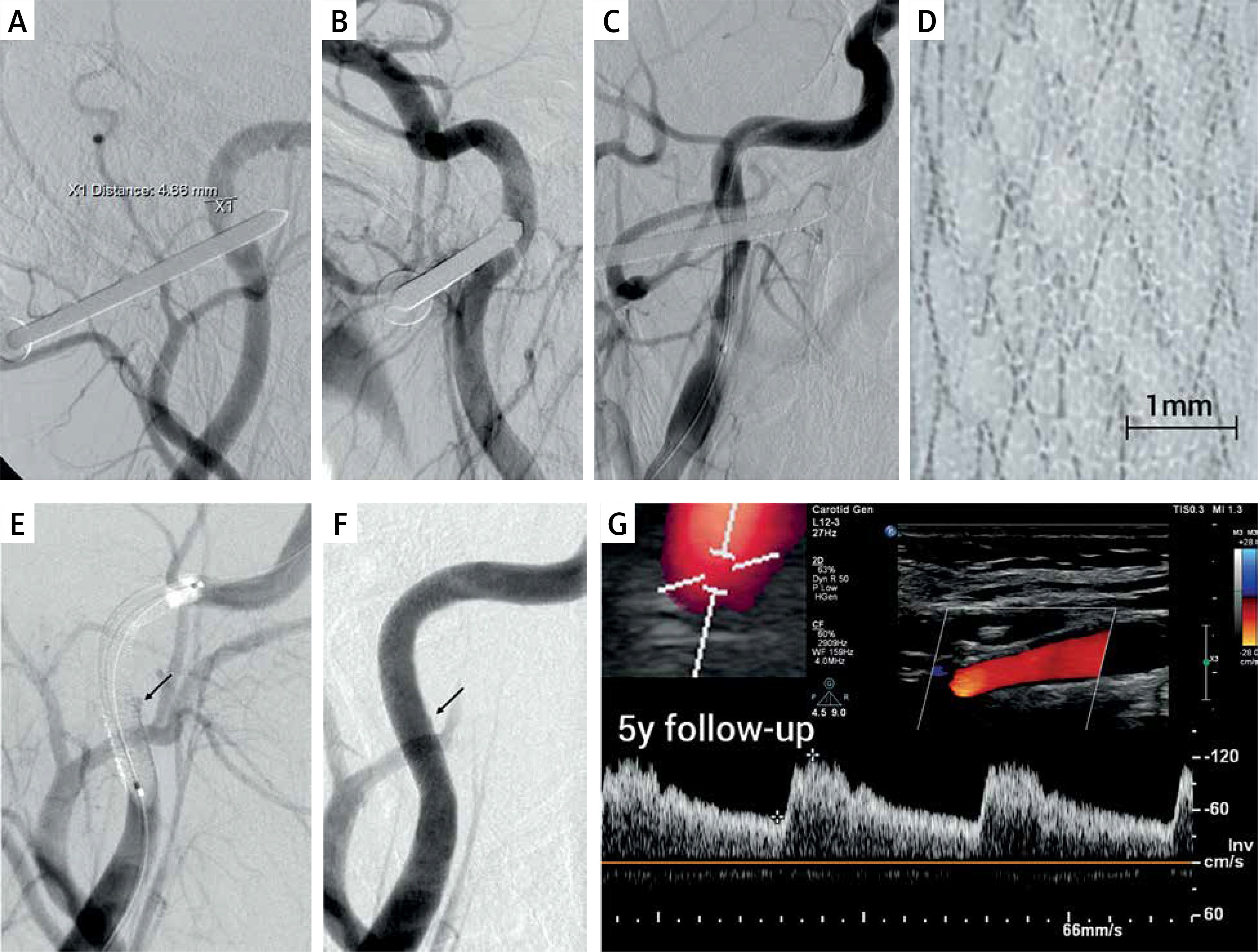
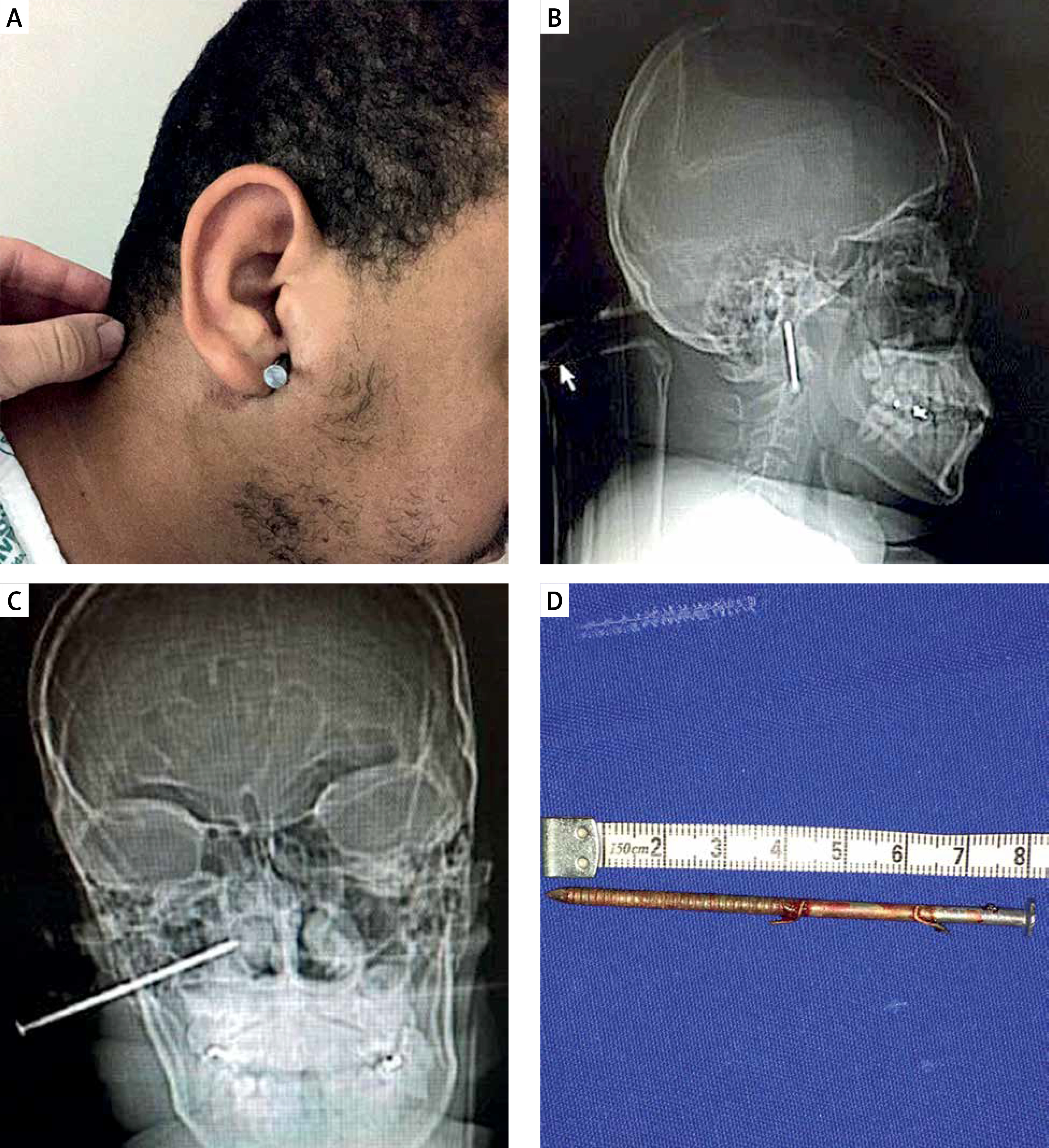
Figure 2
An 8 cm nail at the level of the right ear, causing a perforation of the internal carotid artery: photographs (A, D) and skull X-ray in the lateral (B) and postero-anterior (C) projection
MicroNET-covered stent system consists of a nitinol platform (self-expandable [10–12] or balloon-expandable [13–15]), covered with a highly-adaptable polyethylene terephthalate single-fiber knitted microNET sleeve (Figure 1 D) that is attached to the outward surface of the nitinol stent platform on its edges [10–16]. In a recent prospective multi-centric study of 352 procedures with intravascular imaging in consecutive patients with clinical symptoms or radiologic signs of cerebral injury in association with carotid stenosis, microNET was found to be 100% effective in sealing the atherothrombotic plaque material from the lumen [17, 18]. In addition, similar to microporous membrane stents [19], microNET acts as a flow-diverter that may facilitate aneurysm healing (NCT04434456) while not affecting any high-volume physiologic flow (such as that in the external carotid artery) [16, 20]. Evidence suggests that these microNET properties may translate into a minimized risk of perforation when using the microNET-covered carotid stent to treat the lesions associated with an increased risk of rupture and extravasation (such as highly-calcific stenoses) [21]. However, use of the microNET-covered stent to repair carotid perforation has not yet been reported.
A 28-year-old man using a professional nail-gun tool suffered a work accident when an 8-cm nail was inadvertently shot at the level of his right ear, causing a double (in/out) perforation of the internal carotid artery (Figures 1, 2). The patient did not show any acute neurological deficit and was hemodynamically stable.
Given the difficulty and hazards of the surgical access at the skull base [9], a decision was made to reconstruct the artery using the endovascular route. As no self-expandable stent-graft was available on-shelf (and the carotid self-expandable microNET-covered stent, CGuard [10–12, 22] was not yet commercially available), a balloon-expandable microNET-covered stent (4.0 × 18 mm) was used (Figure 1). The stent was introduced directly, without an additional cerebral protection (such as a distal filter or transient flow reversal that are required when treating atherosclerotic lesions to protect the brain prior to the microNET-exerted embolism prevention [10, 16, 22, 23]). The stent was positioned in the C2-C3 segment so that its middle portion addressed both the “inward” and “outward” vessel perforation site on the course of the nail crossing the artery (Figure 1 C). The nail was removed simultaneously with implanting the stent (3 min inflation at 10 atm) while performing external compression of the neck wound. A reduced dose of unfractionated heparin (3000 IU) was used. Since a suboptimal implantation of the microNET-covered stent may increase the risk of in-stent restenosis [14], the stent was further post-dilated with a 4.5 × 18 mm balloon at 8–10 atm (Figure 1 E) for 5 min despite transient intolerance, maintaining external compression of the wound. No angiographic extravasation was present after stent implantation (Figure 1 F). Acute angiographic result showed a complete, optimal endovascular reconstruction of the perforated vessel (Figure 1 F), and there was absence of any angiographic distal embolism and absence of any neurologic symptoms. The patient received low-dose aspirin (75 mg daily) and clopidogrel (75 mg daily) for 30 days. 5-year clinical follow-up was normal, and Duplex ultrasound demonstrated a normal luminal flow (Figure 1 G). This is consistent with a lasting effect of optimal endovascular reconstruction of carotid artery perforation using a microNET-covered stent, in agreement with a lasting mid-term and long-term effect of microNET-covered stent use to restore normal carotid artery lumen in primary and secondary prevention of carotid atherosclerosis-related stroke [18, 20, 24, 25].
Traumatic carotid artery perforation is a life-threatening emergency [3, 4, 9]. Besides trauma, carotid artery perforation can also occur as a complication of carotid artery stenting, particularly in case of endovascular management of calcified lesions using the 1st generation (single-layer, non-covered) carotid stents [26, 27]. Intraluminal angioplasty has the potential to cause tearing or rupture of the target vessel; a likely under-reported serious complication that may occur especially when using a non-compliant non-undersized balloon [6]. Arterial injuries due to angioplasty and stent placement are somewhat different from those due to carotid trauma in that in the latter a portion of the vessel wall is absent [6]. A single-layer nitinol stent is believed, in general, to reinforce the wall of the artery [6]. With balloon angioplasty, the force is transmitted to the circumference of the vessel wall, with only the weakest portion failing [6]. Speculatively, such angioplasty-inflicted tears may be reinforced by stent placement [6]. Nevertheless, in case of heavy calcification any “aggressive” optimization of a single nitinol layer (non-covered) stent to improve the lumen may cause perforation [28]. It needs to be noted that calcification is the most common cause for post-procedural residual stenosis – a fundamental factor of in-stent restenosis [21, 26]. Therefore, heavy calcifications have been considered a contraindication to endovascular revascularization using single metallic layer (non-covered) stents [26]. While the microNET stent wrap was originally developed to block intraluminal migration of the atherothrombotic plaque material [10, 11, 13], and it was subsequently demonstrated to be effective in this indication [10, 17, 18, 22, 29], recent analysis from the PARADIGM-101 study suggested safety of microNET-covered (CGuard) stent use in optimized endovascular management of highly-calcific carotid artery lesions [21]. This is consistent with the sealing properties of microNET as demonstrated presently (Figure 1). However, it needs to be remembered that the microNET-covered stent is not a stent-graft as it possesses micropores of ≈ 150–180 µm [10–12].
Surgery for carotid perforation may be unavoidable in complex injuries. Surgery, however, is associated with a stroke rate reaching up to 80% [3, 9]; a proportion that may be considered far too high to accept in cases where endovascular repair is feasible. Carotid artery ligation is an independent predictor of stroke (OR = 1.5, p = 0.009) [2, 30]. Coiling, an endovascular equivalent of ligation, is also associated with a significantly greater stroke risk than that seen with reconstructive techniques that maintain vessel patency and thus ipsilateral cerebral supply [9]. Lesions located near the skull base (as in the present case, cf., Figures 1 and 2) are particularly hazardous and difficult to repair surgically [9].
Covered stents (stent-grafts) were demonstrated to be useful as an effective emergency tool to seal both severe perforations and lesser perforations that persist despite prolonged balloon inflation(s) [4, 6–9]. However, the risk of in-stent restenosis with fully covered stents (stent-grafts) placed in the carotid location is as high as 15–40% [9, 31]. Exposing patients to such a high restenosis rate may be acceptable in case of emergency treatment but not in any routine setting [21, 31, 32]. Covered stents show lack of mechanical flexibility [9, 32] and a prohibitively high (in elective use) rate of in-stent restenosis [9, 31]. Furthermore, contrary to typical expectations [32], covered stents fail to prevent lesion-related thrombo-embolism as the embolic material (rather than getting trapped between the stent and the vessel wall) may get squeezed-out and mobilized to blood stream at the covered stent edges [33].
The microNET-covered stent system, by design, respects carotid anatomy [11, 12, 16], and – when properly embedded – shows a minimal risk of restenosis [20, 25, 29]. The microNET-covered stent has been previously reported to be effective in sealing coronary artery perforations [13, 14]. Presently, we report effective and uncomplicated sealing of a traumatic carotid artery perforation using a microNET-covered stent (Figures 1, 2) with a lasting, optimal angiographic and clinical result. This result is consistent with accumulating evidence for optimal short- and long-term outcomes with the microNET-covered stent system in treatment of carotid artery thrombo-atherosclerotic disease in primary and secondary stroke prevention [1–12, 17, 18, 20–22, 25, 28, 34]. Still, operators should be fully aware that the microNET-covered stent system is not fully-covered (i.e., it is not a stent-graft). Thus, in case of potential incomplete microNET sealing of the leak, strategies may involve performing prolonged balloon in-stent inflation or placing another microNET-covered stent (stent-in stent) to increase the sealing effect. Stent-grafts (with their trade-off of a high in-stent restenosis rate that is relevant particularly in the carotid bifurcation use [31]) should continue to be available on-shelf for major ruptures that may not be amenable to sealing with the micro-porous microNET-covered stent.
In conclusion, we have demonstrated a safe and effective use of a microNET-covered stent system (a nitinol stent wrapped in a flexible ultra-thin polymer mesh sleeve on its external surface), to resolve a traumatic carotid artery perforation (Figures 1, 2). There was a 100% sealing efficacy and a lasting endovascular reconstruction. Clinical relevance of the microNET-covered stent sealing properties (Figures 1, 2), taken together with its effective sequestration of thrombo-atherosclerotic material from the lumen [17, 18] and the feasibility of an optimized ‘full’ endovascular restoration of a normal carotid artery lumen [16–18, 23, 24], have opened a new chapter in the history of optimizing carotid artery reconstruction using the endovascular route [30, 34, 35].


