
Introduction
The p21 protein is encoded by the WAF1/CIP1 suppressor gene, induced by oncoprotein p53, a wild-type TP53 gene product, and is involved in inhibiting the cell cycle between G1/S phases. Its contribution to the regulation of DNA repair and replication, as well as apoptosis modulation, has been proven [1–3]. After DNA damage, p21 induced by p53, either leads to cell cycle arrest or it can evoke cell apoptosis. It is believed to be one of the most influential messengers of p53 in cell cycle arrest. Due to the high mutation ratio of TP53 in rectal cancer, a reduced amount of active p53 should be expected, thus a low level of p21 with all its consequences [4, 5]. The contribution of p21 in an intracellular transcription of signals from the growth factor receptors to a cell nucleus by the activation of the kinase cascade and Raf proto-oncogene has also been described [6–8]. It takes part in the process of cell growth inhibition in the process of cell aging or cell damage. Cell growth arrest occurs due to the binding and inhibition of the activity of numerous cyclins and cyclin-dependent kinases (cdk). This p21 interaction does not allow the phosphorylation of proteins regulating a cell cycle. The activity of cyclin-dependent kinases in complex with p21 is especially dependent on the amount of p21 in a nuclear compartment. At low concentrations p21 facilitates the translocation of cdk-cyclin complexes into a cell nucleus, whereas at high concentrations it inhibits kinase activity [9–11]. Another p21 function is to induce cell growth arrest by blocking the nuclear antigen of proliferating cell (PCNA) activity in DNA replication and “mismatch” in DNA repair [12–14]. Numerous studies have confirmed the effect of the RAS gene family mutations as a negative prognostic factor leading to a lack of response to a treatment based on EGFR inhibitors. However, here a well-known association with p21GTP ras protein has been noted, but the relationship of p21AWF1 with the KRAS-BRAF pathway still remains unclear. Despite many studies, their clinical effectiveness has not been unambiguously confirmed [15–17]. Moreover, an alternative perspective was emphasized for a more precise targeted treatment of colorectal cancer with p21 restoration [18]. Therefore, we have decided to test the usefulness of p21 as a cancer biomarker.
The aim of the study in the assessment of the relationship between the p21 immunoactivity and prognosis in rectal cancer.
Material and methods
The study was conducted in the Clinic of General, Oncological and Endocrynological Surgery of Regional Hospital in Kielce, Academic Department of Clinical and Experimental Pathology of Medical and Health Sciences Faculty of Jan Kochanowski University in Kielce and in the Holy Cross Regional Oncological Center in Kielce, Poland. Inclusion criteria involved the following: male and female patients who had undergone a planned rectum resection due to cancer, in whom distant metastasis had been excluded, without any other diagnosed neoplastic disease, who had been qualified for surgery aimed at recovery and had stabilized cardio-respiratory and metabolic load parameters. The endpoints were: overall survival (OS) and recurrence free survival (RFS).
The research methodology was divided into two stages. The first one included retrospective disease analysis in patients qualified for the study, the second stage involved immunohistochemical p21 tests. Applying relevant inclusion and exclusion criteria, the patients in stages I–IV of rectal cancer, according to Union for International Cancer Control (UICC) classification, who had undergone surgery between 2005–2011, qualified for the study. The study involved patients without pre-operative radiotherapy. All patients with adjuvant pre-operative radiotherapy were excluded, and all participant in III–IV UICC stages were treated with a similar chemotherapy regimen based on FOLFOX-4 (oxaliplatin 85 mg/m2, calcium folinate 200 mg/m2, fluorouracil 400 mg/m2, fluorouracil 600 mg/m2). The period was 5 years from the date of surgery. All participants provided written consent to participate in the study. Classic immunohistochemical tests were applied with the use of antibody p21WAF1 (clone H252). All performed tests were fully approved with the aim to be applied in vitro. All reactions were carried out using BenchMark XT (Ventana Medical Systems; Roche Group, Tucson), USA). After a fully automated dewaxing and reirrigation of samples, the process of antigen unmasking by protease K (37°C, 5 minutes) were carried out, followed by an incubation with primary antibodies (dilution 1 : 50, incubation time 20 minutes). The temperature of both antigen collection and the incubation of primary antibodies was closely in compliance with the producer’s recommendations and then a further standard procedure was followed. A universal DAB detection kit DAB Ventana ultra-View was applied. A four-level system describing p21 reactivity was used: 0 – no reaction, 1 – weak reaction, 2 – moderate reaction and 3 – strong reaction (Fig. 1).
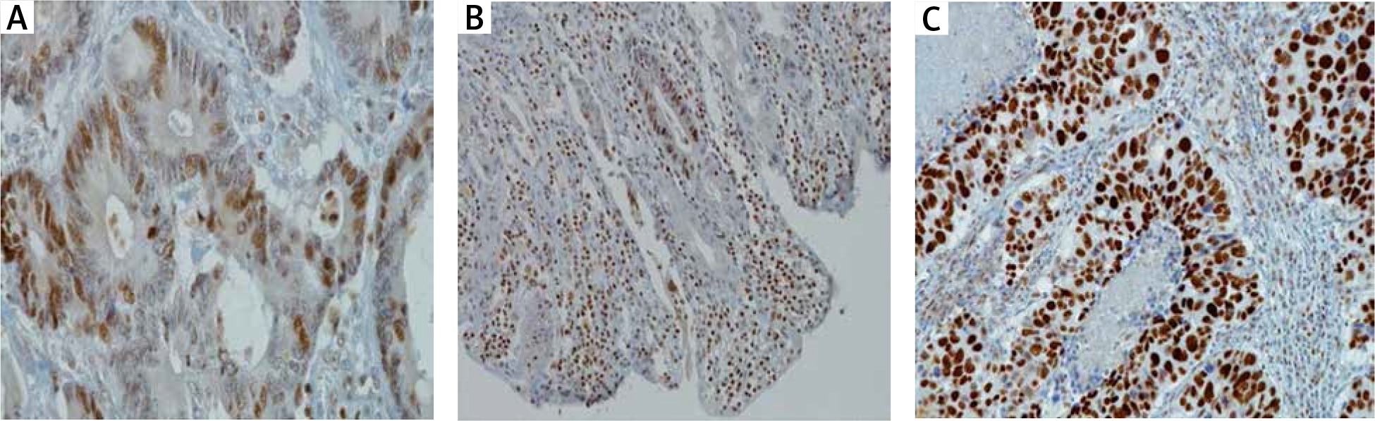
All calculations were done using a digital slide analysis with a slide scanner Hamammatsu NanoZoomer S210 (Hamammatsu®, Hamamatsu City, Shizuoka Pref. Japan). After scanning the entire slide, a dedicated digital image analysis was performed with the use of nuclear application Visiopharm (Visiopharm®, Hoersholm, Denmark). The utilized application allowed us to diversify the intensity of a membrane reaction and avoid subjectivity.
Statistical methods
The collected data were tabulated and then statistically analyzed using the Kaplan-Meier method, the log-rank test, the Cox proportional hazards model and logistic regression. In the applied tests, a significance level of 0.05 was adopted. Statistical analyses were conducted with the use of the SAS 9.3 licensed software.
Results
The study involved 102 patients aged 41–87, including 41 females and 61 males in the UICC stage confirmed by a histopathological examination. At the end of the follow-up period, 56 patients were still alive, i.e. 55%, including one patient with a local recurrence and one with seeding of cancer cells. Cancer-related death was noted in 46 patients, in 7 of them local recurrence was diagnosed and the survival median equaled 26.6 months, while the median without metastasis was 21.6 months. 85% of females (F) and 92% of males (M) survived 1 year; 78% F and 85% M survived 2 years; and 66% F and 67% M survived 3 years. The highest activity of p21, at level 3, was detected in 45 patients, i.e. 44.1%. In 5 patients, i.e. almost 5%, p21 activity was not observed. This group comprised of 53 patients (52%) pT3 stage, in whom p21 activity was at the highest – at level 3 or 2. In most patients – regardless of p21 activity level – the tumor was at G2 grade. There is no statistically significant correlation between G (1; 2; 3) and p21 protein activity p21 (0; 1 vs. 2; 3). There was no statistically significant association between p21 expression and histological cancer type (p = 0.3022). The analysis did not reveal any statistically significant differences in survival curves of patients with protein p21 immunoreactivity in 0; 1; 2; 3 (p = 0.6453 in the log-rank test), leading to the conclusion that the probability of survival is not dependent on p21 expression (Figs. 2–5).
Fig. 2
Survival curves (expressed in months) in four groups of patients, determined based on the immunoreactivity of the p21 expression level
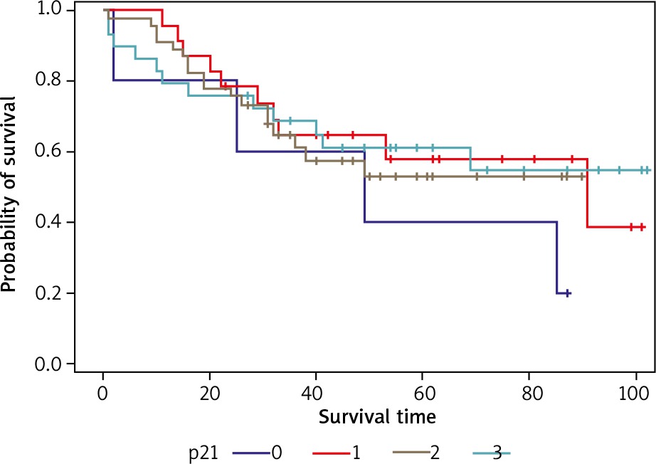
Fig. 3
Survival curves (expressed in months) for patients who did not show immunoreactivity of p21 (grade 0 and 1) and in patients with high immunoreactivity (Grade 2 and 3)
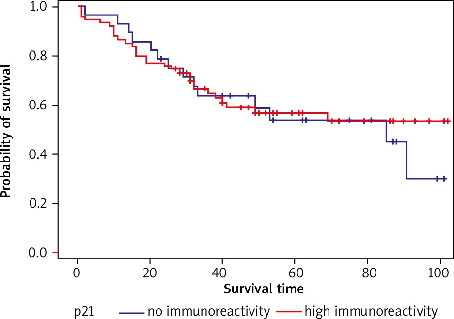
Fig. 4
Curves of cancer-free survival (expressed in months) for patients with no p21 immunoreactivity (0.1) and with high p21 immunoreactivity (2.3). The survival curve of patients in whom the immunoreactivity of p21 (0 and 1) did not differ significantly from the survival curve of patients with high immunoreactivity p21 (2 and 3) (p = 0.7830 in the log-rank test). The survival curve without shedding for patients who did not show immunoreactivity p21 (0; 1) does not differ significantly from the survival curve without spreading for patients with high immunoreactivity p21 (2; 3) (p = 0.6244 in the log-test rank)
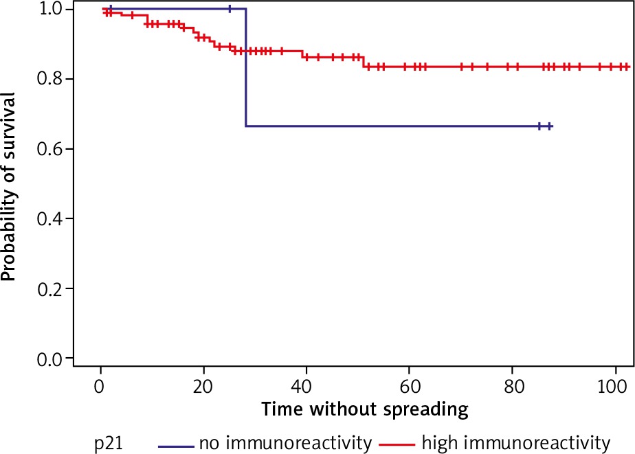
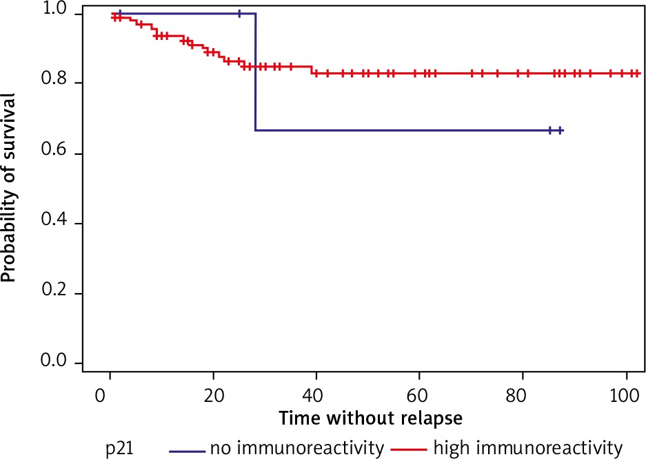
Fig. 5
Curves of cancer-free survival (expressed in months) for patients with no immunoreactivity of p21 (0.1) and with p21 immunoreactivity (2.3). The recurrence-free survival curve (without local recurrence or dissemination) for patients who did not show p21 immunoreactivity does not differ significantly from the survival curve without recurrence for patients with high p21 immunoreactivity (p = 0.7288 in the log-rank test)
The application of the Cox proportional hazards model with a p21 independent variable allows us to conclude that the level of p21 immunoreactivity is not a significant death risk factor (HR = 0.915, p = 0.7842) and is not a significant risk factor of cancer metastasis (HR = 0.94, p = 0.9426). Similarly, the death risk was not confirmed in logistic regression analysis within two years following surgery (p = 0.283 [95% CI 0.4–1.31]).
Discussion
P21 participates in the regulation of several normal cell functions, including proliferation, differentiation and apoptosis. A very close link between p21 and the mutation of the TP53 suppressor gene has led to the conclusion of its clinical usefulness in the final prognosis. In the evaluation of p21 clinical value, accumulation of this protein in the nucleus is usually described qualitatively, and its presence in tumorous cells was between 36% and 71%, and even up to 80% of the cases. Such discrepancies in the results can be explained mostly by the heterogeneity of clinical material, different methods of determination, and even material archiving [19–24]. The most popular method is applied to p21 reaction in relation to its intensity on the intensity scale (4–5 scale). Some studies did not classify staining according to a local or dispersed pattern. The existence of such diversity forces the need to devise more uniform assessments in the determination of these reactivities [25–27]. In our study we utilized a repeatable 4-level scale, which can be digitally analyzed in order to separate a weak and visible reaction. Moreover, the applied solution allowed us to avoid natural subjectivity. In our own study the highest p21 activity was detected in 74 patients, in comparison with 72,5%. In 5 patients, i.e. almost 5%, p21 activity was not detected. The survival curves in the four groups of patients, determined on the basis of p21 expression level, do not show any statistically significant differences (p = 0.6453 in the log-rank test). The survival curve for patients who did not express p21 (0) was not significantly different from the survival curve of patients with p21 overexpression (1; 2; 3) (p = 0.2205). The observation of the course of the recurrence treatment in our own cases encompassed the period up to 102 months after a surgery. In literature there was no clear association between the tumor recurrence and the occurrence of the p21ras protein expression, which is compliant with the observations by other authors [28–30]. In our own study we have also found that the level of p21 is not related to cancer recurrence. Some researchers have found such an association, rather in a limited scope, for example it was related to rectal cancer or a group of patients in stages A and B, according to Dukes [31–33]. There was also not a clear correlation between frequency and RFS and the presence of p21 expression. This conclusion has been confirmed by our own study, as there were no statistically significant differences between RFS in patients with 0, 1; 2; 3 of the expression group, the value of p = 0.9650 in the log-rank test. The obtained results indicate that the p21 accumulation is not an independent prognostic factor in sporadic rectal cancer. Based on the data presented here, we suggest a lack of prognostic significance of p21 in patients with sporadic rectal adenocarcinoma. However, an immunohistochemical evaluation of the p21ras presence may be useful in the determination of a cancer biology [34–36]. The application of the Cox proportional hazards model with a binary independent variable taking the 0 value, when the p21 value equals 0 or 1, and the value of 1, when the level of p21 is 2 or 3, leads to the conclusion that the level of the expression of p21 is not a significant factor of death risk (p = 0.7842), and it is not a significant indicator of cancer spread (p = 0.9426).
The data presented in this paper do not allow us to attribute a higher significance to p21 expression in the detection and monitoring of rectal cancer treatment. However, an immunohistochemical evaluation of the expression of this protein can be useful as one of many factors of a diagnostic and prognostic significance.
Conclusions
The probability of survival is not significantly dependent on p21 immunoreactivity p21 immunoreactivity is not a significant factor of death risk and is not an important factor of cancer metastasis.
Our own study does not confirm the significance of p21 immunoreactivity in detecting and monitoring rectal cancer.
Our study does not confirm the p21 value as a biomarker.


