
Introduction
Gestational trophoblastic disease (GTD) encompasses a spectrum of proliferative disorders ranging from non-neoplastic hydatidiform moles to malignant neoplastic conditions, such as choriocarcinoma (CC). This category encompasses tumours with unclear clinical behaviour, such as placental site trophoblastic tumour (PSTT), and epithelioid trophoblastic tumour (ETT) and tumour-like lesion – exaggerated placental site (EPS) and placental-site nodule (PSN) [1]. Invasive mole, PSTT, ETT, and CC are described as malignant [2, 3].
Placental site trophoblastic tumour is an abnormal trophoblastic proliferation and is an extremely rare neoplasia with a frequency of about 1/50,000–100,000 pregnancies (0.23–3.00% of all GTDs) [4, 5]. There are a minimal number of cases of this disease described in the literature.
Placental site trophoblastic tumour can occur after any type of pregnancy: molar pregnancy, miscarriage, abortion, or a full-term normal pregnancy. It can also develop from several months to years after pregnancy. These tumours develop in the area where the placenta attaches to the endometrium and can invade the muscular layer of the uterus. They evolve slowly and are usually treatable. Although rare, PSTT can metastasize to the lungs or involve organs adjacent to the uterus. Placental site trophoblastic tumours vary significantly in their biological characteristics, clinical behaviour, growth rate, and relative resistance to chemotherapy compared to other forms of GTDs [6]. This requires the development of specific diagnostic and therapeutic strategies.
We present a case of a 35-year-old patient diagnosed with PSTT mimicking an intramural pregnancy.
Case report
A 35-year-old patient was referred to our clinic with a diagnosis of PSTT. The patient had no previous illnesses. Her medical history included an abortion in 2005 and a miscarriage after 8 weeks of gestation in 2015. In 2016, an elective caesarean delivery was performed. In October 2020, an abortion was performed with a diagnosis of blighted ovum (histology was without specific features). Seven days later the procedure was repeated due to ultrasound data suggesting residual tissue, but there was no tissue removed for histological examination. Two months later, the patient had a positive pregnancy test. The ultrasound examination revealed a hypoechoic formation in the posterior uterine wall with a diameter of 30 mm and a peripherally located structure resembling a yolk sac (Fig. 1).
Fig. 1
A hypoechoic formation in the myometrium with a diameter of 30 mm and a peripherally located structure resembling a yolk sac in 2-day differences: (A) first one, (B) second one
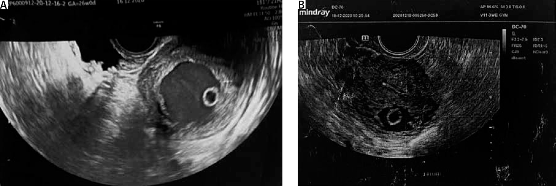
Doppler examination showed well-defined peripheral and central blood flow. β-HCG was 1092 mIU/ml, and an intramural pregnancy was diagnosed. Resection of the described formation was performed by open surgery, and a cyst on the right ovary was removed. The patient was discharged on the 4th day without complications. One week after surgery, β-HCG was 245.7 mIU/ml, and 2 months later it was normal.
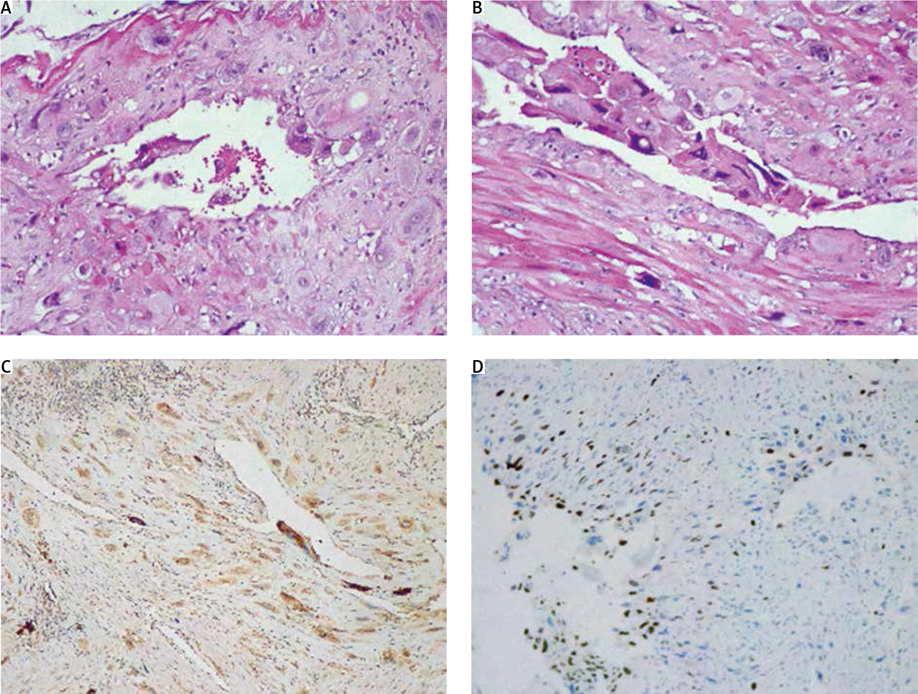
Histological results of the operative intervention showed neoplastic infiltration of myometrial tissue that separated smooth muscle fibres and replaced vascular walls. The cells were large, predominantly mononuclear, with pale cytoplasm and obvious nuclear pleomorphism. Scattered multinucleated cells could also be seen. Multifocal areas of necrosis were present alongside intravascular tumour cells. There were also fragments of chorion and amnion, as well as single chorionic villi with fibrosis and trophoblastic proliferation – which may be residue from the patient’s previous pregnancy. Immunohistochemical exam showed hPL (+) in tumour cells, hCG (–) in invasive trophoblastic cells and positive in villous trophoblast, p63 (–), and Ki67 labelling index 10–30%. The cyst was described as a usual functional cyst.
On the basis of the clinical data, histology, and immunohistochemical findings, the patient was diagnosed with PSTT that developed possibly after a missed abortion. The Ki67 labelling index absolutely excludes the possibility of endoscopic papillary abnormalities and stone recognition (Fig. 2).
Fig. 2
Histological findings. (A) Myometrial invasion of large eosinophilic mononuclear pleomorphic cells and replacement of vascular wall. HE × 20, (B) replacement of vascular wall and intravascular tutor cells. HE × 20, (C) all neoplastic cells show cytoplasmic positivity for hPL × 10, (D) Ki67 labelling index 10–30% × 10
The patient was referred to our clinic for radical surgical treatment, performed 4 months after the first surgical intervention. A preoperative positron emission tomography (PET) scan was performed, which showed no specific features. A total hysterectomy with adnexa was performed. The patient was discharged on the 5th day without complications.
The histological examination revealed no evidence of disease persistence.
During 10 months of follow-up (in the 6th month a PET scan was performed) no relapse of the disease has been found.
Discussion
Gestational trophoblastic disease continues to be a diagnostic challenge for pathologists because of its rarity, broad differential diagnosis, and histological overlap with common gynaecological tumours [7]. Diagnosis of GTD and tumour-like lesions is histologic, but information about the clinical history, hCG serum levels, and disease spread is essential. Close collaboration between pathologists and gynaecologists is crucial for correct diagnosis. Elevated serum levels of hCG, degree of elevation, and clinical data of previous pregnancies and abortions help pathologists eliminate other nontrophoblastic gynaecological neoplasias. Gestational trophoblastic tumours (GTT) have one of the highest overall survival rates (98%) among solid tumours [8], but they have different sensitivity to chemotherapy, varying from highly sensitive (CC) to chemoresistant (ETT) [9]. That is why the differentiation of the correct GTT is of great importance for the patient’s therapeutic management.
Placental site trophoblastic tumour is the rarest form of GTT, accounting for up to 3% of all GTDs [5]. The malignant potential of this tumour was first described in 1981 by Scully and Young [10].
Placental site trophoblastic tumour can occur at any reproductive age and after any type of pregnancy, and at different times after pregnancy [11, 12]. In 61% of patients it occurs after a normal pregnancy, in 12% it develops after a molar pregnancy, 9% after a miscarriage, 8% after voluntary abortion, 3% after an ectopic pregnancy, stillbirth, or premature birth, and in 7% the cause is not clear [13].
Placental site trophoblastic tumour is derived from the implantation site intermediate trophoblast. In contrast to hydatidiform mole and CC, intermediate trophoblast (IT) lesions have only been recognized relatively recently, and therefore their behaviour has not been well characterized [2]. Placental site trophoblastic tumours possess some typical histopathologic features: an infiltrative growth pattern consisting of sheets or nests of large, polyhedral, predominantly mononuclear cells with abundant amphophilic, eosinophilic, or clear cytoplasm, scattered multinucleated cells, nuclear pleomorphism, replacement of vascular wall by tumour cells, focal haemorrhages, and necrosis [1, 7, 14]. These histopathological criteria are typical of PSTTs but not unique to this tumour and therefore are not sufficient for an accurate diagnosis. Luckily, some immunohistochemical markers are beneficial in this aspect. The best markers for PSTT are hPL [1, 7, 14] and Ki67 labelling indices varying from > 5% [7, 14] to 10–30% [1, 7]. Negativity or only focal positivity of hCG together with mildly to moderately elevated serum levels of hCG (5–26000 mIU/ml) helps to exclude CC [1, 7, 14] while negativity of p63 assists in excluding ETT [1, 7].
The time frame for the development of PSTT after pregnancy is broad – it ranges from weeks to years. A study from 2018 by Alexander et al. reported a median time from antecedent pregnancy to diagnosis of 12 months (range 0–240) [15].
Abnormal vaginal bleeding and elevated β-hCG levels are common clinical features in these rare cases, but the clinical picture may vary [16]. In PSTT, β-hCG levels are generally lower than invasive moles and CC, with a mean serum β-hCG level of 132 IU/l when PSTT is diagnosed [17]. However, cases with higher serum levels of β-hCG [3, 18] and those with normal levels [5] have also been reported. Rarely, PSTT can be presented with preeclampsia, β-hCG triggered hyperthyroidism, and unspecific symptoms such as nausea or haemoptysis, enlargement of the uterus, and lutein-cysts of the ovaries [13].
The diagnosis of PSTT is usually confirmed by transvaginal ultrasound. Zhou et al. [19] classified the ultrasound findings into 3 types:
type I – heterogeneous solid mass in the uterine cavity with a degree of vascularization on colour Doppler imaging from minimal to moderate,
type II – heterogeneous solid mass deepening in the myometrium and coexisting with a degree of vascularization from minimal to high,
type III – cystic lesions within the myometrium with a high degree of vascularization (lacunar-type lesions).
Myometrial invasion can be determined by magnetic resonance imaging (MRI), and the presence of metastases by computed tomography (CT) or PET scan [20, 21]. It should be noted that ultrasound is sufficiently informative for the presence of liver metastases, and MRI can be used to detect brain metastases [22].
The International Federation of Gynaecology and Obstetrics (FIGO) anatomical staging system for trophoblastic tumours (2000) is used for the staging of the disease [7] (Table 1).
Table 1
International Federation of Gynaecology and Obstetrics anatomical staging of trophoblastic tumours [6]
A scoring system for the staging of GTD developed by FIGO in 2000 is based on prognostic factors (Table 2). Based on this system, GTD are divided into low and high risk. This system is also used for staging PSTTs, although some authors believe it is inappropriate in these cases [5] as it has been mostly used for gestational malignant tumours deriving from villi, namely invasive molar disease and CC.
Table 2
World Health Organization scoring system based on prognostic factors
De Nola et al. [5], based on an analysis of the scientific literature, determined the following poor prognostic factors for PSTT:
long interval since the last pregnancy ≥ 24–48 months,
age under 35 years,
deep myometrium invasion,
invasion of the serous membrane,
vascular invasion,
anatomical FIGO stage ≥ III,
extensive coagulative necrosis,
cells with clear cytoplasm,
high mitotic index,
β-HCG level > 1000 mIU/ml and/or persistent high post-operative β-HCG levels.
The primary treatment for PSTT is hysterectomy with removal of all suspected lymph nodes. The ovaries may be preserved except in postmenopausal women or those with a family history of ovarian cancer [8, 23–26]. The role of lymphatic dissection in overall survival has not been investigated, but it is estimated that in 5.9% of PSTT cases there are lymphatic metastases at diagnosis or recurrence [27]. Some authors suggest the use of lymph dissection for PSTT stage I in the presence of risk factors such as a myometrium invasion > 50% and in all cases of stage II [28].
Patients with risk factors, such as a high mitotic rate or metastatic disease, will need multiagent adjuvant chemotherapy, either etoposide and platinum alternating with etoposide, methotrexate-folinic acid rescue, actinomycin-D (EP/EMA) or paclitaxel, cisplatin/paclitaxel, etoposide (TE/TP) [6, 11, 29].
If the patient has not fulfilled her fertility plans and meets certain conditions, i.e. clinical stage I without poor prognostic factors, then the patient is eligible for organ-preserving surgery, namely uterine evacuation [6], hysteroscopy resection, and combined hysteroscopic/laparoscopic resection [26]. Another method for local uterine resection is a laparotomy with the modified Strassman approach. This procedure has a 20% success rate [30, 31].
Fertility sparing surgery can lead to the omission of microscopic multifocal uterine disease or disseminated disease, compromising overall survival [11].
Placental site trophoblastic tumours have a very good prognosis when diagnosed in the first stage of the disease and treated surgically. Unfortunately, advanced cases have poor prognosis due to poor chemosensitivity compared to other forms of GTD [11]. However, there are too many ambiguities about PSTT behaviour, mainly due to its rarity and relatively recent differentiation into a separate group.
According to the last edition (2020) of the World Health Organization (WHO) classification of GTD, PSTT is determined as a malignant neoplasm of the placental implantation site, but the tumour is coded (ICD-O coding 9104/1) as a neoplasm of uncertain and unknown potential [1]. Approximately 25–30% of patients develop recurrent disease or metastases [1, 7], and about half of those patients die of the tumours [1]. Other researchers have found that about 10–15% of PSTTs are clinically malignant [14]. In addition, few poor prognostic features have been listed: tumour cells with clear cytoplasm, deep myometrial invasion, large tumour size, necrosis, and high mitotic count > 5 MF/10HPF/1/. The information is quite confusing for both pathologists, who have to assess the malignant potential of the tumour, and gynaecologists, who have to consider the best treatment options for each patient.
In our case, PSTT occurred after pregnancy, which ended as a blighted ovum. β-hCG was not very high, and the patient had no complaints. The diagnosis was found after resection of the formation, which was accepted for intramural pregnancy. As we know, this is the first such case described in the literature. The hysterectomy performed afterwards confirmed the absence of a residual tumour after conservative intervention. The lack of distant metastases, confirmed by PET-CT scan, allowed us to perform a surgery that was not too radical. The patient was classified as low risk according to the WHO scoring system.
Conclusions
Placental site trophoblastic tumour is a rare malignant tumour (despite its WHO coding) from the group of GTDs. It does not present with a classic clinical picture, and its clinical diagnosis is challenging. However, clinicians should consider a diagnosis of PSTT in the case of unclear events after any type of pregnancy. Further refinement of histological criteria and their prognostic significance is needed to better define the clinical behaviour of this tumour and choose a beneficial strategy, especially for patients who want to preserve their fertility.


