
INTRODUCTION
Pemphigus refers to a group of autoimmune acantholytic disorders characterized by the formation of blisters and erosions involving the skin and mucous membranes. The pathological process results from the presence of IgG autoantibodies directed against desmogleins 1 and 3, leading to disruption of intercellular adhesion between keratinocytes. Consequently, intraepidermal blisters develop in pemphigus vulgaris (PV), whereas subcorneal blisters occur in pemphigus foliaceus [1]. Current expert opinion emphasizes the predilection of PV for areas adjacent to natural body orifices, which may represent a unifying clinical feature despite variable involvement of other anatomical sites [2]. PV affects men and women with similar frequency, with an average age of onset typically between 40 and 60 years. The incidence is estimated at 1–5 new cases per 1,000,000 people annually [1]. Sepsis remains the most common cause of mortality in these patients [3, 4].
Treatment remains challenging and is based on high-dose systemic glucocorticosteroids combined with immunosuppressive agents. One of the novel therapies for PV, approved by the Food and Drug Administration (FDA) in 2018 and European Medicines Agency (EMA) in 2019, is rituximab (RTX), a chimeric anti-CD20 monoclonal antibody. Its use is recommended by both the Polish Dermatological Society (PDS) and the European Academy of Dermatology and Venereology [5, 6]. However, in clinical practice, the effective and safe administration of RTX may be limited by severe disease-related complications, including sepsis, which can temporarily preclude further immunosuppression.
OBJECTIVE
The aim of this report is to present a patient with exceptionally severe PV complicated by sepsis and pulmonary embolism, in whom bridging therapy was required before RTX safe administration and disease control achievement.
CASE REPORT
A 65-year-old man with PV was transferred from a regional dermatology ward to a tertiary clinical center for treatment intensification due to his deteriorating clinical status.
The first blistering skin lesions had appeared approximately 1 year earlier. PV was diagnosed based on the direct immunofluorescence examination of a skin biopsy, which revealed in vivo-bound IgG antibodies in an intraepidermal “fishnet” pattern. Serum analysis confirmed the presence of pemphigus antibodies at a titer of 1 : 1000. Further evaluation excluded paraneoplastic, infectious, and drug-induced etiologies.
The patient’s medical history included chronic hypertension and benign prostatic hyperplasia. His long-term medications comprised amlodipine, indapamide, ramipril, dutasteride, and tamsulosin.
Initial treatment for PV consisted of prednisone, 30 mg/day with gradual tapering, doxycycline 100 mg/ day, and subcutaneous methotrexate (15 mg/week); however, disease control was not achieved. Subsequently, a methylprednisolone pulse therapy at a dose of 500 mg/day was administered for 3 consecutive days, prednisone was increased to 1 mg/kg body weight, and azathioprine was introduced at 50 mg/day, with planned escalation to 100 mg/day. This approach resulted only in transient stabilization.
During prednisone tapering, the patient’s local condition rapidly deteriorated, with numerous new lesions. Methylprednisolone pulses were therefore repeated, and intravenous third-generation cephalosporin therapy was initiated. Given the lack of clinical improvement and the escalation of inflammatory markers, including C-reactive protein and procalcitonin, the patient was transferred to our clinical center in severe general condition.
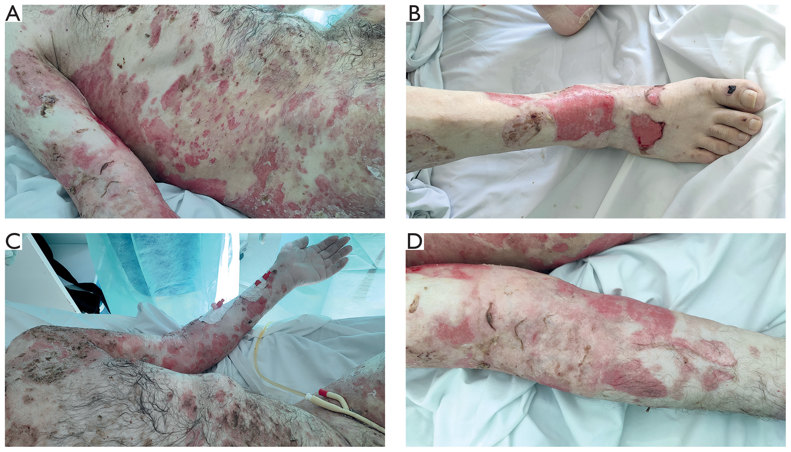
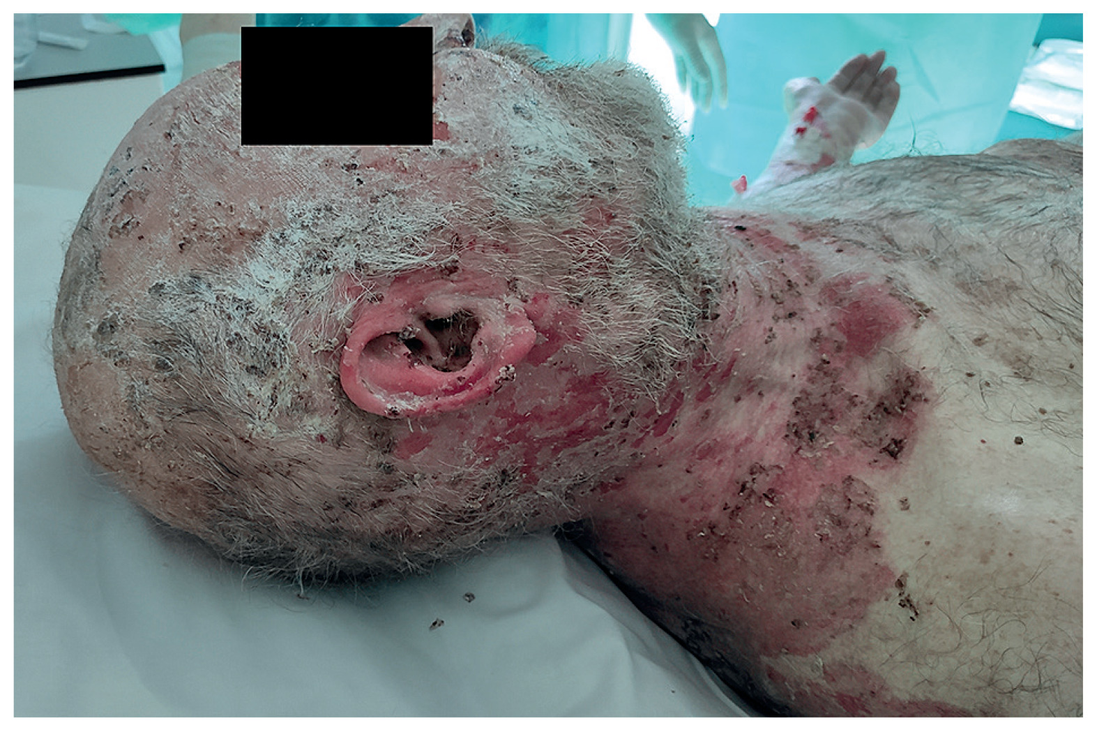
Upon admission, dermatological examination revealed involvement of approximately 80% of the body surface area, with concomitant lesions affecting the conjunctivae, oral mucosa, and genital region. The skin lesions manifested as confluent erosions covered with crusts. Additionally, oozing eruptions with peripheral epidermal detachment were present on the back, buttocks, extensor surfaces of the elbows, and lower legs (fig. 1). Erosions covered with hemorrhagic crusts were also observed on the scalp, auricles, and face (fig. 2). The Nikolsky sign was positive in the immediate vicinity of the lesions. Disease severity assessed with the Pemphigus Disease Area Index (PDAI) was 196 points.
Figure 1
Extensive confluent erosions covered with crusts, with oozing lesions and peripheral epidermal detachment on the trunk, flanks, extensor surfaces of the elbows, and lower legs
Further serologic assessment using enzyme-linked immunosorbent assay for autoimmune blistering diseases showed strongly positive antibodies against desmoglein 1 and desmoglein 3, with negative results for BP180, BP230, envoplakin, and collagen VII (“Dermatology Profile” test kit, Euroimmun).
Because of the patient’s critical condition and increasing inflammatory markers, he was transferred to the intensive care unit. Management included five sessions of plasmapheresis followed by fresh frozen plasma replacement, correction of fluid and electrolyte disturbances, multimodal analgesia, and passive oxygen therapy.
Blood cultures were positive for extended-spectrum β-lactamase (ESBL)-producing Klebsiella pneumoniae and Corynebacterium striatum, necessitating broad-spectrum antibiotic therapy with meropenem/vaborbactam, and vancomycin, while prednisone and azathioprine were continued. Subsequent blood and urine cultures revealed Acinetobacter baumannii, prompting further modification of the antimicrobial treatment with the addition of colistin.
Due to recurrent fever, oxygen desaturation, and chest pain, computed tomography pulmonary angiography was performed and confirmed bilateral pulmonary embolism. Emergency treatment was promptly initiated.
The patient was repeatedly evaluated in the pain management clinic and treated with opioid analgesics. Psychological support and active rehabilitation were also provided.
As PV remained uncontrolled, the patient was qualified for RTX therapy once antibiotic treatment had been completed and his general condition stabilized.
After transfer back to the clinical department of dermatology, two intravenous infusions of RTX, 1000 mg each, were administered 2 weeks apart without complications. The total duration of hospitalization preceding the first RTX infusion was 15 weeks.
During the maintenance phase, methylprednisolone and azathioprine, at the previous dose of 100 mg/day, were continued, resulting in remission of most lesions.
Because of persistent erosions on the scalp and face and steroid-dependent nature of the disease (i.e., relapse when attempting to reduce the methylprednisolone dose below 12 mg/day), a subsequent infusion of 1000 mg RTX was administered after 6 months, and the azathioprine dose was increased to 150 mg/day.
The patient currently remains under follow-up at our dermatology outpatient clinic, as well as geriatric, urologic, and mental health clinics. Psychiatric care was introduced because of post-traumatic stress disorder and mood disturbances that developed in connection with the disease.
DISCUSSION
PV was first described in 1788 by Stephen Dickson, who observed bullous lesions with a predilection for oral mucosal involvement [7]. In 1964, Beutner and Jordon demonstrated the presence of autoantibodies directed against keratinocyte antigens, confirming the autoimmune etiology of the disease [8]. Pemphigus is usually classified into three primary types: PV, pemphigus foliaceus (PF), and paraneoplastic pemphigus. Diagnosis is based on immunopathological examination, particularly direct immunofluorescence (DIF), supported by serologic tests detecting antibodies against specific target antigens [9]. While drugs remain the most frequent triggers of the disease, a significant role is also attributed to foods rich in proteins containing numerous thiol groups [10, 11].
Prior to the introduction of glucocorticosteroids into dermatological therapy, the majority of PV patients died within 2–5 years of diagnosis. The severity of the disease is determined by its progressive course, intensive catabolism with loss of fluids and proteins, and susceptibility to bacterial and viral infections, which can lead to sepsis. In many cases, severe mucosal lesions occur, further compromising the patients’ daily functioning and quality of life [12]. Despite the introduction of glucocorticosteroids and immunosuppressive agents, mortality remains high due to potential contraindications and therapy-related complications [6].
B lymphocytes contribute to pemphigus pathogenesis through antigen presentation, regulation of inflammatory responses, and production of pathogenic autoantibodies. RTX reduces IgG antibody titers and limits the activity of autoreactive helper T lymphocytes [13]. These mechanisms explain its high efficacy in pemphigus treatment.
According to the PDS guidelines, RTX is particularly recommended for steroid-dependent pemphigus, defined as the need for a prednisone-equivalent dose > 10 mg/day combined with an immunosuppressive agent, and in steroid-resistant pemphigus, defined as a lack of control after 3 months of full-dose prednisone and azathioprine. It is also considered a first-line option in paraneoplastic pemphigus [5]. Treatment efficacy appears to depend partly on disease duration, with better outcomes and fewer relapses reported when RTX is introduced early [14]. Patients with scalp involvement are at increased risk of severe course and may therefore particularly benefit from this approach [15].
In a 2017 multicenter study, Joly et al. demonstrated that RTX combined with short-term prednisone treatment was more effective than long-term glucocorticosteroid monotherapy in patients with newly diagnosed pemphigus, significantly reducing the incidence of adverse effects [16]. Similar outcomes were obtained in a recent prospective multicenter study, where complete remission was achieved in 89% of patients treated with RTX and prednisone, compared to 34% in the prednisone monotherapy group.
Comparable findings have been reported by Polish centers. In a series of 11 patients with severe autoimmune bullous diseases, including 8 with PV, Spałek et al. showed that RTX administered in a 2 × 1000 mg regimen allowed glucocorticosteroid doses to be reduced by 65.05%, 73.99%, and 76.93% after 2, 6, and 12 months, respectively [17]. During the same intervals, anti-desmoglein 1 antibody titers decreased by 43.29%, 75.86%, and 54.02%, whereas anti-desmoglein 3 antibody titers decreased by 27.88%, 14.48%, and 5.09%. Within 1 year, relapse occurred in 18.18% of patients, while the remaining patients achieved partial or complete remission. Adverse effects were reported in 36.36% of patients and were mild and transient [17]. In an 8-year follow-up study by Szymański et al., complete remission was achieved in 45.5% of patients with PV; among 28 participants, 1 patient developed sepsis and 1 died [18].
Rashid et al. evaluated the impact of RTX on quality of life in 47 patients with PV or PF using Dermatology Life Quality Index (DLQI), visual analogue scale (VAS), Hospital Anxiety and Depression Scale (HADS), and Treatment of Autoimmune Bullous Disease Quality of Life (TABQOL) questionnaire. Treatment consisted of 2 RTX infusions of 1000 mg, followed by 500 mg maintenance doses at 6 and 12 months. Marked improvement was observed in DLQI, TABQOL, and VAS scores, highlighting the beneficial effect of the drug on patients’ functioning and well-being. However, no significant improvement was found in anxiety and depression, underscoring the need to integrate psychological and psychiatric care into the treatment plan for pemphigus patients [19].
The most common adverse effects of RTX occur during infusion and include headaches, chills, fever, hypotension, pruritus, and erythema. These reactions typically resolve after slowing or temporarily interrupting the infusion, and their risk can be reduced by premedication with analgesics, antihistamines, and glucocorticoids [20]. Serious complications are rare, and the drug is generally well tolerated. However, RTX increases the risk of serious infections and is strictly contraindicated during active infection [21]. Consequently, the initiation of RTX may be delayed in patients with a severe and complicated disease course.
Anandan et al., conducted an open-label prospective study between 2013 and 2015, enrolling 20 patients with pemphigus treated with RTX according to the standard protocol of 2 intravenous infusions of 1000 mg administered 2 weeks apart [22]. During 3–23 months of follow-up, most adverse events were manageable; reported complications included pulmonary tuberculosis, erythema nodosum, or herpes simplex reactivation [22]. In the present case, RTX was well tolerated, and no delayed complications were observed.
In a 2023 global cohort study, Kridin et al. compared 963 pemphigus patients initiating RTX with a control group treated with azathioprine or mycophenolate mofetil [23]. During the first year of therapy, RTX was associated with an increased risk of COVID-19, parasitic, and cytomegalovirus infections, as well as pneumonia, osteomyelitis, and viral infections, with some risks persisting beyond 12 months. These findings support infection risk assessment and appropriate vaccination, including pneumococcal vaccination, before treatment initiation. No increased risk was confirmed for tuberculosis, hepatitis B virus reactivation, Pneumocystis jirovecii pneumonia, or progressive multifocal leukoencephalopathy [23].
CONCLUSIONS
This case demonstrates that the successful use of RTX in PV complicated by severe infections requires prior stabilization and careful assessment of contraindications to immunosuppression. Bridging therapy with plasmapheresis enabled safe initiation of biological therapy, ultimately leading to disease control. This experience highlights the necessity of individualized management and close multidisciplinary supervision in similar clinical scenarios.












