
A 75-year-old woman with hypertension, diabetes mellitus type 2, permanent atrial fibrillation, and chronic kidney disease was admitted to the hospital due to exacerbation of chronic heart failure, vertigo, and syncopal events. Transoesophageal and transthoracic echocardiography revealed severe stenosis and calcification of the prosthetic aortic valve (Trifecta 21 mm), mid-range left ventricle ejection fraction (LVEF) of 49%, moderate insufficiency of the mitral valve, and tricuspid insufficiency with a high probability of pulmonary hypertension. Regarding multiple comorbidities and a high peri-operative risk (logistic EuroScore 48.6%), valve-in-valve (ViV) transcatheter aortic valve implantation (TAVI) was proposed by the heart team.
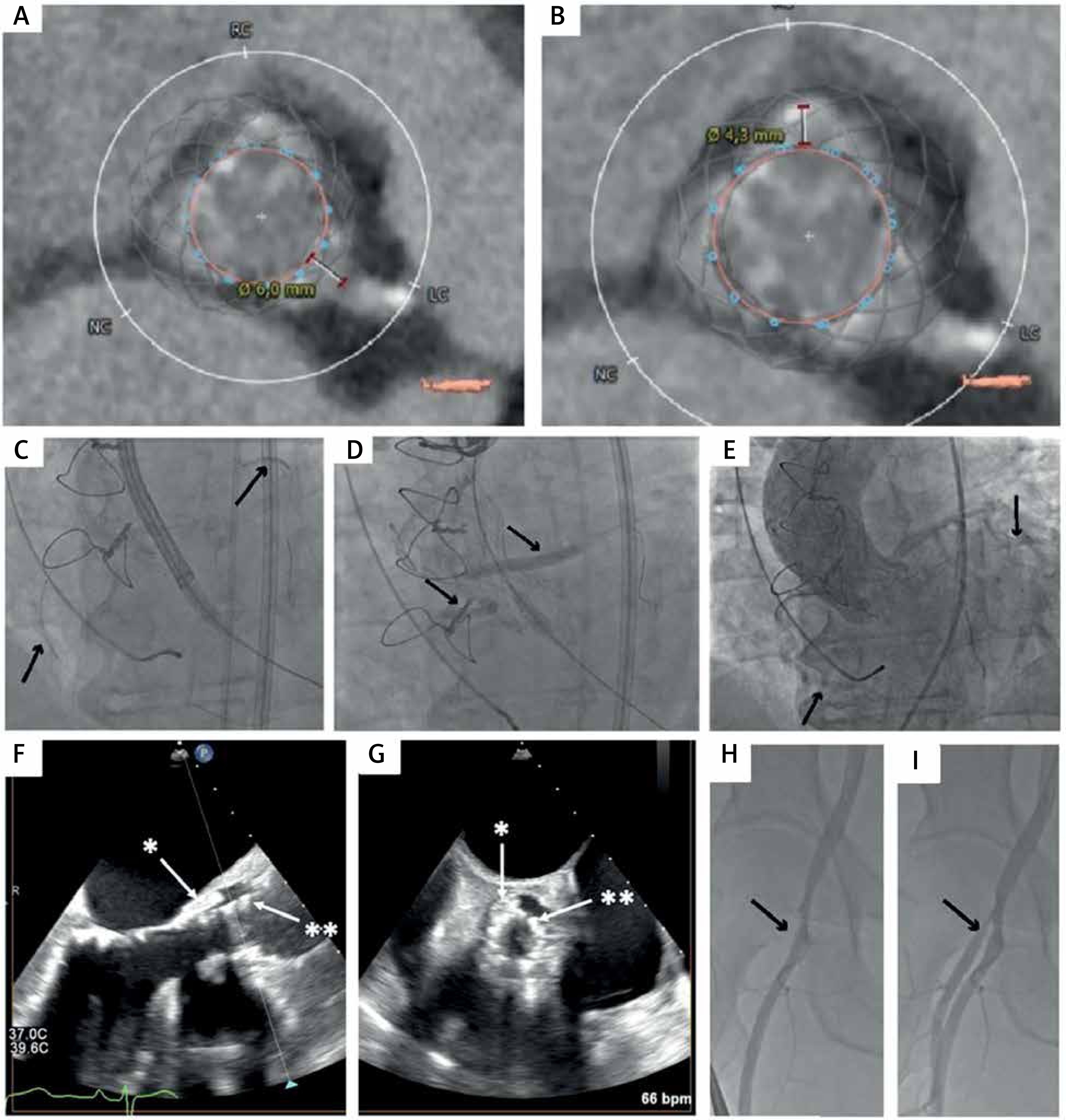
The pre-operative computed tomography (CT) revealed a very short distance from the bioprosthesis to the coronary ostia (Figures 1 A, B: 3.0 mm and 3.9 mm to the left and right coronary artery, respectively). TAVI was performed via the right femoral artery access, using transoesophageal echocardiography (TOE) guidance. The pig-tail catheter for aortography was inserted via the left radial artery. Considering the high risk of coronary obstruction, the ostia of the right and left coronary arteries were engaged with JR 4.0 and JR 3.5 guiding catheters via the right femoral artery and left radial artery, respectively. Two undeployed drug-eluting stents (4.0 × 28 mm and 3.0 × 28 mm) were inserted into the median segments of the left anterior descending (LAD) and right coronary artery (RCA; Figure 1 C; Video S1). This technique has previously been suggested as a feasible way of protecting coronary arteries in high-risk cases [1–3]. The Evolut R bioprosthesis was chosen due to lower postoperative gradients and possible haemodynamic advantage over other models due to its supra-annular design [4]. Following the implantation of the Evolut R 23 mm, the patient experienced ventricular fibrillation and sudden cardiac arrest. Both stents were immediately moved to the ostia of the RCA and left main and deployed using the double-chimney technique, allowing restoration of coronary blood flow (Figure 1 D; Video S2). After several defibrillation attempts, the haemodynamically stable rhythm returned. Control aortography showed normal coronary blood flow (Figure 1 E; Video S3) and a aortic valve peak-to-peak gradient of 10–12 mm Hg. TOE showed a fully expanded Evolut R valve, with no evidence of paravalvular leak (Figures 1 F, G). Following closure of the femoral artery with the ProGlide system, an occlusion of the deep femoral artery was visible on routine angiography check-up (Figure 1 H), which was successfully treated with a percutaneous angioplasty (compliant balloon 5.0 × 15 mm; Figure 1 I). Following the procedure, the patient’s condition improved, showing no symptoms of overt heart failure and no neurological defects. Control echocardiography showed appropriate function of the prosthetic valve and LVEF of 47%.
Figure 1
A, B – Distance between the bioprosthesis and coronary arteries; C – undeployed stents in the mid segments of left anterior descending and right coronary arteries; D – deployed stents after Evolut R implantation, complicated with sudden cardiac arrest; E – patent coronary tree; F, G – degenerated Trifecta bioprosthesis (*) and newly implanted, fully expanded Evolut R valve (**), with no evidence for paravalvular leak in the long-axis and short-axis views in transoesophageal echocardiography; H – occluded deep femoral artery; I – patent deep femoral artery following balloon angioplasty
Considering the growing popularity of TAVI and the ongoing demographic changes, there is a need for the development of guidelines that will enable safe TAVI in specific subpopulations of patients, such as those with low coronary arteries or undergoing ViV procedures, which have an increased risk of potentially fatal coronary obstruction [5].
In our patient, a thorough CT-based preparation enabled the deployment of coronary stents as a life-saving treatment of coronary artery obstruction following bioprosthesis implantation. Furthermore, this case highlights that TAVI carries a risk of vascular site complications, especially if the puncture site is not optimal, as in our patient. Ultrasonography-guided puncture might help to choose the optimal puncture site.


