eISSN: 1897-4295
ISSN: 1734-9338
Advances in Interventional Cardiology/Postępy w Kardiologii Interwencyjnej
Current issue Archive Manuscripts accepted About the journal Editorial board Abstracting and indexing Subscription Contact Instructions for authors Publication charge Ethical standards and procedures
Editorial System
Submit your Manuscript
SCImago Journal & Country Rank
4/2022
vol. 18
 
Share:
Share:
Review paper

Single-photon emission computed tomography as a fundamental tool in evaluation of myocardial reparation and regeneration therapies

Łukasz Tekieli
1, 2
,
Wojciech Szot
3
,
Ewa Kwiecień
1
,
Adam Mazurek
1
,
Eliza Borkowska
3
,
Łukasz Czyż
1
,
Maciej Dąbrowski
4
,
Anna Kozynacka
5
,
Maciej Skubera
1
,
Piotr Podolec
1
,
Marcin Majka
6
,
Magdalena Kostkiewicz
3
,
Piotr Musiałek
1

1.
Department of Cardiac and Vascular Diseases, John Paul II Hospital, Jagiellonian University, Krakow, Poland
2.
Department of Interventional Cardiology, John Paul II Hospital, Jagiellonian University, Krakow, Poland
3.
Department of Radiology, John Paul II Hospital, Krakow, Poland
4.
Department of Interventional Cardiology and Angiology, National Institute of Cardiology, Warsaw, Poland
5.
Department of Coronary Artery Disease and Heart Failure, John Paul II Hospital, Jagiellonian University, Krakow, Poland
6.
Department of Transplantation, Jagiellonian University, Krakow, Poland
Adv Interv Cardiol 2022; 18, 4 (70): 326–339
Online publish date: 2023/01/23
Article file
- single-photon.pdf  [0.18 MB]
Get citation


Introduction

Despite on-going improvements in interventional and pharmacological therapy, ischemic heart disease remains a leading cause of death in modern societies, with chronic ischaemic heart failure (CIHF) as an important cause of reduced quality of life and disability [14].

The pathophysiology of acute and chronic heart failure is similar and is primarily associated with irreversible loss of viable myocardium, leading to impairment of contractile function. Although wide access to primary percutaneous coronary intervention (PCI) decreased the rate of “direct” myocardial infarction-related death, the number of CIHF patients has been increasing worldwide and their prognosis remains poor [2, 3, 5].

Optimal pharmacotherapy may prolong life and improve its quality [6, 7].

However, overall benefit arising from conservative treatments is likely to be “biologically” limited because, as of today, there is no clinically available feasibility for any meaningful myocardial tissue regeneration. One of the directions towards restoration of functionally effective heart muscle is through progenitor cell-mediated repair and regeneration. Progenitor cells have a self-renewal capability and they were found to be able to differentiate into specialized cells, including (at least in some conditions) cardiac myocytes and endothelial cells. A key concept in the regeneration hypothesis is to effectively deliver progenitor cells to the area of infarction and to keep them alive so that they can promote endogenous repair and regeneration by a paracrine effect and/or to direct them to transform into fully functional myocardial tissue. Thus, cell uptake and retention are fundamental for any effect of cell therapy.

There is evidence that progenitor cells may stimulate cardiac repair and regeneration by producing wide range of cytoprotective, anti-inflammatory and angiogenesis promoting factors leading to oxidative stress reduction, ventricle remodeling inhibition and recruitment of endogenous progenitor cells [810].

There are several types of cells that have been investigated in this field, including skeletal myoblasts, embryonic stem cells, cardiosphere-derived autologous stem cells, endothelial progenitor cells, bone marrow-derived mesenchymal stem cells (BMSCs), stromal vascular fraction containing primitive stem cells and pluripotent stem cells [1114]. Although there is evidence that all of these cell lines may have regenerative potential, the type of cell, method of delivery and time from ischemic damage to cell transplantation have not been established clearly, though they seem to be key factors influencing the final pro-regenerative effect. To better understand the fate of progenitor cells after implantation, two main focus points of cell-derived regeneration therapy have been evaluated: a) cells’ biodistribution, including local retention, i.e. the amount of cells that are captured by myocardial tissue after transplantation (the higher the retention, the greater the chance for the activation regeneration process; and b) quantitatively measured long-term effects of cell-derived regeneration (i.e. left ventricle ejection fraction and volumes). Those parameters may be evaluated with different imaging methods including magnetic resonance imaging (MRI), positron emission tomography (PET) and single-photon emission computed tomography (SPECT) [15]. Each of these modalities has its strengths and limitations. This review focuses on SPECT – a technique that, amongst other techniques, may offer an optimal balance between advantages and limitations in the context of cell uptake imaging, evaluation of myocardial perfusion, and study of evolution of global and regional function.

SPECT, applied in everyday clinical practice, is widely available, easy to perform and provides feasibility to estimate precisely and repetitively left ventricle parameters [16]. Moreover, with the use of different tracers, SPECT enables one to track progenitor cells in-vivo in the early phase after transplantation [17].

It has been shown that as few as 2900 cells can be detected by SPECT without significant viability loss due to radiation [18]. SPECT may provide detailed information concerning localization and homing (Figure 1) and – using tracers with a long half-life – also migration of the transplanted cells [17]. Specifically , if the tracer used has a sufficiently long half-time (i.e. 111In with half-life of 2.8 days), it is also possible to track cells at several time points [1921]. However, a long half-life of the tracer is inextricably linked to an important drawback of SPECT – radiotoxicity [2224]. Another important limiting factor is time-dependent efflux (leak) of the label from the cells (Table I). For these reasons, SPECT may not be suitable for long-term tracking of progenitor cells (i.e. half-lives 99mTc, t1/2 = 6 h; 111In, t1/2 = 2.8 d). As compared to computed tomography or magnetic resonance imaging, clinical SPECT has somewhat lower spatial resolution (~7–15 mm) that can be an issue in precise signal localization [15].

Table I

Advantages and limitations of direct cell labelling
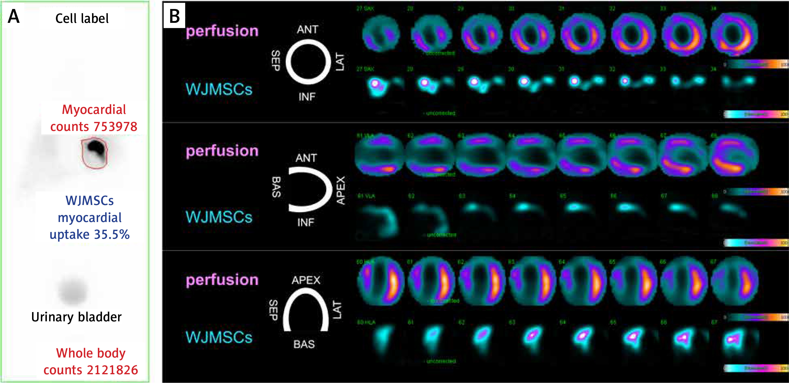
SPECTMRI
Advantages
  • Has high sensitivity and good spatial resolution

  • Provides dynamic tracking imaging and the tissue distribution of transplanted stem

  • Allows one to determine in vivo cell homing

  • With long half-time allows one to track cells at several time points

  • Requires lower regulatory barrier for clinical application compared to indirect methodologies

  • Does not require genetic cell modification

  • Is relatively simple, fast, and inexpensive

  • Has high spatial resolution (1–2 mm)

  • Has no radiation exposure

  • Provides excellent tissue contrast

  • Requires lower regulatory barrier for clinical application compared to indirect methodologies

  • Has low toxicity

  • Does not require genetic cell modification

  • Is relatively simple, fast, and inexpensive

Limitations
  • Radiolabel-related:

  • –Diluted when cells divide

  • –Radiolabel decay in time

  • –Radiolabel signal detected even after efflux/cell death

  • Causes cell dysfunction/death

  • Provides no data on cell viability and biological status

  • Does not allow one to perform imaging at very late time points

  • Has lower spatial resolution (~7–15 mm; vs. MRI)

  • Has relatively low sensitivity (~105 cells with MRI vs. ~103 cells with SPECT)

  • Tracer signal is detected even after efflux/cell death

  • May cause cell dysfunction/death

  • Requires long incubation periods for cell labelling

  • Provides no data on cell viability and biological status

  • Is not suitable for patients with intracardiac defibrillators or pacemakers

  • Does not allow one to perform imaging at very late time points

[i] MRI – magnetic resonance imaging, SPECT – single-photon emission computed tomography.

Figure 1

Essential role of single-photon emission computed tomography (SPECT) in evaluating the magnitude of cardiac uptake of multipotent stem cells and in determining the zone(s) of early cell homing in relation to the infarct injury: use of standardized Wharton jelly mesenchymal stem cells (WJMSCs, umbilical cord stem cells) as an advanced technology medical product (WJMSCs-ATMP). A 62-year-old man was admitted due to anterior ST-segment elevation acute myocardial infarction. Left anterior descending coronary artery proximal occlusion was treated successfully with thrombus aspiration (to minimize distal embolization and myocardial microcirculatory obstruction in the infarct zone) [104106] and primary angioplasty with stent implantation. Six days later, consistent with the CIRCULATE-AMI Pilot Study Protocol, 30 × 106 standardized Wharton jelly pluripotent stem cells (50% labeled with 99mTc-sestamibi) were administered via the infarct-related-artery using a dedicated system for transcoronary delivery of cells and cell-based products (CIRCULATE Catheter, Protected Design No 72837, Patent Office of the Republic of Poland) [107]. Whole-body scintigraphy (A) performed 60 min after transcoronary WJMSCs-ATMP transplantation revealed a large-magnitude (35.5%) myocardial uptake of the WJMSCs-ATMP (red-line delineation; note that this WJMSCs-ATMP uptake exceeds, by ~7-fold, the uptake of prior-tested cell types such as CD34+ cells [32, 57, 72]. B shows the areas of WJMSCs-ATMP early homing (bottom rows labeled ‘WJMSCs-ATMP’ in the top, middle and bottom panel) in relation to regional myocardial perfusion by SPECT (top rows [“perfusion”] in the top, middle and bottom panel). Note WJMSCs-ATMP homing to the areas of severe perfusion defect in the anterior wall, septum and apex of the LV myocardium (myocardial infarct zones), consistent with a role of biologic mechanisms attracting WJMSCs to ischaemia-injured myocardium but not to normal myocardium [108]

/f/fulltexts/PWKI/49993/PWKI-18-49993-g001_min.jpg

Cell labeling protocols

The principles of labelling are similar for all nuclides. After harvesting cells are incubated with so-called ‘linker’ allowing the tracer to penetrate the cell membrane. For cell tracking in the heart, [In-111]oxine [19, 23, 25, 26], [In-111]tropolone [27, 28], and [Tc-99m] hexamethylpropyleneamine oxime [2932] have been adopted.

99m-Technetium radioactive isotope bound to hexamethylpropyleneamine oxime (99mTc-HMPAO, 99mTc-extametazime, CERETEC) is the most widely used compound for viable cell labelling. This complex is lipophilic and can easily cross the cell membrane. When inside, in an alkaline environment, it changes into the hydrophilic ionic form and remains sequestered inside the cell. The complex efflux is constant, but this phenomenon prolongs the time of possible gamma camera acquisition. Nevertheless, cells labelled with 99mTc-HMPAO can be observed only in the first 24 h after administration, due to the relatively short half-time of 99mTc of 6 h. 99mTc-HMPAO that is released by cells is taken up by the liver and excreted via the intestines. Labelling cells with 99mTc requires expertise in handling blood-derived products, and does not significantly affect cell viability and functionality while reaching a high level of binding capacity (~40–69%) [33, 34]. For in-vivo tracking, the cultured cells are first trypsinized and incubated with Tc-99 with HMPAO linker for a 10–30 min period, then the cells are washed to eliminate any unbound radioactivity. Finally they are injected into the host. The time of incubation is a compromise between the need for a high rate of labelling efficiency and cell viability, and it usually does not exceed 30 min with cell viability deterioration < 2% [3436].

The 111In-oxine is a complex of indium and three molecules of 8-hydroxyquinoline (oxine). The complex is lipid-soluble, and, similarly to 99mTc-HMPAO, and penetrates the cell external membrane by passive diffusion. After binding cytoplasmatic proteins, particles of 8-hydroxyquinoline are liberated and released out. Due to the long half-life of 111In (2.8 days) it is possible to track the transplanted cell up to 2 weeks. On the other hand, labelling cells with long half-life 111In-oxine may significantly impair the viability, proliferation and differentiation [23, 37]. Moreover, in-vivo cell tracking with 99mTc and 111In labelling is applicable for short-term analysis as it is difficult to detect whether the radiation intensity decrease is caused by radionuclide efflux from viable cells or it is associated with cell death or cell transfer to a remote location.

Cell tracking

Progenitor cell SPECT tracking feasibility has been tested in numerous animal models of myocardial infarction [19, 21, 25, 28, 3852] that are summarized in Table II.

Table II

SPECT evaluation of myocardial uptake for cell therapies in preclinical studies of myocardial infarction

CellsModelConditionDelivery methodSPECT tracerTimepoint of SPECT evaluationUptake (%)Author
CD34(+) Human HPCsRatMIIntra-ventricular111In-oxine1, 24, 48, 96 h1%Brenner et al.
MSCsPorcineMIiv111In-oxine1, 2, 7, 14 daysNo visible accumulation in the myocardiumChin et al.
Human EPCRatMIiv, intra-ventricular111In-oxine1, 24, 48, 96 h1–2%Aicher et al.
Rat MSCsRatMIiv99mTc-HMPAO6 hFocal cardiac uptakeGarikipati et al.
Rat BMSCsRat1 month after MIim111In oxine2 days< 1%Tran et al.
Human IPScPorcineMIim123I5 min up to 15 days~2%Templin et al.
Rat ESCsRatMIim111In-oxine2, 24, 48, 72, 96 hNot specifiedZhou et al.
Canine BM-MSCsCanineMIiv111In-oxine1–8 daysFocal cardiac uptake
2.56 ±1.9% (ant.m.)
1.95 ±1.4% (inf.m.)
Kraitchman et al.
Rat ESCsRat mMIim111In-oxine30 min.
Not specified
Not specifiedShen et al.
Canine BM-MSCsCanineMIim111In-tropolone1 dayNot specifiedBlackwood et al.
Rat ADSCsRatMIim99mTc-HMPAOImmediately26.8%Danoviz et al.
Canine EPCsCanineMIEpicardial, endo-cardial111In-tropoloneSame dayEpicardial 56.7 ±6.0 %
Endocardial 59.5 ±5.5%
Mitchel et al.2013
Rat BM-MSCsRat4 months after MIim111In-oxine48 hNot specifiedMaureira et al.
Porcine BM-MSCsPorcineMIim, ic111In-oxine2 and 24 hNot specifiedMakela et al.
BM-MSCsPorcineMIic, iv99mTc1 and 24 h6 ±1.7% (ic)
no cardiac homing (iv)
Forest et al.
Canine EPCsCanineMIEpicardial, endo-cardial111In -tropolone30–40 minEpicardial 57 ±15%
Endocardial 54 ±26%
Mitchel et al.2010
Rat BM-MSCsRatMIiv and intra-ventricular99mTc-HMPAONo data0.9 ±0.32%(Intra LV)
0.2 ±0.02 % (iv)
Barbash et al.
Porcine BM-MSCsPorcineMIic no balloon (ic noB), ic with balloon (ic/B) im111In-tropoloneImmediately, 1–24 hAt 1 h:
4.1 ±1.1% (IC/noB)
6.1 ±2.5% (IC/B)
20.7 ±2.3% (im)
At 24 h:
3.0 ±0.6% (IC/noB)
3.3 ±0.5% (IC/B) 15.0 ±3.1%
Tossios et al.

[i] 111In – indium-111, 99mTc – technetium 99m, HMPAO – hexamethylpropyleneamine oxime, 123I – iodine-123, MSCs – mesenchymal stem cells, IPSc – induced pluripotent stem cells, EPCs – endothelial stem cells, ESCs – embryonic stem cells, BM-MSCs – bone marrow mesenchymal stem cells, ADSCs – adipose tissue stem cells, HPCs – hematopoietic cells, iv – intravenous, im – intramyocardial, ic – intracoronary, ant.m. – anterior myocardium, inf.m. – inferior myocardium.

Brenner et al. demonstrated the feasibility of in vivo method for monitoring myocardial homing of transplanted cells in a rat myocardial infarction model using 111In-oxine-labelled CD34(+) hematopoietic cells. They found that viability of radiolabelled hematopoietic cells (HPCs) was impaired by 30% after 96 h, whereas proliferation and differentiation of cells were nullified after 7 days. The overall radioactivity detected in the heart was only about 1% [25].

Chin et al. examined the feasibility of 111In-oxine labelling of mesenchymal stem cells (MSCs) and single photon emission computed tomography imaging after intravenous administration in a porcine model of myocardial infarction. High initial MSC localization occurred in the lungs and no appreciable accumulation occurred in the myocardium. The authors conclude that 111In-oxine radiolabelling of MSCs is feasible, and in vivo imaging with SPECT provides a non-invasive method for sequentially monitoring cell trafficking with good spatial resolution [47].

Aicher et al. observed that after administration of 111In-oxine-labeled endothelial progenitor cells, the heart-to-muscle radioactivity ratio increased significantly in a myocardial infarction rat model, indicating increased homing of transplanted EPCs [48].

Garikipati et al., using pinhole gated SPECT-CT in a rat model, observed focal uptake of 99mTc-labeled fC-MSCs in the region of myocardial infarction. The uptake was associated with significant improvement in left ventricular ejection fraction 4 week after cell transplantation [31].

Tran et al. tracked 111In-oxine-labeled autologous BMSCs injected directly at the 1-month-old infarction site. One week later the myocardial retention of BMSCs was definitely higher in myocardial infarction than in the normal myocardial area (retention at 2 h: 63% vs. 25%, p < 0.001) and the estimated cardiac retention values were unchanged in both groups during the 7 days of follow-up [19].

Wisenberg et al. recorded an effective biological clearance half-life from the injection site of ~5 days for 111In-tropolone labelled bone marrow monocytes and stromal cells in a canine model [53].

Templin et al. applied dual isotope SPECT-CT imaging: 123I to follow donor cell survival and distribution, and 99mTC-tetrofosmin for perfusion imaging in a pig model of myocardial infarction. Additionally, sodium iodide symporter (NIS) transgene imaging was evaluated as an approach to follow in vivo survival, engraftment, and distribution of human-induced pluripotent stem cells. In vivo, viable NIS(pos)-hi pluripotent stem cells (PSCs) could be visualized for up to 15 weeks. Immunohistochemistry demonstrated that hiPSC-derived endothelial cells contributed to vascularization. Up to 12 to 15 weeks after transplantation, no teratomas were detected [50].

Dual isotope SPECT 111In-labeled/99mTc-sestamibi enables the imaging of both cells and perfusion deficit in the infarcted region simultaneously. Zhou et al. found that the 111In signal from the labelled stem cells overlaps the perfusion deficits identified from the 99mTc-sestamibi images. The 111In signal associated with the radiolabelled stem cells could be detected with SPECT of the heart for 96 h after engraftment [51].

Shen et al. used dual-tracer small-animal SPECT images to detect successfully 111In-labeled stem cells in the region of perfusion deficit assessed with 99mTc-sestamibi tracer. SPECT regional perfusion deficit coincided with the akinetic region on the MR image [52].

In several studies, combined, hybrid tomography has been applied for co-registration of structural and functional information within a single study. It has been demonstrated that the hybrid SPECT/CT system allows the combination of the exquisite anatomic details provided by CT with the functional, physiologic or metabolic information provided by molecular imaging.

The hybrid SPECT/CT system used by Sabondjian et al. was able to detect a signal of endothelial progenitor cells labelled with ¹¹¹In-tropolone within the zone of reduced perfusion delineated on first-pass perfusion CT in a canine model [27].

Kraitchman et al. detected focal and diffuse uptake of 111In-oxine-labeled mesenchymal stem cells in the infarcted myocardium in SPECT/CT images in the first 24 h after injection. The activity persisted until 7 days after injection. In contrast, MRI was unable to demonstrate targeted cardiac localization of MSCs, in part because of the lower sensitivity of MRI (~103 cells with SPECT versus ~105 cells with MRI) [21, 53, 54].

With the use of novel, nanoparticle(NP)-based labels (including iron oxide NPs, gadolinium-based NPs, manganese-based NPs, 19F-based NPs and SPIONs), MRI aims to reach high sensitivity, high spatial resolution and penetration depth for in vivo cell tracking [55].

Blackwood et al. developed a quantitative method to assess transplanted cell survival in myocardium using SPECT and 111In. The authors found that the measured half-time for transplanted cells was 74.3 h, and, when appropriate corrections (related to radiolabel leakage and extracellular 111In (e.g., after cell death)) were applied, the time was 71.2 h [28, 56].

Our group introduced SPECT-cMRI hybrid imaging, involving SPECT (a highly sensitive cell label) and magnetic resonance imaging (infarct) to evaluate the relationship between infarct size and progenitor cell uptake, including determination of the early homing zones [57].

How to enhance therapeutic cell retention

The low retention rate in most animal studies gave rise to a question of the mechanisms of cell washout and potential solutions leading to engraftment improvement.

Danoviz et al. injected directly in the infarct zone 99mTc-labeled adipose tissue-derived stem cells 24 h after MI using fibrin or collagen as the vehicle. The collagen group showed the highest radioactivity retention (26.8%) as compared to the fibrin group (13.7%) and control group (4.84%). The authors suggest that the low retention rate may be a result of washing out of cells from the myocardium through the lymphatic vessels and veins from the left ventricle into the lungs. Another finding was that intramyocardial injection of ASCs mitigates the negative cardiac remodeling and preserves post-MI ventricular function in rats, and these beneficial effects can be further enhanced by administering co-injection of ASCs with biopolymers [38].

Mitchell et al. reported that transplantation of 111In-tropolone-labeled endothelial progenitor cells into sustained occlusion infarcts resulted in a slower cell clearance half-life of 77.1 h (n = 18) versus reperfused – 59.4 h (n = 21). Sustained occlusion infarcts had longer cell retention in comparison to reperfusion whereas the timing of injection did not affect clearance rates [43].

Maureira et al. using dual 111In/99mTc-Sestamibi imaging observed cell engraftment in the MI area 48 h after stem cell transplantation. Interestingly, the authors also found that perfusion enhancement was sustained during the 6-month follow-up in the non-engrafted MI-areas from treated rats, whereas the engrafted ones, as well as the MI areas from control rats, exhibited progressive deterioration over time, suggesting a distant paracrine effect of transplanted cells [40].

Routes of cell administration to myocardium

During the first animal studies on cell retention, along with different cell types and methods of labelling, the way of transplantation was also examined. It has been found in numerous studies that cell delivery route can significantly influence the level of retention.

Forest et al. found that a significant 99mTc-labeled bone marrow stem cell fraction remained within the heart after intracoronary injection (6 ±1.7% of injected radioactivity at 24 h). With peripheral intravenous cell injection, no cardiac homing was observed at 24 h and cells were mainly detected within the lungs [42].

Although feasible, epicardial cell implantation appeared to be a complex and time-consuming procedure, requiring surgical technique. To simplify direct cell injection, Mitchell et al. proposed an endocardial approach. The authors found no significant difference between the endocardial (retention: 54%) and epicardial (retention: 57%) injection methods or the clearance kinetics, indicating that the injection strategies are comparable [39].

Barbash et al. examined different ways of cell delivery. They found that delivery by left ventricular cavity infusion results in drastically lower lung uptake, better uptake in the heart, and specifically higher uptake in infarcted compared with sham-MI hearts [44].

Hou et al. found significantly higher retention of 111In-oxine-labeled human peripheral blood mononuclear cells injected directly into myocardium (11%) as compared to intracoronary (2.6%) and retrograde coronary venous (3.2%) delivery [58].

Tossios et al. were another group investigating role of cell delivery method. They found that retention of 111In-labeled bone marrow cells after intracoronary infusion with or without balloon occlusion does not differ significantly (4.1% vs. 6.1% respectively) as opposed to direct intramyocardial injection (20.7%). Interestingly, dynamic SPECT during intracoronary injections showed rapid (20%) cell loss during balloon inflation and rapid (37%) cell loss after balloon deflation. After intramyocardial injection only slow linear cell loss was observed (9.7% per h) [45].

Kupatt et al. found, in a pig model of ischemia, that 1 h after reperfusion, 99Tc-HMPAO-labelled endothelial progenitor cells (eEPCs) engrafted to a 6-fold higher extent in the ischemic myocardium after retroinfusion than after intravenous application. Moreover, compared with medium-treated animals, retroinfusion of eEPCs decreased infarct size (35% vs. 52%) and improved regional myocardial reserve of the apical LAD region (SES 31% vs. 7%), whereas intravenous application displayed a less pronounced effect (infarct size 44%; SES 12%). Retroinfusion of an equal amount of neonatal coronary endothelial cells (rat) did not affect infarct size or regional myocardial reserve. Interestingly, the eEPC-dependent effect was detected at 24 h of reperfusion, suggesting an important role for enzyme-mediated cardioprotection [30].

It was hypothesized that a low engraftment rate may be influenced by a hostile environment for transplanted cells (hypoxia, inflammation etc.). Chan et al. investigated the theory of protective features of hydrogels. They found that hyaluronic acid-serum hydrogels markedly increase acute intramyocardial retention (∼6 fold), and promote in vivo viability, proliferation, engraftment of encapsulated stem cells and angiogenesis. The authors conclude that hyaluronic acid-serum hydrogels serve as ‘synthetic stem cell niches’ that rapidly restore the metabolism of encapsulated stem cells and promote stem cell engraftment and angiogenesis [59].

As shown in Table II, direct, intramyocardial cell injection provides the highest rate of in-tissue retention immediately after administration (up to ~60%). On the other hand, this type of cell implantation is not physiological and may be of limited value in terms of further cell survival. However, myocardial injections may cause myocardial damage [60]. Data from the Congestive Heart Failure Cardiopoietic Regenerative Therapy (CHART-1) trial indicated an association between an increase in the number of endomyocardial injections to deliver therapeutic cells and a reduction in the therapeutic effect of mesenchymal cell transplantation in patients with chronic ischemic heart failure [61].

Human studies

With encouraging results of animal model studies, SPECT has been adopted to investigate biodistribution of 99mTc and 111In-labelled progenitor cells in a clinical setting.

The results of numerous important studies in humans confirm observations from animal model concerning cell retention and distribution. Table III provides the most relevant data from studies in humans in which SPECT was used [29, 34, 6271].

Table III

SPECT evaluation of myocardial uptake for cell therapies in clinical studies of myocardial infarction

CellsConditionDelivery methodSPECT TracerTimepoint of SPECT evaluationUptake (%)Author
BM-MSCsCHFic (stop flow)111In-oxine2, 12 h6.9–8% (2h)
2.3–3.2% (12h)
Caveliers et al.
BM-MSCsAcute and chronic anterior MIic (LAD)99mTc-HMPAO2 h1.31–5.1% (acute)
1.1–3.0% (chronic)
Penicka et al.
BM-MSCsAMIic99mTc-HMPAO30 min,
2.5 h,
24 h
7.8%
6.8%
3.2%
Karpov et al.
BM-MNCsAMIic111In-oxine24 h2.6–11%Kurpisz et al.
PB-MNCsCHFic111In-oxine2-12 h6.9–8.0% (2 h)
2.3–3.2% (12 h)
Schots et al.
BM-MSCsCHFic99mTc-HMPAO1, 24 h9.2% (1 h)
6.8% (24 h)
Goussetis et al.
Proangiogenic progenitor cells (PB-MSCs)AMI and CHFic111In-oxine1, 24 h6.9% (1 h)
2% (after 3–4 days)
Schachinger et al.
BM-MNCsAMIic, IRCV99mTc-HMPAO4, 24 h4 h:
16% (ic)
4% (IRCV)
24 h:
10% (ic)
3% (IRCV)
Silva et al.
BM-MNCsAMIic (OTW and PC)99mTc-HMPAO1 h4.86 ±0.49% (OTW)
5.05 ±0.48% (PC)
Musialek et al. 2011
BM-MNCsAMIic99mTc-HMPAO1 h5.2%Musialek et al. 2013
BM-MNCsCHF (nonischemic)ic, transendocardial99mTc-HMPAO18 h4.4% (ic)
19.2% (transendocardial)
Vrtovec et al.
PB-MSCsCHF (nonischemic)transendocardial99mTc-HMPAO2, 18 h11.4%Haddad et al.
WJMSCsAMIic99mTc-HMPAO1 h30.2 ±5.3%Musialek et al. 2017
WJMSCsCHFic99mTc-HMPAO1 h40.3 ±6%Kozynacka et al.

[i] 111In – indium-111, 99mTc – technetium 99m, HMPAO – hexamethylpropyleneamine oxime, CHF – chronic heart failure, AMI – acute myocardial infarction, BM-MSCs – bone marrow mesenchymal stem cells, PB-MSCs – peripheral blood mononuclear cells, BM-MNCs – bone marrow mononuclear cells, WJMSCs – Wharton’s jelly mesenchymal stem cells, ic – intracoronary, LAD – left anterior descending artery, IRCV – interstitial retrograde coronary venous, OTW – over-the-wire (stop-flow technique), PC – perfusion catheter (perfusion technique).

Work from our group, using SPECT, has addressed several critical questions in cardiac regenerative medicine including optimizing transcoronary cell delivery, determination of the zone(s) of myocardial cell uptake, and late functional improvement in relation to the magnitude of cell uptake [29, 32, 72, 73].

Caveliers et al. investigated homing of 111In-oxine-labeled peripheral blood stem cells in chronic ischemic heart disease conditions. The cells were infused intracoronarily through a balloon catheter with stop-flow technique. Fused 99mTc-sestamibi/111In SPECT images demonstrated the regional distribution of the transplanted cells within the no/low perfusion zone, as delineated by the flow tracer. The radioactivity retention in the heart was 6.9–8% after 1–2 h and 2.3–3.2% after 12 h [64].

Penicka et al. investigated the kinetics of myocardial engraftment of 99mTc-HMPAO-labeled bone marrow-derived mononuclear cells after intracoronary (LAD) injection in patients with acute and chronic anterior myocardial infarction. At 2 h after infusion, myocardial activity was observed in all patients with acute (range: 1.31–5.10%) and in all but 1 patient with chronic infarction (range: 1.10–3.0%). At 20 h, myocardial engraftment was noted only in 3 patients with acute IM [65].

Karpov et al. performed a randomized controlled study including 44 patients with acute myocardial infarction. It was found that intracoronary injection of bone marrow mononuclear cells is safe, ensures fixation of the injected cells in the myocardium, reduces blood levels of IL-1β and TNF-α, increases the content of insulin-like growth factor, and does not provoke malignant arrhythmias [66].

Goussetis et al. examined biodistribution of CD133+ and CD133-CD34+ 99mTc-hexamethylpropylenamineoxime-labeled selected autologous bone marrow progenitor cells infused into the infarct-related artery in patients with chronic ischemic cardiomyopathy. One and 24 h after transplantation the radioactivity in the infarcted area was 9.2% and 6.8% respectively; the remaining activity was distributed mainly to the liver and spleen, similarly to other studies [68].

Schächinger et al. transplanted circulating proangiogenic progenitor cells labelled with 111In-oxine in patients with previous myocardial infarction and a revascularized infarct vessel at various stages after infarction (5 days to 17 years). Similarly to other studies, 1 h after cell administration, 6.9% of total radioactivity was detected in the heart, which declined to 2% after 3 to 4 days. The authors also found that average activity within the first 24 h was highest among patients with acute myocardial infarction. Moreover, proangiogenic progenitor cell homing was influenced by low viability of the infarcted myocardium and reduced coronary flow reserve [74].

Silva et al. investigated the safety and feasibility of autologous bone marrow mononuclear cell (BMMNC) transplantation in ST elevation myocardial infarction (STEMI), comparing anterograde intracoronary artery (ICA) delivery with the retrograde intracoronary vein (ICV) approach. 1% of cells were labelled with 99mTc-hexamethylpropylenamineoxime. Cell distribution was evaluated 4 and 24 h after injection. The authors observed exceptionally high retention in the intracoronary group (early 16%, late 10%) as compared to the retrograde intracoronary group (early 4%, late 3%). Early and late retention of radiolabelled cells was higher in the ICA than in the ICV group [70].

As the stop-flow technique has never been shown to be mandatory in intracoronary cell transplantation, our group focused on comparing two methods of autologous 99Tc-extametazime-labeled bone marrow CD34+ cell delivery with insight into patterns of cell retention. We found that the effectiveness of the perfusion technique (side-holed perfusion catheter, cell injections under maintained coronary flow) was not different from that seen with the over-the-wire (OTW)-balloon method (stop-flow technique, 5.0% vs. 4.9% respectively). It was also found that retention of progenitor cells occurs preferentially in the (viable) peri-infarct zone, suggesting that the infarct zone is largely inaccessible to transcoronary-administered cells [29].

Moreover, the coronary-non-occlusive method delivery of Wharton’s jelly mesenchymal stem cells (WJMSCs – combining high angiogenic and cardiogenic potential with low immunogenicity) showed a high and reproducible retention rate (30%) of 99Tc-labeled WJMSCs in the peri-infarct zone in humans after recent myocardial infarction [62].

There is a significant amount of data showing a very high level of agreement between G-SPECT and other imaging techniques when considering measurement of left ventricle volumes and ejection fraction. With the most widely used, quantitative gated SPECT (QGS, Cedars-Sinai Medical Center, Los Angeles, CA), the correlation coefficients of this tool with gold standard magnetic resonance imaging reach 0.72–0.94 for left ventricle ejection fraction, r = 0.81–0.97 for end-diastolic volume and r = 0.87–0.99 for end-systolic volume [75]. Moreover, it was demonstrated that SPECT distinguishes itself from other imaging tools with its outstanding reproducibility [76, 77]. Considering regional wall motion abnormalities, gated SPECT also showed excellent (83%) agreement with MRI [78]. Apart from volumes and ejection fraction evaluation, gated SPECT allows assessment of regional LV function. The perfusion and wall motion defects have been widely adopted as outcome parameters of the human cardiac cell therapy.

Tables IV and V provide detailed data on end-point parameters acquired by SPECT in animal [47, 7982] and human [8398] models of trials concerning the clinical effect of progenitor cell transplantation. In contrast to encouraging outcomes in cell therapy in animals, the results of randomized trials in humans investigating the potential effect of progenitor cells transplanted into myocardium show no or a minimal effect on cardiac function. In fact, this subtle positive effect of cells transplantation on quantitative, measurable end-point parameters (ejection fraction, myocardial perfusion, regional wall motion index) is visible only in large-scale meta-analyses [99101].

Table IV

SPECT-tracked effect of cell transplantation on myocardium function in animal model of ischemia

CellsModelConditionDelivery methodCardiac function evaluationSPECT tracerEffectAuthor
Small Animal Models:
 Allogeneic CSCsRat modelAMIIntramyocardialSPECT cell tracking99mTcCells detected as a perfusion deficit at day 6 post-injectionTerrovitis 2008
Large Animal Models:
 Mesenchymal stem cells labeled with 111In oxinePorcine modelAMIIntravenousSPECT cell tracking99mTc-sestamibiSPECT useful for semiquantitative and non-invasive traffickingChin 2003
 Adipose tissue-derived stem cells (porcine)Porcine modelAMIIntracoronary injectionSPECT99mTc-sestamibiLV function improvement
↑ Myocardial perfusion
Alleviation of LV remodeling
Valina 2007
 Wharton’s jelly mesenchymal stem cells (human)Porcine modelAMIPeri-infarct (intramyocardial) injectionsSPECT
Echo
99mTc-sestamibiLV function improvement
↑ Infarct area wall thickening
↑ Myocardial perfusion
Zhang 2013
 Human umbilical cord-derived mesenchymal stem cellsPorcine modelHFIntravenousSPECT
Echo
99mTc-MIBIImprovement of myocardial perfusion and collateral vesselsLiu 2016
 Umbilical cord derived multipotent mesenchymal stromal cells (porcine)Porcine modelAMIIntravenousSPECT
PET
Echo
99mTc-sestamibiTendency to LVEF improvement
Reduction of LV nonviable myocardium area after MI
Reduction of total perfusion defect and tendency to improvement of myocardial blood flow
Lim 2018

[i] AMI – acute myocardial infarction, HF – heart failure, LVEF – left ventricle ejection fraction, SPECT – single-photon emission-computed tomography, PET – positron emission tomography, Echo – echocardiography, BM MNSCs – bone marrow mononuclear stem cells, BMCs – bone marrow-derived cells, CPCs – circulation progenitor cells, SSS – summed stress score, SRS – summed rest score, WJMSCs – Wharton’s jelly mesenchymal stem cells, ADRC – adipose-derived regenerative cells, Tc-technetium, Tl – thallium, MIBI – methoxyisobutylisonitrile.

Table V

SPECT-tracked effect of cell transplantation on myocardium function in clinical studies of myocardial ischemia

CellsConditionDelivery methodEvaluationSPECT TracerEffectAuthor
BMCs/CPCsAMIIntracoronarySPECT
PET
201Tl↑ Signal intensity
↑ LVEF (on PET)
Dobert 2004
BM MNSCsAMIIntracoronarySPECT
Echo, MRI
99mTc-sestamibi↑ EF 0.6% SPECTLunde 2006
CPCsAMIIntracoronarySPECT
PET
99mTc-tetrofosmin↓ Number of segments with mismatched perfusion – viabilityKendziorra 2008
BM MNCsAMIIntracoronarySPECT
Echo
99mTc↑ LVEFCao 2009
BM MNCsAMIIntracoronarySPECT99mTc-sestamibi↑ Regional perfusionLipiec 2009
BMCsAMIIntracoronarySPECT
PET, Echo
99mTc-sestamibi↑ Cardiac function in patients with high sestamibi uptakeKaminek 2010
BMCsAMIIntracoronarySPECT99mTc-sestamibi↑ Wall motionGrajek 2010
BM MNSCs CD 133+Ischemic cardiomyopathyIntracoronarySPECT99mTcMIBI↓ Infarct sizeKurbonov 2013
BM MSCsAMIIntramyocardialSPECT
Echo
99mTc-tetrofosmin↑ SSS
↑ SRS
↓ Number of ischemic segments
↑ LVEF
Rodrigo 2013
BM MSCsAMIIntracoronarySPECT
Echo
18FDG
99mTc-sestamibi
↑ Myocardial viability perfusion – no changeGao 2013
BM MSCsAMIIntracoronarySPECT
Echo
99mTc-sestamibi↑ LVEFJun-Won Lee 2013
Allogenic MSCsAMIIntravenousSPECT
Echo
MRI
↑ LVEF (Echo)
No significant differences in perfusion
Chullicana 2014
ADRCIschemic cardiomyopathyTransendocardialSPECT
Echo
MRI
↓ Inducible ischemia
↑ Wall motion score index
Perin 2014
WJMSCsAMIIntracoronarySPECT
PET
Echo
99mTc↑ LVEF
↓ LVEDV, LVESV
↑ Perfusion
Gao 2015
BM MSCsIschemic cardiomyopathyIntramyocardialSPECT
Echo
201Tl↓ SSS
Viable segments – no change
Guijarro 2016
BM MSCsAMIIntracoronarySPECT
Echo
99mTc-sestamibi↑ LVEFKim 2018

[i] AMI – acute myocardial infarction, HF – heart failure, LVEF – left ventricle ejection fraction, SPECT – single-photon emission-computed tomography, PET – positron emission tomography, Echo – echocardiography, BM MNSCs – bone marrow mononuclear stem cells, BMCs – bone marrow-derived cells, CPCs – circulating progenitor cells, SSS – summed stress score, SRS – summed rest score, WJMSCs – Wharton’s jelly mesenchymal stem cells, ADRC – adipose-derived regenerative cells, MIBI – methoxyisobutylisonitrile.

Thus, further optimal types of cells, ways of administration and uptake, and mechanisms of regeneration are still to be investigated [102].

Progenitor cells have been recently examined as a transfer vehicle for non-viral gene delivery systems for tissue repair and regeneration therapies. In fact, gene-corrected CD34+ stem cells have already been successfully adopted for treatment of inherited diseases – progenitor cells are harvested, transduced ex-vivo with a viral vector, and then reinfused into the patient [103]. The same technology may be used in the future for therapeutic cell modification or local gene transfer into damage tissue.

In conclusion, SPECT is a technique that offers high-sensitivity, quantitative cell tracking on top of its ability to evaluate myocardial perfusion and function on both a cross-sectional and a longitudinal basis. SPECT, with its direct relevance to routine clinical practice, plays a fundamental role in evaluation of myocardial reparation and regeneration therapies (Table VI).

Table VI

Cell therapies to stimulate myocardial repair and regeneration

Why important
  • Growing medical and societal problem of chronic heart failure

  • No effective myocardium regeneration therapies available today

Challenges
  • Cell homing, retention and viability improvement

  • Cell regeneration capacity enhancement

  • Better understanding of repair and regeneration mechanisms

Future perspective
  • Progenitor cell gene correction

  • Local gene transfer via cells

  • “Cocktails” of cells and growth factors

  • Cells and scaffolds

Acknowledgments

Supported by K/ZDS/005644 (Jagiellonian University Medical College) and and a research grant (STRATEGMED2/265761/10/NCBR/2015) from the National Center for Research and Development (Poland).

Conflict of interest

The authors declare no conflict of interest.

References

1 

Naghavi M, Abajobir AA, Abbafati C, et al. Global, regional, and national age-sex specifc mortality for 264 causes of death, 1980-2016: a systematic analysis for the Global Burden of Disease Study 2016. Lancet 2017; 390: 1151-210.

2 

Pearson J, Sipido KR, Musialek P, Van Gilst WH. The Cardiovascular Research community calls for action to address the growing burden of cardiovascular disease. Cardiovasc Res 2019; 115: E96-8.

3 

Challenges and Opportunities for Cardiovascular Disease Research Challenges and Opportunities for Cardiovascular Disease Research Strategic Research Agenda for Cardiovascular Diseases (SRA-CVD). Accessed December 21, 2022. https://www.era-cvd.eu/

4 

Tsao CW, Aday AW, Almarzooq ZI, et al. Heart Disease and Stroke Statistics-2022 Update: a report from the American Heart Association. Circulation 2022; 145: E153-639.

5 

Jones NR, Roalfe AK, Adoki I, et al. Survival of patients with chronic heart failure in the community: a systematic review and meta-analysis. Eur J Heart Fail 2019; 21: 1306-25.

6 

McDonagh TA, Metra M, Adamo M, et al. 2021 ESC Guidelines for the diagnosis and treatment of acute and chronic heart failure. Eur Heart J 2021; 42: 3599-726.

7 

Heidenreich PA, Bozkurt B, Aguilar D, et al. 2022 AHA/ACC/HFSA Guideline for the management of heart failure: a report of the American College of Cardiology/American Heart Association Joint Committee on Clinical Practice Guidelines. Circulation 2022; 145: E895-1032.

8 

Dimmeler S, Burchfield J, Zeiher AM. Cell-based therapy of myocardial infarction. Arterioscler Thromb Vasc Biol 2008; 28: 208-16.

9 

Broughton KM, Wang BJ, Firouzi F, et al. Mechanisms of cardiac repair and regeneration. Circ Res 2018; 122: 1151-63.

10 

Poch CM, Foo KS, De Angelis MT, et al. Migratory and anti-fibrotic programmes define the regenerative potential of human cardiac progenitors. Nat Cell Biol 2022; 24: 659-71.

11 

Nguyen PK, Riegler J, Wu JC. Stem cell imaging: from bench to bedside. Cell Stem Cell 2014; 14: 431.

12 

Nguyen PK, Lan F, Wang Y, Wu JC. Imaging: guiding the clinical translation of cardiac stem cell therapy. Circ Res 2011; 109: 962.

13 

Qiao H, Zhang H, Yamanaka S, et al. Long-term improvement in postinfarct left ventricular global and regional contractile function is mediated by embryonic stem cell-derived cardiomyocytes. Circ Cardiovasc Imaging 2011; 4: 33-41.

14 

Huang NF, Niiyama H, Peter C, et al. Embryonic stem cell-derived endothelial cells engraft into the ischemic hindlimb and restore perfusion. Arterioscler Thromb Vasc Biol 2010; 30: 984-91.

15 

Li X, Hacker M. Molecular imaging in stem cell-based therapies of cardiac diseases. Adv Drug Deliv Rev 2017; 120: 71-88.

16 

Szot W, Kwiecien E, Tekieli Ł, et al. Objective, observer-independent evaluation of myocardial perfusion and function: role of SPECT. Adv Interv Cardiol 2022; 18: 366-72.

17 

Bengel FM, Schachinger V, Dimmeler S. Cell-based therapies and imaging in cardiology. Eur J Nucl Med Mol Imaging 2005; 32 Suppl 2: S404-16.

18 

Jin Y, Kong H, Stodilka RZ, et al. Determining the minimum number of detectable cardiac-transplanted 111In-tropolone-labelled bone-marrow-derived mesenchymal stem cells by SPECT. Phys Med Biol 2005; 50: 4445-55.

19 

Tran N, Li Y, Maskali F, et al. Short-term heart retention and distribution of intramyocardial delivered mesenchymal cells within necrotic or intact myocardium. Cell Transplant 2006; 15: 351-8.

20 

Palmowski M, Goedicke A, Vogg A, et al. Simultaneous dual-isotope SPECT/CT with 99mTc- and 111In-labelled albumin microspheres in treatment planning for SIRT. Eur Radiol 2013; 23: 3062-70.

21 

Kraitchman DL, Tatsumi M, Gilson WD, et al. Dynamic imaging of allogeneic mesenchymal stem cells trafficking to myocardial infarction. Circulation 2005; 112: 1451-61.

22 

Park BN, Shim W, Ahn YH, et al. High-dose 111In induces G1 cell cycle arrest and cell death in rat bone marrow mesenchymal stem cells. Nucl Med Mol Imaging 2012; 46: 81-8.

23 

Gildehaus FJ, Haasters F, Drosse I, et al. Impact of indium-111 oxine labelling on viability of human mesenchymal stem cells in vitro, and 3D cell-tracking using SPECT/CT in vivo. Mol Imaging Biol 2011; 13: 1204-14.

24 

Chen MF, Lin CT, Chen WC, et al. The sensitivity of human mesenchymal stem cells to ionizing radiation. Int J Radiat Oncol Biol Phys 2006; 66: 244-53.

25 

Brenner W, Aicher A, Eckey T, et al. In-labeled CD34 hematopoietic progenitor cells in a rat myocardial infarction model. J Nuclear Med 2004; 45: 512-8.

26 

Cussó L, Mirones I, Peña-Zalbidea S, et al. Combination of single-photon emission computed tomography and magnetic resonance imaging to track 111in-oxine-labeled human mesenchymal stem cells in neuroblastoma-bearing mice. Mol Imaging 2014; 13. doi: 10.2310/7290.2014.00033.

27 

Sabondjian E, Mitchell AJ, Wisenberg G, et al. Hybrid SPECT/cardiac-gated first-pass perfusion CT: locating transplanted cells relative to infarcted myocardial targets. Contrast Media Mol Imaging 2012; 7: 76-84.

28 

Blackwood KJ, Lewden B, Wells RG, et al. In vivo SPECT quantification of transplanted cell survival after engraftment using 111In-tropolone in infarcted canine myocardium. J Nucl Med 2009; 50: 927-35.

29 

Musialek P, Tekieli L, Kostkiewicz M, et al. Randomized transcoronary delivery of CD34+ cells with perfusion versus stop-flow method in patients with recent myocardial infarction: early cardiac retention of 99mTc-labeled cells activity. J Nucl Cardiol 2011; 18: 104-16.

30 

Kupatt C, Kinkel R, Lamparter M, et al. Retroinfusion of embryonic endothelial progenitor cells attenuates ischemia-reperfusion injury in pigs: role of phosphatidylinositol 3-kinase/AKT kinase. Circulation 2005; 112 (9 Suppl.): I117-22.

31 

Garikipati VNS, Jadhav S, Pal L, et al. Mesenchymal stem cells from fetal heart attenuate myocardial injury after infarction: an in vivo serial pinhole gated SPECT-CT study in rats. PLoS One 2014; 9: e100982.

32 

Musialek P, Kostkiewicz M, Banys RP, et al. Early myocardial engraftment of autologous CD34+ cells administered transcoronary via a physiological cell-delivery system. Eur J Nucl Med Mol Imaging 2008; 35: 1929-30.

33 

Gratz S, Rennen HJJM, Boerman OC, et al. 99mTc-HMPAO-labeled autologous versus heterologous leukocytes for imaging infection. J Nucl Med 2002; 43: 918-24.

34 

Musialek P, Tekieli L, Kostkiewicz M, et al. Infarct size determines myocardial uptake of CD34+ cells in the peri-infarct zone: results from a study of 99mTc-extametazime-labeled cell visualization integrated with cardiac magnetic resonance infarct imaging. Circ Cardiovasc Imaging 2013; 6: 320-8.

35 

Srivastava SC, Straub RF. Blood cell labeling with 99mTc: progress and perspectives. Semin Nucl Med 1990; 20: 41-51.

36 

Papós M, Láng J, Rajtár M, Csernay L. Leukocyte labeling with 99mTc-HMPAO. The role of the in vitro stability of HMPAO on the labeling efficacy and image quality. Nucl Med Biol 1994; 21: 893-5.

37 

Nowak B, Weber C, Schober A, et al. Indium-111 oxine labelling affects the cellular integrity of haematopoietic progenitor cells. Eur J Nucl Med Mol Imaging 2007; 34: 715-21.

38 

Danoviz ME, Nakamuta JS, Marques FLN, et al. Rat adipose tissue-derived stem cells transplantation attenuates cardiac dysfunction post infarction and biopolymers enhance cell retention. PLoS One 2010; 5: e12077.

39 

Mitchell AJ, Sabondjian E, Sykes J, et al. Comparison of initial cell retention and clearance kinetics after subendocardial or subepicardial injections of endothelial progenitor cells in a canine myocardial infarction model. J Nucl Med 2010; 51: 413-7.

40 

Maureira P, Marie PY, Liu Y, et al. Sustained therapeutic perfusion outside transplanted sites in chronic myocardial infarction after stem cell transplantation. Int J Cardiovasc Imaging 2013; 29: 809-17.

41 

Mäkelä J, Ylitalo K, Lehtonen S, et al. Bone marrow-derived mononuclear cell transplantation improves myocardial recovery by enhancing cellular recruitment and differentiation at the infarction site. J Thorac Cardiovasc Surg 2007; 134: 565-73.

42 

Forest VF, Tirouvanziam AM, Perigaud C, et al. Cell distribution after intracoronary bone marrow stem cell delivery in damaged and undamaged myocardium: Implications for clinical trials. Stem Cell Res Ther 2010; 1: 4.

43 

Mitchell AJ, Sabondjian E, Blackwood KJ, et al. Comparison of the myocardial clearance of endothelial progenitor cells injected early versus late into reperfused or sustained occlusion myocardial infarction. Int J Cardiovasc Imaging 2013; 29: 497-504.

44 

Barbash IM, Chouraqui P, Baron J, et al. Systemic delivery of bone marrow-derived mesenchymal stem cells to the infarcted myocardium: feasibility, cell migration, and body distribution. Circulation 2003; 108: 863-8.

45 

Tossios P, Krausgrill B, Schmidt M, et al. Role of balloon occlusion for mononuclear bone marrow cell deposition after intracoronary injection in pigs with reperfused myocardial infarction. Eur Heart J 2008; 29: 1911-21.

46 

Gyongyosi M, Hemetsberger R, Wolbank S, et al. Imaging the migration of therapeutically delivered cardiac stem cells. JACC Cardiovasc Imaging 2010; 3: 772-5.

47 

Chin BB, Nakamoto Y, Bulte JWM, et al. In oxine labelled mesenchymal stem cell spect after intravenous administration in myocardial infarction. Nucl Med Commun 2003; 24: 1149-54.

48 

Aicher A, Brenner W, Zuhayra M, et al. Assessment of the tissue distribution of transplanted human endothelial progenitor cells by radioactive labeling. Circulation 2003; 107: 2134-9.

49 

Garikipati VNS, Jadhav S, Pal L, et al. Mesenchymal stem cells from fetal heart attenuate myocardial injury after infarction: an in vivo serial pinhole gated SPECT-CT study in rats. PLoS One 2014; 9: e100982.

50 

Templin C, Zweigerdt R, Schwanke K, et al. Transplantation and tracking of human-induced pluripotent stem cells in a pig model of myocardial infarction: assessment of cell survival, engraftment, and distribution by hybrid single photon emission computed tomography/computed tomography of sodium iodide symporter transgene expression. Circulation 2012; 126: 430-9.

51 

Zhou R, Thomas DH, Qiao H, et al. In vivo detection of stem cells grafted in infarcted rat myocardium. J Nucl Med 2005; 46: 816-22.

52 

Shen D, Liu D, Cao Z, et al. Coregistration of magnetic resonance and single photon emission computed tomography images for noninvasive localization of stem cells grafted in the infarcted rat myocardium. Mol Imaging Biol 2007; 9: 24-31.

53 

Wisenberg G, Lekx K, Zabel P, et al. Cell tracking and therapy evaluation of bone marrow monocytes and stromal cells using SPECT and CMR in a canine model of myocardial infarction. J Cardiovasc Magn Reson 2009; 11: 11.

54 

Hung TC, Suzuki Y, Urashima T, et al. Multimodality evaluation of the viability of stem cells delivered into different zones of myocardial infarction. Circ Cardiovasc Imaging 2008; 1: 6-13.

55 

Ni JS, Li Y, Yue W, et al. Nanoparticle-based cell trackers for biomedical applications. Theranostics 2020; 10: 1923-47.

56 

The Krakow Myocardial Regeneration Team impact on the global progress in the field of pre-clinical and clinical progenitor cell therapy : inventions and innovations in 2002-2015. Accessed December 21, 2022. https://ruj.uj.edu.pl/xmlui/handle/item/137307

57 

Musialek P, Tekieli L, Kostkiewicz M, et al. Infarct size determines myocardial uptake of CD34+ cells in the peri-infarct zone: results from a study of 99mTc-extametazime-labeled cell visualization integrated with cardiac magnetic resonance infarct imaging. Circ Cardiovasc Imaging 2013; 6: 320-8.

58 

Hou D, Youssef EAS, Brinton TJ, et al. Radiolabeled cell distribution after intramyocardial, intracoronary, and interstitial retrograde coronary venous delivery: implications for current clinical trials. Circulation 2005; 112 (9 Suppl.): I150-6.

59 

Chan AT, Karakas MF, Vakrou S, et al. Hyaluronic acid-serum hydrogels rapidly restore metabolism of encapsulated stem cells and promote engraftment. Biomaterials 2015; 73: 1-11.

60 

Drabik L, Mazurek A, Dzieciuch-Rojek M, et al. Trans-endocardial delivery of progenitor cells to compromised myocardium using the “needle technique”and risk of myocardial injury. Adv Interv Cardiol 2022; 18: 423-30.

61 

Teerlink JR, Metra M, Filippatos GS, et al. Benefit of cardiopoietic mesenchymal stem cell therapy on left ventricular remodelling: results from the Congestive Heart Failure Cardiopoietic Regenerative Therapy (CHART-1) study. Eur J Heart Fail 2017; 19: 1520-9.

62 

Musialek P, Mazurek A, Kwiecien E, et al. P4027Safety and high-grade myocardial uptake of Whartons jelly plurioptent stem cells transcoronary transfer in acute myocardial infarction in man. Eur Heart J 2017; 38 (Suppl_1): EHX504.P4027.

63 

Kozynacka A, Kwiecien E, Mazurek A, et al. P774 Transcoronary transfer of Wharton’s jelly mesenchymal pluripotent stem cells in patients with chronic ischaemic heart failure shows safety and unprecedented high-grade myocardial uptake. Eur Heart J 2019; 40 (Suppl_1): EHZ747.0374.

64 

Caveliers V, De Keulenaer G, Everaert H, et al. In vivo visualization of 111In labeled CD133+ peripheral blood stem cells after intracoronary administration in patients with chronic ischemic heart disease. Q J Nucl Med Mol Imaging 2007; 51: 61-6.

65 

Penicka M, Lang O, Widimsky P, et al. One-day kinetics of myocardial engraftment after intracoronary injection of bone marrow mononuclear cells in patients with acute and chronic myocardial infarction. Heart 2007; 93: 837-41.

66 

Karpov RS, Popov SV, Markov VA, et al. Autologous mononuclear bone marrow cells during reparative regeneratrion after acute myocardial infarction. Bull Exp Biol Med 2005; 140: 640-3.

67 

Kurpisz M, Czepczyński R, Grygielska B, et al. Bone marrow stem cell imaging after intracoronary administration. Int J Cardiol 2007; 121: 194-5.

68 

Goussetis E, Manginas A, Koutelou M, et al. Intracoronary infusion of CD133+ and CD133− CD34+ selected autologous bone marrow progenitor cells in patients with chronic ischemic cardiomyopathy: cell isolation, adherence to the infarcted area, and body distribution. Stem Cells 2006; 24: 2279-83.

69 

Schächinger V, Erbs S, Elsässer A, et al. Intracoronary bone marrow-derived progenitor cells in acute myocardial infarction. N Engl J Med 2006; 355: 1210-21.

70 

Silva SA, Sousa ALS, Haddad AF, et al. Autologous bone-marrow mononuclear cell transplantation after acute myocardial infarction: comparison of two delivery techniques. Cell Transplant 2009; 18: 343-52.

71 

Vrtovec B, Poglajen G, Lezaic L, et al. Effects of intracoronary CD34+ stem cell transplantation in nonischemic dilated cardiomyopathy patients: 5-year follow-up. Circ Res 2013; 112: 165-73.

72 

Musiałek P, Tracz W, Kostkiewicz M, et al. Visualisation of early engraftment of transcoronary applied CD34+ cells in the infarct border zone. Kardiol Pol 2008; 66: 73-7.

73 

Musialek P, Tekieli L, Kostkiewicz M, et al. Infarct size determines myocardial uptake of CD34+ cells in the peri-infarct zone: results from a study of (99m)Tc-extametazime-labeled cell visualization integrated with cardiac magnetic resonance infarct imaging. Circ Cardiovasc Imaging 2013; 6: 320-8.

74 

Schächinger V, Aicher A, Döbert N, et al. Pilot trial on determinants of progenitor cell recruitment to the infarcted human myocardium. Circulation 2008; 118: 1425-32. 2

75 

Germano G, Kiat H, Kavanagh PB, et al. Automatic quantification of ejection fraction from gated myocardial perfusion SPECT. J Nucl Med 1995; 36: 2138-47.

76 

Verberne HJ, Dijkgraaf MGW, Somsen GA, van Eck-Smit BLF. Stress-related variations in left ventricular function as assessed with gated myocardial perfusion SPECT. J Nucl Cardiol 2003; 10: 456-63.

77 

De Winter O, De Bondt P, Van De Wiele C, et al. Day-to-day variability of global left ventricular funtional and perfusional measurements by quantitative gated SPECT using Tc-99m tetrofosmin in patients with heart failure due to coronary artery disease. J Nucl Cardiol 2004; 11: 47-52.

78 

Bax JJ, Lamb H, Dibbets P, et al. Comparison of gated single-photon emission computed tomography with magnetic resonance imaging for evaluation of left ventricular function in ischemic cardiomyopathy. Am J Cardiol 2000; 86: 1299-305.

79 

Terrovitis J, Kwok KF, Lautamäki R, et al. Ectopic expression of the sodium-iodide symporter enables imaging of transplanted cardiac stem cells in vivo by SPECT or PET. J Am Coll Cardiol 2008; 52: 1652.

80 

Valina C, Pinkernell K, Song YH, et al. Intracoronary administration of autologous adipose tissue-derived stem cells improves left ventricular function, perfusion, and remodelling after acute myocardial infarction. Eur Heart J 2007; 28: 2667-77.

81 

Liu CB, Huang H, Sun P, et al. Human umbilical cord-derived mesenchymal stromal cells improve left ventricular function, perfusion, and remodeling in a porcine model of chronic myocardial ischemia. Stem Cells Transl Med 2016; 5: 1004-13.

82 

Lim M, Wang W, Liang L, et al. Intravenous injection of allogeneic umbilical cord-derived multipotent mesenchymal stromal cells reduces the infarct area and ameliorates cardiac function in a porcine model of acute myocardial infarction. Stem Cell Res Ther 2018; 9: 129.

83 

Döbert N, Britten M, Assmus B, et al. Transplantation of progenitor cells after reperfused acute myocardial infarction: evaluation of perfusion and myocardial viability with FDG-PET and thallium SPECT. Eur J Nucl Med Mol Imaging 2004; 31: 1146-51.

84 

Lunde K, Solheim S, Aakhus S, et al. Intracoronary injection of mononuclear bone marrow cells in acute myocardial infarction. N Engl J Med 2006; 355: 1199-209.

85 

Lee JW, Lee SH, Youn YJ, et al. A randomized, open-label, multicenter trial for the safety and efficacy of adult mesenchymal stem cells after acute myocardial infarction. J Korean Med Sci 2014; 29: 23-31.

86 

Chullikana A, Majumdar A Sen, Gottipamula S, et al. Randomized, double-blind, phase I/II study of intravenous allogeneic mesenchymal stromal cells in acute myocardial infarction. Cytotherapy 2015; 17: 250-61.

87 

Perin EC, Sanz-Ruiz R, Sánchez PL, et al. Adipose-derived regenerative cells in patients with ischemic cardiomyopathy: the PRECISE Trial. Am Heart J 2014; 168: 88-95.e2.

88 

Gao LR, Chen Y, Zhang NK, et al. Intracoronary infusion of Wharton’s jelly-derived mesenchymal stem cells in acute myocardial infarction: double-blind, randomized controlled trial. BMC Med 2015; 13: 162.

89 

Guijarro D, Lebrin M, Lairez O, et al. Intramyocardial transplantation of mesenchymal stromal cells for chronic myocardial ischemia and impaired left ventricular function: results of the MESAMI 1 pilot trial. Int J Cardiol 2016; 209: 258-65.

90 

Kim SH, Cho JH, Lee YH, et al. Improvement in left ventricular function with intracoronary mesenchymal stem cell therapy in a patient with anterior wall ST-segment elevation myocardial infarction. Cardiovasc Drugs Ther 2018; 32: 329-38.

91 

Kendziorra K, Barthel H, Erbs S, et al. Effect of progenitor cells on myocardial perfusion and metabolism in patients after recanalization of a chronically occluded coronary artery. J Nucl Med 2008; 49: 557-63.

92 

Cao F, Sun D, Li C, et al. Long-term myocardial functional improvement after autologous bone marrow mononuclear cells transplantation in patients with ST-segment elevation myocardial infarction: 4 years follow-up. Eur Heart J 2009; 30: 1986-94.

93 

Lipiec P, Krzemińska-Pakuła M, Plewka M, et al. Impact of intracoronary injection of mononuclear bone marrow cells in acute myocardial infarction on left ventricular perfusion and function: a 6-month follow-up gated 99mTc-MIBI single-photon emission computed tomography study. Eur J Nucl Med Mol Imaging 2009; 36: 587-93.

94 

Kamínek M, Meluzín J, Panovský R, et al. Long-term results of intracoronary bone marrow cell transplantation. The potential of gated sestamibi SPECT/FDG PET imaging to select patients with maximum benefit from cell therapy. Clin Nucl Med 2010; 35: 780-7.

95 

Grajek S, Popiel M, Gil L, et al. Influence of bone marrow stem cells on left ventricle perfusion and ejection fraction in patients with acute myocardial infarction of anterior wall: randomized clinical trial: impact of bone marrow stem cell intracoronary infusion on improvement of microcirculation. Eur Heart J 2010; 31: 691-702.

96 

Kurbonov U, Dustov A, Barotov A, et al. Intracoronary infusion of autologous CD133(+) cells in myocardial infarction and tracing by Tc99m MIBI scintigraphy of the heart areas involved in cell homing. Stem Cells Int 2013; 2013: 582527.

97 

Rodrigo SF, Van Ramshorst J, Hoogslag GE, et al. Intramyocardial injection of autologous bone marrow-derived ex vivo expanded mesenchymal stem cells in acute myocardial infarction patients is feasible and safe up to 5 years of follow-up. J Cardiovasc Transl Res 2013; 6: 816-25.

98 

Gao LR, Pei XT, Ding QA, et al. A critical challenge: dosage-related efficacy and acute complication intracoronary injection of autologous bone marrow mesenchymal stem cells in acute myocardial infarction. Int J Cardiol 2013; 168: 3191-9.

99 

Lipinski MJ, Biondi-Zoccai GGL, Abbate A, et al. Impact of intracoronary cell therapy on left ventricular function in the setting of acute myocardial infarction. a collaborative systematic review and meta-analysis of controlled clinical trials. J Am Coll Cardiol 2007; 50: 1761-7.

100 

Fisher SA, Dorée C, Brunskill SJ, et al. Bone marrow stem cell treatment for ischemic heart disease in patients with no option of revascularization: a systematic review and meta-analysis. PLoS One 2013; 8: e64669.

101 

Fisher SA, Zhang H, Doree C, et al. Stem cell treatment for acute myocardial infarction. Cochrane Database Syst Rev 2015; 2015; CD006536.

102 

Kooreman NG, Ransohoff JD, Wu JC. Tracking gene and cell fate for therapeutic gain. Nat Mater 2014; 13: 106-9.

103 

Staal FJT, Aiuti A, Cavazzana M. Autologous stem-cell-based gene therapy for inherited disorders: state of the art and perspectives. Front Pediatr 2019; 7: 443.

104 

Musiałek P, Tekieli Ł, Pieniazek P, et al. How should i treat a very large thrombus burden in the infarct-related artery in a young patient with an unexplained lower GI tract bleeding? Eurointervention 2011; 7: 754-5.

105 

Musialłek P. TASTE-less endpoint of 30-day mortality (and some other issues with TASTE) in evaluating the effectiveness of thrombus aspiration in STEMI: not the “evidence” to change the current practice of routine consideration of manual thrombus extraction. Kardiol Pol 2014; 72: 479-87.

106 

Zalewski J, Zmudka K, Musialek P, et al. Detection of microvascular injury by evaluating epicardial blood flow in early reperfusion following primary angioplasty. Int J Cardiol 2004; 96: 389-96.

107 

Bilewska A, Abdullah M, Mishra R, et al. Safety and efficacy of intra-coronary delivery of human neonatal stem cells using a novel system (CIRCULATE catheter) in a swine model of acute myocardial infarction. Adv Interv Cardiol 2022; 18: 431-8.

108 

Majka M, Sułkowski M, Badyra B, Musiałek P. Concise review: mesenchymal stem cells in cardiovascular regeneration: emerging research directions and clinical applications. Stem Cells Transl Med 2017; 6: 1859.

Copyright: © 2023 Termedia Sp. z o. o. This is an Open Access article distributed under the terms of the Creative Commons Attribution-NonCommercial-ShareAlike 4.0 International (CC BY-NC-SA 4.0) License (http://creativecommons.org/licenses/by-nc-sa/4.0/), allowing third parties to copy and redistribute the material in any medium or format and to remix, transform, and build upon the material, provided the original work is properly cited and states its license.
 
Quick links
© 2024 Termedia Sp. z o.o.
Developed by Bentus.