
We present a case of a 70-year-old man with a medical history of exacerbated coronary artery disease, arterial hypertension, diabetes mellitus and hypercholesterolemia. After diagnostic coronary angiography the patient was qualified to undergo angioplasty of significant calcified stenosis in the right coronary artery (RCA). The distal RCA lesion was successfully predilated, but predilatations of the medial and proximal parts of the RCA at maximal pressure of 18 atm did not bring the expected result. Due to the undilatable character of the lesion rotational atherectomy (RA) was applied with several runs of the 1.5 mm burr. Unfortunately, the 3.5 × 20 mm non-compliant (NC) balloon was not fully opened again. The next step was the use of a 1.75 mm burr, but at this point the visualisation of the artery using optical coherence tomography (OCT) was introduced. Unexpectedly, OCT revealed highly calcified lesions with extensive dissection in the proximal RCA (Figures 1 A, B) which were not clearly visible in angiography.
Figure 1
A, B – OCT showing dissection in RCA; white arrows show dissection range; longitudinal OCT showing long dissection, C – cracks of calcium after scoring balloon inflations, D – final OCT with stent struts placed in calcified wall of the artery
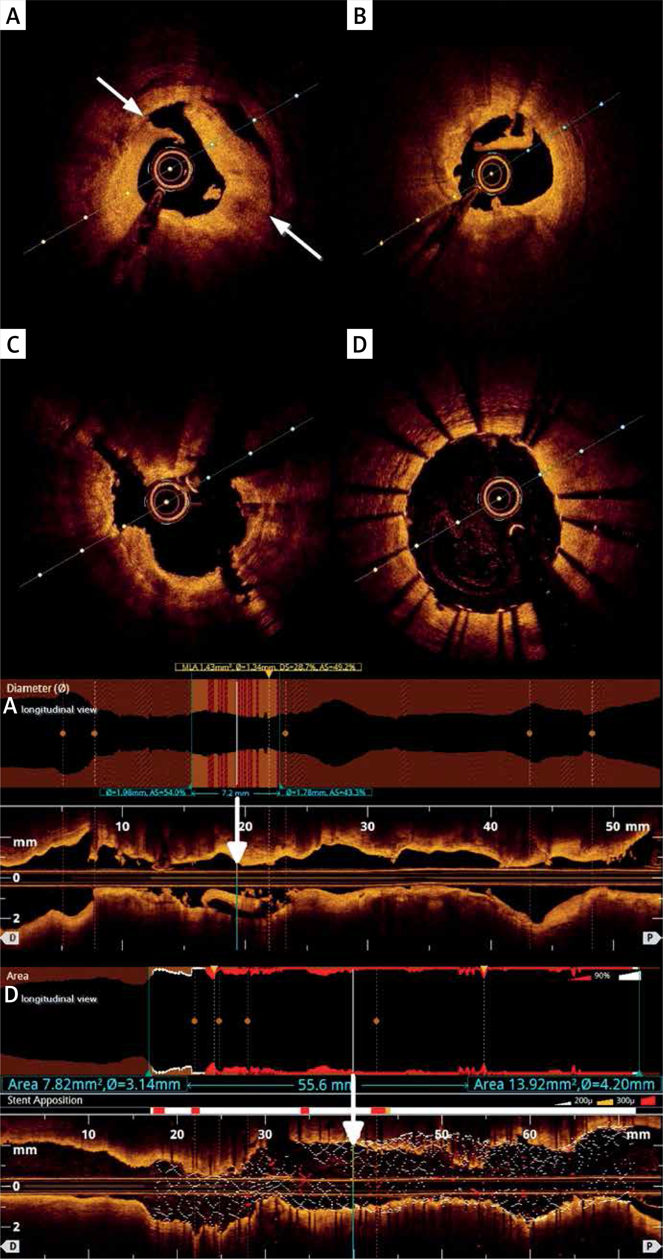
In this situation the treatment strategy had to be rearranged, because such dissection is one of the contraindications to perform RA. We decided to use scoring balloons and as a result several 3.5 × 15 mm balloon inflations were made. Repeated OCT showed cracks in calcified plaques (Figure 1 C). The proximal and medial RCA was successfully covered by 3.5 × 38 mm and 4.0 × 15 mm zotarolimus-eluting stents. For optimal stent expansion the NC 4.0 × 15 mm balloon was finally used with an optimal effect. A reassessment with OCT confirmed appropriate apposition of stent struts (Figure 1 D).
In the report we indicated the importance of intravascular imaging during complex procedures with RA that can impact the operator’s decision making [1–3]. Both intravascular ultrasound (IVUS) and OCT are useful during optimization of percutaneous treatment of highly calcified coronary lesions, with some superiority of the latter. OCT-guided RA proved to be associated with better final stent expansion and may improve clinical outcomes compared to IVUS-guided RA [2].
An extensive dissection revealed in OCT, which was not visible in angiography and is a contraindication to perform RA [1], made us change our treatment strategy. In this way we prevented even life-threatening complications of RA, including extension of the dissection, perforation or no-flow phenomenon. There is no consensus on the management of lesions which are still resistant after first rotablation-burr passage. The two main strategies are aggressive post-dilatation with non-compliant/cutting balloons versus continuing to rotablate with larger burrs. Another novel device in this field is intravascular lithotripsy, which is a method of plaque modification that uses sonic waves to selectively fracture intimal and medial calcium deposits without any damage to soft vascular tissue. It might have been helpful in our case as well, but we succeeded using scoring balloons. More common use of intravascular imaging could result in better in-hospital and long-term outcomes, not only in RA cases [2, 4].


