
INTRODUCTION
Superficial epithelioma with sebaceous differentiation (SESD) is a rare benign skin tumor [1]. It is poorly documented in the literature and it lacks standardized nomenclature, which may lead to underdiagnosed or misclassified cases [2]. The broad clinical presentation and histological features of this tumor make diagnosis challenging [1].
We report a rare case of SESD and discuss its main differential diagnoses.
CASE REPORT
A 68-year-old man with no relevant comorbidities presented to the dermatology department with a skin lesion on the forehead that had persisted for 1 year. On examination, there was a brownish nodule with a central crust. Basal cell carcinoma was considered and the lesion was surgically excised.
On gross examination, the specimen consisted of a well-demarcated pigmented nodule with central umbilication, measuring 2.2 cm in diameter.
Microscopic evaluation showed a superficial, multilobular epithelial proliferation broadly connected to the overlying epidermis and arranged in a plate-like pattern with well-defined pushing borders (fig. 1 A). The neoplasm was composed predominantly of basaloid cells with pale basophilic nuclei, scant eosinophilic cytoplasm, and indistinct borders. Focal peripheral palisading was present, without stromal retraction. Cytologic atypia was absent, and mitotic activity was minimal (< 1 mitosis per 10 high-power fields). Occasional squamous eddies were also observed (fig. 1 B).
Figure 1
Histopathologic features of superficial epithelioma with sebaceous differentiation. A – Dermal, plate-like proliferation of basaloid cells with numerous broad attachments to the overlying epidermis (hematoxylin and eosin (HE), ×50). B – Basaloid cells with pale basophilic nuclei and scant eosinophilic cytoplasm; focal squamous eddies are visible (arrow) (HE, ×400). C – High-power view showing the sebaceous differentiation composed of clusters of mature sebocytes (asterisk) (HE, ×400). D – Diffuse melanin deposition and cystic degeneration (arrows) (HE, ×100)
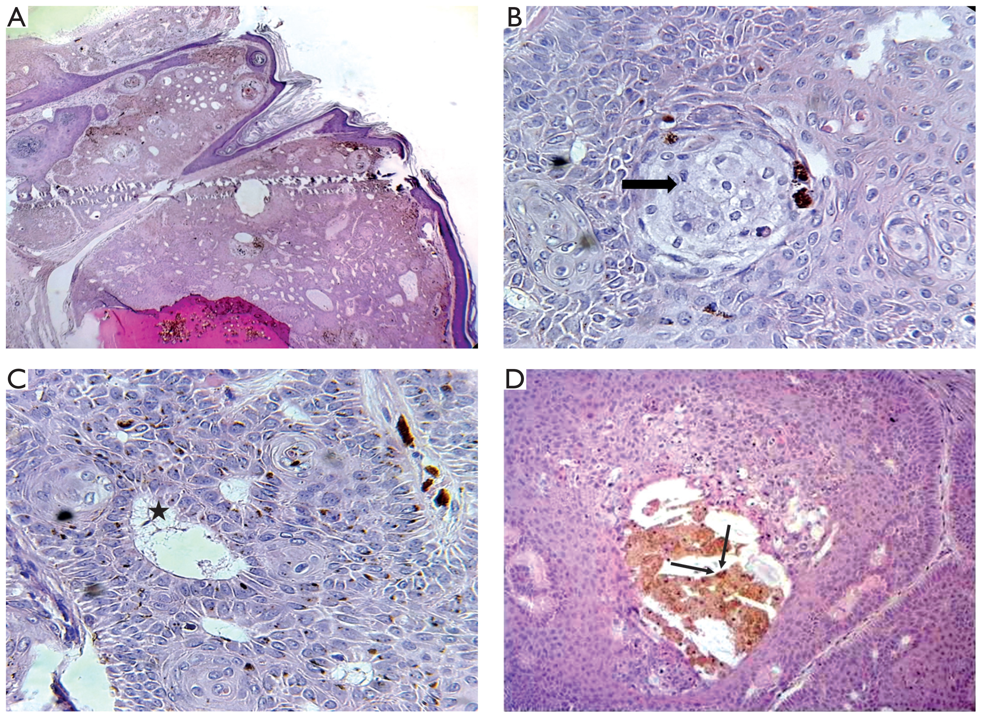
Mature sebocytes were seen mainly in the lower portions of the lobules, either in small clusters or as isolated cells partially replacing the basaloid component. They had microvesicular cytoplasm and centrally located scalloped nuclei (fig. 1 C). Diffuse melanin deposition and cystic degeneration were also noted (fig. 1 D).
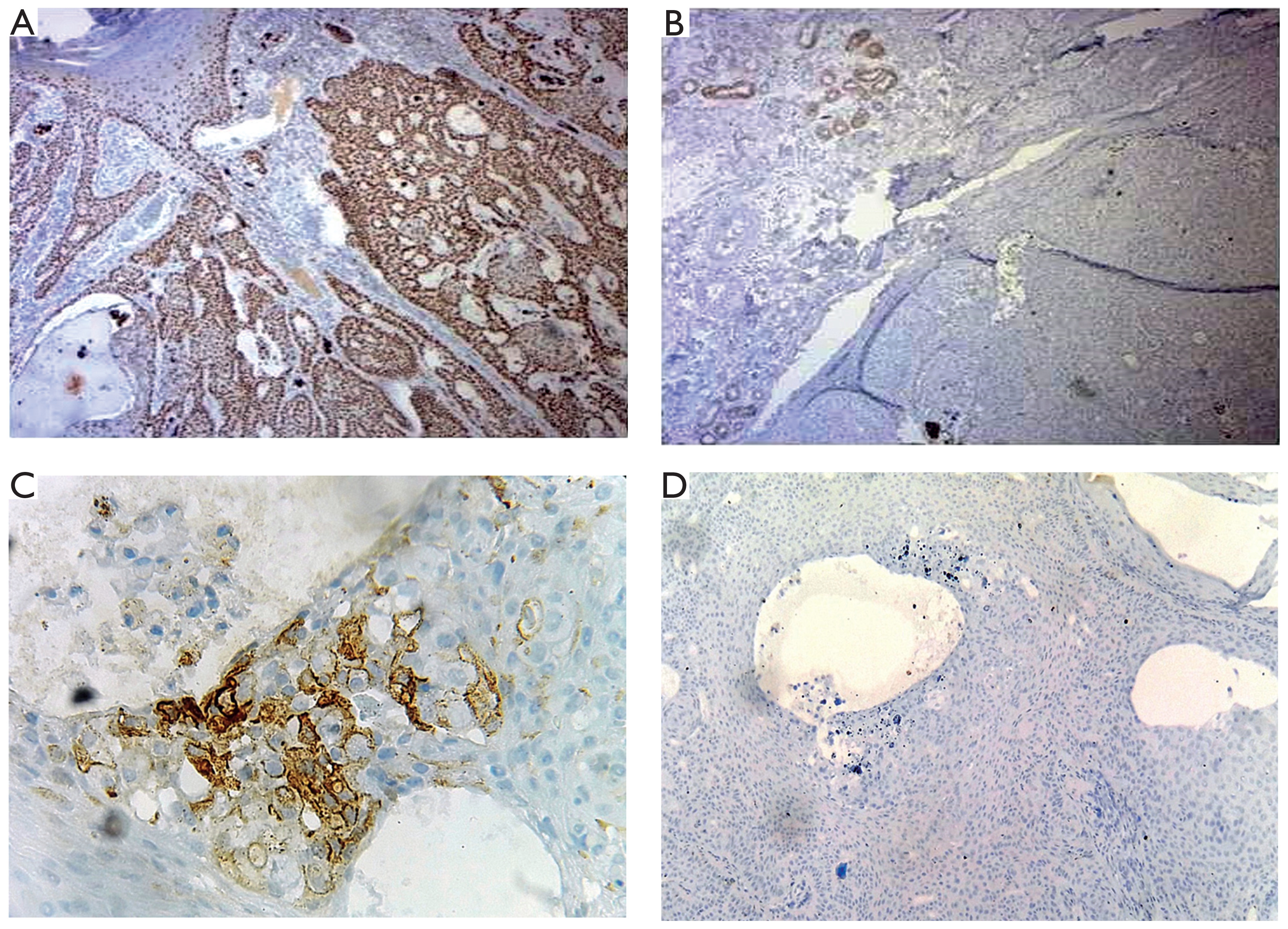
Immunohistochemistry showed cytokeratin 5/6 expression in the neoplastic cells (fig. 2 A), whereas Ber-EP4 was negative (fig. 2 B). Epithelial membrane antigen (EMA) highlighted scattered sebocytes (fig. 2 C). The Ki-67 proliferation index was low (< 1%) (fig. 2 D). These findings supported a final diagnosis of superficial epithelioma with sebaceous differentiation. No recurrence was observed after 6 months of follow-up.
DISCUSSION
SESD is a rare benign cutaneous tumor. To the best of our knowledge, fewer than 21 cases have been described in the literature [1].
The entity was first reported by Rothko et al. in 1980 [3]. Its nomenclature remains inconsistent, and the term SESD has been questioned by some authors, which may have contributed to underrecognition and misclassification [2]. Previously used names include reticulated acanthoma with sebaceous differentiation, acanthomatous superficial sebaceous hamartoma, sebomatricoma, sebocrine adenoma, and poroma with sebaceous differentiation [2, 4].
SESD occurs mainly in adults, with a mean age of approximately 60 years and a slight female predominance [2, 5]. The most frequent location is the face, followed by the back [3, 5]. Clinically, it may present as a papule, nodule, or plaque of variable color consistency [6, 7]. Because of this nonspecific appearance, the differential diagnosis is broad and includes both benign lesions, such as seborrheic keratosis, and malignant tumors, particularly basal cell carcinoma [2]. Apart from the original case of Rothko et al., which described multiple lesions, subsequent cases have been solitary.
The tumor size is usually smaller than 20 mm [8]. In the present case, the lesion measured 22 mm, making it one of the largest reported to date.
The diagnosis relies primarily on morphology. SESD typically presents as a superficial, plate-like proliferation of basaloid to squamoid cells, with broad connections to the overlying epidermis. Mature sebocytes are usually arranged as single cells or small clusters, most often at the periphery of the proliferation [3, 7]. Additional findings may include horn cysts, squamous eddies, cystic degeneration, focal peripheral palisading, verrucous or reticulated architecture, and melanin deposition [9]. Mitoses may be present, but atypical mitotic figures are absent. Cytologic atypia and necrosis are not expected [1].
Immunohistochemistry may support the diagnosis but is not required in typical cases. The epithelial component usually expresses keratinocyte markers, consistent with sebofollicular differentiation [2]. EMA highlights the sebaceous component [8]. However, some authors have suggested an origin from epidermal pluripotent cells rather than the pilosebaceous unit [2].
The presence of sebocytes raises several differential diagnostic considerations, including seborrheic keratosis with sebaceous differentiation (SKSD), sebaceoma, sebaceous adenoma, sebaceous carcinoma, tumor of the follicular infundibulum, and basal cell carcinoma with sebaceous differentiation [8] (table 1). The plate-like architecture of SESD may resemble acanthotic seborrheic keratosis; however, horn cysts and squamous whorls are more typical of SKSD, in which sebaceous elements are usually more limited [1, 2].
Table 1
Differential diagnosis of superficial epithelioma with sebaceous differentiation
Sebaceoma is composed predominantly of basaloid sebocytes arranged in lobules and is usually located deeper in the dermis, with only occasional epidermal attachment [9].
Sebaceous adenoma is a benign neoplasm that consists of mature sebaceous cells arranged in a multilobular pattern, with small basaloid cells at the periphery of lobules. It also tends to involve the mid or deep dermis, unlike SESD [8].
Tumor of the follicular infundibulum has a reticulated pattern and is connected to the epidermis by thin anastomosing bands. In contrast to SESD, sebaceous differentiation is only occasional, and the basaloid cells typically have paler cytoplasm with more prominent peripheral palisading [1, 8].
Basal cell carcinoma with sebaceous differentiation is a particularly important distinction because of its more aggressive behavior. It usually shows a lobular rather than plate-like architecture, higher cellularity, nuclear atypia, numerous mitoses, peripheral palisading, and stromal clefting. Sebaceous differentiation is generally limited. In SESD, the lack of Ber-EP4 expression in basaloid cells may help to exclude basal cell carcinoma [9–11].
SESD typically follows a benign clinical course after total excision or electrodesiccation. Local recurrence had been reported in 2 cases after partial resection [1].
CONCLUSIONS
SESD is an extremely rare benign tumor that remains poorly recognized and may be misdiagnosed. Because of its broad clinical and histologic spectrum and the absence of standardized nomenclature, diagnosis can be challenging for both dermatologists and pathologists. Awareness of this entity and careful clinicopathologic correlation are essential to avoid confusion with malignant adnexal tumors or basal cell carcinoma.












