
INTRODUCTION
Pigmentary diseases include a wide range of conditions characterized by changes in skin color, either as hyperpigmentation or hypopigmentation. These disorders can significantly impact an patients’ quality of life due to their visible nature and potential psychological effects [1]. Among the most common pigmentary disorders are vitiligo, melasma, and post-inflammatory hyperpigmentation.
Vitiligo is an autoimmune disorder that affects about 0.5–2% of the population. This disease is characterized by the destruction and dysfunction of melanocytes, resulting in depigmented patches on the skin. Melasma, on the other hand, is characterized by brown or gray-brown patches, often due to hormonal changes and sun exposure. Recent research has improved our understanding of pigmentary disease pathogenesis and highlighted the interplay between genetic and environmental factors, alongside immune system dysregulation [1–5].
The skin is exposed to many environmental factors, one of which is the skin microbiome. A number of microorganisms, including viruses, fungi, and bacteria, make up the skin microbiome. The immune system of the host and these microbes interact and may affect immune responses. The gut microbiome can also affect skin immunity through the gut–skin axis and alternations in immune chemokines and reactivity [6].
Multiple studies have demonstrated the role of microbiome in skin immune homeostasis, skin barrier function, and inflammatory skin diseases, as well as the influence of microbiota on immune homeostasis, oxidative stress, and skin barrier integrity – factors known to be involved in the pathogenesis of pigmentary disorders. However, findings across studies remain heterogeneous [6]. Therefore, this systematic review aims to summarize and synthesize the available evidence regarding the relationship between the microbiota and pigmentary diseases, particularly vitiligo and melasma, to better understand the underlying mechanisms and identify potential microbiota-based interventions.
METHODS
Search strategy
This review was conducted according to the Preferred Reporting Items for Systematic Reviews and Meta-Analyses (PRISMA) guidelines [7].
We searched for English- and Persian-language manuscripts related to pigmentary diseases and their association with the host immune system and microbiota. PubMed, Scopus, Web of Science, Science Direct, Embase, and Magiran were thoroughly searched up to December 2024.
Search terms included combinations of keywords such as “microbiota”, “microbiome”, “gut-skin axis”, “skin microbiota”, “vitiligo”, “melasma”, “hypopigmentation”, “hyperpigmentation”, and “pigmentary disorders”. No language restrictions were applied initially; however, only studies with full texts available in English were included in the final analysis. Additionally, manual searches of reference lists from relevant publications were performed to identify further eligible studies.
Selection criteria and data extraction
Studies investigating interactions between pigmentary diseases and skin or gut microbiota were included in this review, whereas studies with insufficient data, studies published in languages other than English and Persian, and studies that failed quality assessments were excluded. Review articles, case reports and editorials were also excluded.
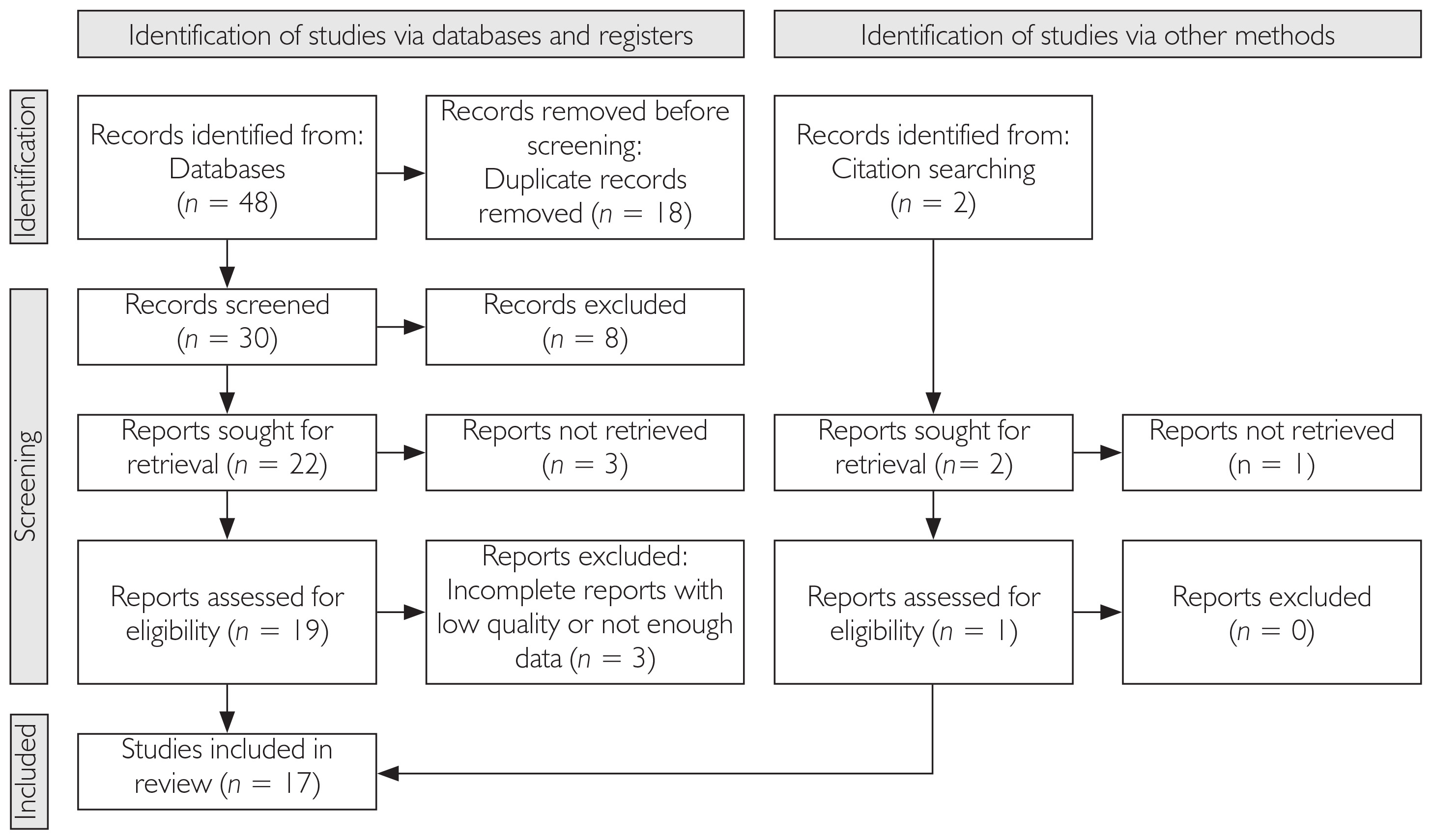
After removing duplicates, titles and abstracts were screened. Subsequently, the full text of the eligible studies was retrieved and assessed. Two independent reviewers evaluated the quality of the final manuscripts, and the data were eventually extracted.
A flowchart of the study selection process is presented in figure 1.
Quality assessment of included articles
NIH quality assessment forms were used to evaluate the scientific quality of the selected manuscripts. According to these protocols, the following domains were assessed: 1) appropriate selection of the study group, 2) control of confounding factors, and 3) outcome assessment. The papers were then categorized as good, fair, or poor quality. Studies assessed as poor quality were excluded from this review. The process of study selection and quality assessment was conducted by 3 independent reviewers [8].
The quality assessment of the included studies is presented in table 1.
Table 1
| Author | Year | Country | Disease | Human/ Animal | Microbial community and details | NIH quality |
|---|---|---|---|---|---|---|
| Ganju et al. [9] | 2016 | India | Vitiligo | Human | Actinobacteria and Firmicutes were more abundant in vitiligo skin, whereas Proteobacteria and Bacteroidetes were less abundant. Variations were observed at the genus level, although they were less pronounced than those at the phylum level. Higher richness and diversity were observed in the non-lesional microbiome. | Good |
| Ni et al. [10] | 2020 | China | Vitiligo | Human | Decreased Bacteroidetes-to-Firmicutes ratio in the gut microbiota of patients with vitiligo. Corynebacterium, Ruminococcus, Jeotgalibaca, and Psychrobacter showed a significant negative correlation with disease duration and a positive correlation with serum IL-1β levels in vitiligo patients. | Fair |
| Dellacecca et al. [11] | 2020 | USA | Vitiligo | Mice | Ampicillin-induced depigmentation was associated with gut, but not skin, dysbiosis, together with a decrease in T cells at both sites. Treatment led to an increased fecal bacterial population dominated by Bacteroides. Reduced diversity became particularly evident after ampicillin treatment, during which Pseudomonas species dominated the gut. Neomycin induced intestinal T-cell reorganization and accumulation of regulatory T cells in the skin. | Good |
| Lu et al. [12] | 2021 | China | Vitiligo | Human | Streptomyces and Streptococcus were enriched in the skin of patients with active vitiligo compared with stable vitiligo. No significant difference in alpha diversity was observed between the two groups. Lipid biosynthesis may contribute to vitiligo development through its effects on the innate immune system. | Good |
| Bzioueche et al. [13] | 2021 | France | Vitiligo | Human | Lesional-site swabs showed reduced Staphylococcus levels compared with non-lesional-site swabs. The microbiota obtained from lesional-skin biopsy samples was characterized by a marked deficiency in protective genera, such as Bifidobacterium and Bacteroides, together with increased abundances of Proteobacteria, Streptococcus, and Mycoplasma. Vitiligo was associated with reduced alpha diversity and an increased Firmicutes-to-Bacteroidetes ratio in the gut microbiome. Mitochondrial DNA was associated with immune responses. | Good |
| Zanchetta et al. [14] | 2022 | France | Lentigo | Human | Healthy skin showed higher microbial diversity and higher abundances of Cutibacterium, Prevotella, Staphylococcus aureus, and Streptococcus. Healthy skin was dominated by common skin commensals, such as Staphylococcus and Corynebacterium. | Good |
| Liu et al. [15] | 2022 | China | Melasma | Human | Actinobacteria, including Collinsella and Actinomyces, as well as Bacteroides and Firmicutes, were more abundant in the melasma group. The gut microbiome may influence estrogen metabolism, as β-glucuronidase released by gut bacteria can enhance intestinal estrogen reabsorption. | Good |
| Yasmeen Ali et al. [16] | 2022 | USA | Vitiligo | Human | Higher abundance of Bacteroides was observed in the control group compared with vitiligo patients. | Good |
| Luan et al. [17] | 2023 | China | Vitiligo | Human | The alpha diversity of the intestinal microbiome was lower in vitiligo patients than in controls. In vitiligo patients, the abundance of Staphylococcus thermophiles decreased, whereas Bacteroides fragilis abundance increased compared with controls. | Good |
| Wu et al. [18] | 2023 | China | Vitiligo | Human | The Firmicutes-to-Bacteroidetes ratio was higher in vitiligo patients. Megamonas, Bifidobacterium, and Psychrobacter were more abundant in vitiligo patients, whereas Akkermansia, Paraprevotella, and Parabacteroides were less abundant. A protective role of Bacteroides in depigmentation was suggested. | Fair |
| Mao et al. [19] | 2023 | China | Vitiligo | Human | Vitiligo was causally associated with the family Ruminococcaceae. Lactobacillus showed a protective role in vitiligo. | Good |
| Kuroda et al. [20] | 2024 | Japan | Vitiligo | Human | Gammaproteobacteria, Staphylococcus spp., and Corynebacterium spp. were more abundant in vitiligo. Staphylococcus spp. were more abundant during the stable phase in the forehead region. A lower proportion of Malassezia sympodialis was observed in vitiligo patients, whereas Malassezia globosa was more abundant during the progressive phase on the dorsal aspect. Enterococcus spp. were less prevalent during the stable phase and even less prevalent during the progressive phase. No dysbiosis was observed in vitiligo lesions; however, both lesional and non-lesional skin in patients showed higher organism diversity. | Good |
| Kim et al. [21] | 2024 | South Korea | Vitiligo | Human | Higher rates of vitiligo were observed in infants exposed to antibiotics, potentially due to dysbiosis. | Good |
| Touni et al. [22] | 2024 | USA | Vitiligo | Mice | Topical application of Neosporin significantly reduced depigmentation and showed effects beyond the localized treatment area, whereas bacitracin ointment had no observable effect. Stool samples indicated that Neosporin administration was associated with reduced abundance of the genus Alistipes in the gastrointestinal tract, whereas changes in the skin microbiome were less evident. Antibiotic intervention reduced MR1 expression, which may limit mucosal-associated invariant T-cell activation, whereas Neosporin-treated skin showed a selective and substantial decrease in CD8+ T-cell abundance. | Good |
| Sekino et al. [23] | 2024 | Japan | – | In vitro | Following screening of commensal skin bacteria, Corynebacterium tuberculostearicum was found to inhibit tyrosinase activity. | Good |
| Yuan et al. [24] | 2020 | China | Vitiligo | Human | Significant microbial dysbiosis was observed in lesional skin compared with non-lesional skin. NB-UVB treatment reduced microbial differences between lesional and non-lesional sites, suggesting a potential role in restoring microbiota balance in vitiligo-affected skin. | Good |
| Piyavatin et al. [25] | 2021 | Thailand | Melasma | Human | A 12-week oral synbiotic supplement significantly improved melasma severity, as indicated by reductions in the modified Melasma Area and Severity Index (mMASI) and melanin indices. | Good |
RESULTS
Study characteristics
A total of 17 studies were included in this systematic review, comprising primarily human observational studies (13), with additional animal studies (3) and 1 in vitro study. Among these, 15 focused on vitiligo and 2 on melasma. The studies varied in terms of microbiota analysis, with some assessing both gut and skin microbiota. The characteristics of the included studies are summarized in table 1 [9–25].
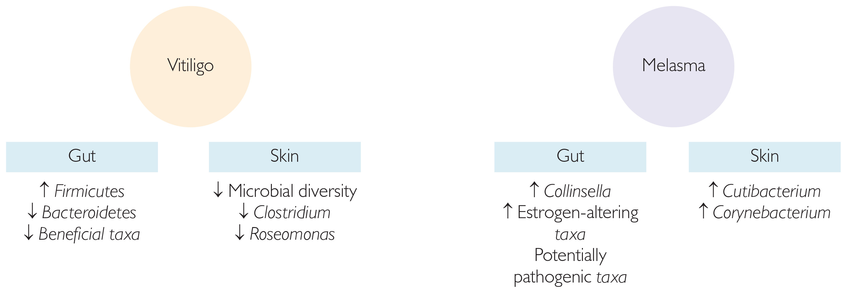
Studies investigating the gut microbiota in vitiligo patients reported significant alterations in microbial diversity. One key finding was a disrupted Firmicutes to Bacteroidetes ratio, with Bacteroidetes being less abundant than Firmicutes [10, 18], suggesting a potential protective role of Bacteroidetes. Animal studies have also shown that antibiotic-induced gut dysbiosis may lead to depigmentation, whereas probiotic interventions could help protect against this effect [11].
In studies focusing on skin microbiota, vitiligo patients exhibited reduced microbial diversity in lesional skin compared to non-lesional or healthy skin. For instance, Actinobacteria and Firmicutes were more abundant in the skin of vitiligo patients, whereas Proteobacteria and Bacteroidetes were less abundant [9]. Notably, non-lesional skin demonstrated greater microbial richness and diversity than lesional skin. Additionally, the microbiota within vitiligo lesions exhibited a notable deficiency in protective genera, such as Bifidobacterium and Bacteroides, while showing increased abundances of Proteobacteria and Streptococcus [9, 13].
Melasma patients showed distinct gut microbiota profiles, compared to healthy controls. For example, higher abundances of Collinsella, Actinomyces, and Bacteroides were observed in the gut microbiota of melasma patients [15]. It has been suggested that gut microbial alterations may indirectly influence pathways related to estrogen metabolism, which plays a role in the development of melasma. However, this association remains circumstantial, as no studies have directly demonstrated altered estrogen metabolism in relation to microbiota shifts. Only alterations in β-glucuronidase activity and the regulation of estrogen synthesis or metabolism have been reported [15, 25].
Key microbial patterns observed in patients with vitiligo and melasma are summarized in figure 2.
DISCUSSION
Pigmentary diseases, such as vitiligo and melasma, are common dermatological conditions characterized by abnormal pigmentation of the skin. These conditions can have profound psychological and social implications for affected individuals because of their visible nature [3]. While the pathogenesis of both conditions remains multifactorial, recent evidence suggests that disruptions in the microbiome may play a significant role in their development and progression [6].
Recent studies have highlighted the potential connection between the microbiota and pigmentary diseases, suggesting that microbial imbalances, or dysbiosis, may contribute to the onset and progression of conditions such as vitiligo and melasma. In vitiligo, alterations in both the skin and gut microbiome have been observed. For example, Ganju et al. (2016) reported an increased abundance of Actinobacteria and Firmicutes, while Proteobacteria and Bacteroidetes were reduced in vitiligo skin lesions [9]. Similarly, Ni et al. (2020) highlighted a decrease in the Bacteroidetes to Firmicutes ratio in the gut microbiome of vitiligo patients, along with a correlation between microbial composition and disease duration [10]. In melasma, Liu et al. (2022) identified specific microbial taxa, including Collinsella and Bacteroides, which were present in greater abundance in the gut microbiota of melasma patients [15]. These findings support the hypothesis that microbiota imbalances may affect the immune system, oxidative stress, and skin barrier integrity, all of which are implicated in the pathogenesis of these diseases.
Microbial diversity plays a critical role in maintaining skin and gut health. In the context of vitiligo, studies have demonstrated that lesional skin exhibits significantly reduced microbial diversity compared to non-lesional areas. Bzioueche et al. (2021) found that lesional skin in vitiligo patients was characterized by a decreased presence of beneficial microbes, such as Bifidobacterium and Bacteroides, which are typically associated with protective immune responses. In contrast, pathogenic genera, such as Proteobacteria and Streptococcus were more abundant in affected skin [13]. These shifts in microbial diversity could contribute to a breakdown in immune tolerance, leading to the development and exacerbation of vitiligo lesions. The diversity of microbial species also appears to be altered in melasma, with certain gut microbes potentially influencing estrogen metabolism, further suggesting a role of the microbiota in regulating skin pigmentation and disease progression. Alternatively, these alterations may represent a secondary consequence of disease-related skin changes. Thus, whether dysbiosis is a cause or a consequence of vitiligo remains to be clarified.
Several microbial species have been consistently associated with either protective or non-protective roles in pigmentary diseases. In vitiligo, Bifidobacterium and Lactobacillus are often considered protective because of their ability to promote regulatory T cell (Treg) function and stimulate the production of anti-inflammatory cytokines, such as interleukin 10 (IL-10) [6]. For instance, Mao et al. (2023) highlighted the protective role of Lactobacillus in modulating immune responses in vitiligo [19]. In contrast, species like Proteobacteria, Streptococcus, and Mycoplasma are frequently associated with non-protective roles, as they may promote inflammation and dysregulated immune responses in both vitiligo and melasma [9, 12, 14]. Furthermore, the development of hypopigmentation in other dermatoses, such as lichen striatus (LS), has also been linked to microbial alterations. Cutibacterium acnes was found to be more abundant in patients with LS–associated hypopigmentation than in those without hypopigmentation [26]. These findings suggest that targeting specific microbial taxa may represent a potential therapeutic strategy for modulating immune responses and reducing disease severity.
In melasma, Liu et al. (2022) observed that genera such as Collinsella and Bacteroides were enriched in the gut microbiota of melasma patients, potentially influencing estrogen metabolism, which may be involved in the pathogenesis of melasma [15].
In addition to microbiota imbalances, other factors including diet, medications, and therapeutic interventions, may influence the progression of pigmentary diseases. A growing body of evidence suggests that dietary factors, particularly the intake of prebiotics and probiotics, may help restore microbiota balance. Piyavatin et al. (2021) demonstrated that synbiotic supplementation significantly improved melasma severity [25]. Likewise, Touni et al. (2024) found that antibiotic treatment could alter the gut microbiome, which subsequently influenced skin depigmentation in vitiligo [22]. Certain probiotics, such as malassezin and Pediococcus acidilactici, have also been shown to be effective in the treatment and prognosis of hyperpigmentation [27, 28]. These findings highlight the potential role of dietary and microbiota-targeted interventions in the management of pigmentary disorders. However, further studies are required to determine the precise mechanisms through which these factors influence skin pigmentation.
Additionally, short-chain fatty acids (SCFAs) produced by gut bacteria have been shown to influence immune tolerance and the balance between Treg and Th17 cells, which appears to be crucial in diseases such as vitiligo [6].
Another proposed mechanism is oxidative stress, which plays a central role in vitiligo. Altered microbiota composition may exacerbate oxidative damage to melanocytes, leading to melanocyte death and the formation of depigmented patches. Wu et al. (2023) examined how Bacteroides fragilis may protect against oxidative stress, suggesting that this genus may exert a protective effect in autoimmune diseases such as vitiligo [18].
The gut–skin axis has emerged as an important pathway, with Piyavatin et al. (2021) exploring the potential of synbiotics in improving skin conditions such as melasma. Their study suggested that modulation of the gut microbiota could influence skin immunity by reducing inflammation and improving skin pigmentation [25]. Additionally, Liu et al. (2022) found that microbial shifts in melasma patients may influence estrogen metabolism, which may be involved in the development of melasma [15].
Piyavatin et al. (2021) demonstrated that a 12-week oral synbiotic supplement significantly reduced melasma severity, improved gut microbiota balance, and decreased inflammation [25]. In line with these findings, Touni et al. (2024) found that topical Neosporin, an antibiotic, reduced vitiligo-associated depigmentation in mice by modulating gut and skin microbiota, notably decreasing the abundance of Alistipes [22]. These studies provide preliminary interventional evidence highlighting the therapeutic potential of microbiota modulation. However, the limited number of such studies underscores the need for further research to validate and expand these findings, particularly through larger human studies aimed at establishing efficacy and optimal intervention strategies. Current evidence is constrained by small sample sizes, short follow-up periods, heterogeneous formulations, and the absence of standardized protocols. As the causal role of the microbiota remains unclear, these findings cannot yet be directly translated into clinical practice. Larger randomized controlled trials are therefore needed to confirm therapeutic efficacy and define reliable intervention strategies.
In summary, this systematic review highlights the growing evidence supporting the role of the microbiome in the pathogenesis of pigmentary diseases, such as vitiligo and melasma. Alterations in both skin and gut microbiota, particularly shifts in microbial diversity and abundance of specific microbial taxa, suggest that dysbiosis may contribute to the immune dysfunction and oxidative stress associated with these conditions. While certain genera, such as Bifidobacterium, Lactobacillus, and Bacteroides, appear to have protective roles in modulating immune responses and reducing inflammation, pathogenic taxa such as Proteobacteria and Streptococcus have been linked to disease exacerbation.
Moreover, this review underscores the potential therapeutic applications of microbiota modulation, particularly through dietary interventions such as probiotics and synbiotics. However, the current body of research is still limited, with only a few studies directly assessing the impact of probiotics on pigmentary diseases. In contrast, there were little data on other pigmentary diseases other than melasma and vitiligo. Most available research focuses predominantly on vitiligo and melasma, with minimal exploration of other pigmentary conditions such as post-inflammatory hyperpigmentation, lentigines, or lichen striatus. This limits the applicability of our findings across the full spectrum of pigmentary disorders and highlights the need for broader investigation. Given the promising results observed in preliminary studies, further research is needed to determine the effectiveness of probiotics and other microbiota-based therapies in managing vitiligo and melasma.
CONCLUSIONS
Understanding the intricate relationship between the microbiome and pigmentary diseases could open new avenues for therapeutic interventions, potentially offering more personalized treatment options. Further studies should focus on elucidating the precise mechanisms by which microbiota influence these diseases, as well as exploring the potential of microbiota-modulating therapies in clinical practice.












