
Introduction
Male breast cancer (MBC) is a rare disease that accounts for less than 1% of male cancer cases in the United States and 1.1% of cases in Germany [1–3]. The exact aetiology of this disease is still largely unknown; described risk factors include age, BRCA1 and 2 mutations, obesity, Klinefelter’s syndrome, and radiation exposure [4].
More than 90% of MBC cases are oestrogen receptor (ER)-positive, and 80–90% progesterone receptor (PR)-positive. Compared to female breast cancer (FBC), these frequencies are higher [5–7]. HER-2/neu is expressed in 5–29% of MBC cases compared to 15–25% in FBC cases [8–10]. The most common histologic type of MBC is the invasive carcinoma of no special type [10]. Compared to women, breast cancers of lobular histology are less frequently encountered, which is most probably due to the significantly lower number of lobules in male breast tissue [5]. Due to its rarity, the biology of MBC is still not fully understood, and cell culture model systems are still in development.
The G protein-coupled oestrogen receptor 1 (GPER-1) was first identified in 1997 [11]. Regarding transcription modulation and rapid non-genomic action, GPER-1, together with classical oestrogen receptors (ERs), appears to mediate the action of oestrogen [12, 13]. In hormone-sensitive tumours, such as breast and ovarian cancer, GPER-1 is involved in proliferative oestrogen signalling [14, 15]. The role of GPER-1 as a potential tumour suppressor is a subject of debate. In cell culture studies, activation of GPER-1 by the specific agonist G-1 resulted in reduced cell proliferation and cell death in ER-positive as well as ER-negative cell lines [16–18]. However, in FBC patients, the situation seems more complicated because GPER-1-positive cases exhibited a shorter relapse-free survival [14], but this depended on anti-oestrogen treatment in ER-positive cases. In ER-positive BC, the agonistic influence of tamoxifen on GPER-1 seems to favour the development of acquired tamoxifen-resistance [13, 15, 19].The question of whether a blockade of the GPER-1 signalling pathway could overcome tamoxifen resistance, thus being a target in the therapy of oestrogen-related tumours, still needs to be answered [12, 20].
To date, the literature contains no data on the expression of GPER-1 in MBC or its potential impact on patients’ survival. In this study, we investigated the expression of GPER-1 in 161 patients with primary MBC and correlated these data with clinical and pathological parameters comprising patients’ survival.
Material and methods
Patients
We investigated MBC cases included in the German prospective cancer registry of MBC. This trial was registered at the German Clinical Trials Register DRKS under the number DRKS00009536. Male patients, diagnosed with histologically confirmed primary breast cancer, and older than 18 years were included in the study. This cancer registry contains data on the age at diagnosis, patient and tumour characteristics, operative, neo-, and/or adjuvant treatment, date and localization of relapse, date and cause of death, secondary cancer, and comorbidities. The database was compiled between 2009 and 2018. The last update was in May 2020. Other studies conducted on the basis of this database have already been published [7, 21, 22]. Formalin-fixed, paraffin-embedded tumour samples from 161 patients were available for this retrospective analysis.
Materials
Immunostaining was performed on 3-µm slices [14] using a Benchmark XT automatic staining system (Ventana, Unterhaching, Germany) in batches of about 30 slides. As positive control, an FBC sample with known histochemistry score was included into each batch. The staining procedure was essentially as described in [14].
The stained slides were evaluated by pathologists of the Department of Pathology, Magdeburg, Germany, and the Medical University of Innsbruck, Austria using light microscopy (SS, PC). Staining intensity and staining extensity were scored as previously described [19]. Staining intensity was scored as follows: 0 (no staining), 1 (weak), 2 (moderate), and 3 (strong response). Staining extensity was scored numerically as 1 (< 10% positive cells), 2 (10–50% positive cells), or 3 (> 50% positive cells). Both scores were multiplied for each sample, thus forming the immunoreactive score (IRS) [14]. Therefore, this score included values from 0 to 9.
Statistics
Statistical calculations were performed using IBM SPSS Statistics Version 26.0 for Microsoft Windows (Armonk, NY, USA) or IBM SPSS Statistics Subscription for Apple Mac-OS (IBM SPSS, Armonk, NY, USA). Computer scripts were programmed in the language R (https://www.R-project.org). Overall survival (OS) was defined as the period between the date of diagnosis and date of death from any cause. Relapse-free survival (RFS) was defined as the time span between the date of diagnosis and the date of loco-regional and/or distant relapse. Loco-regional relapse included the recurrence in ipsilateral breast, chest wall, or regional lymph nodes. Distant recurrences consisted of distant lymph node metastases (beyond the ipsilateral axillary, infra- and/or supraclavicular, internal mammary area), as well as of metastases in bone (including bone marrow), brain, liver, lung (including pleura and lymphangitic carcinomatosis), and other organs (including peritoneum, other organs not elsewhere classified, and skin tumours not affecting the breast and chest wall). The paired Wilcoxon signed rank test was used to compare GPER-IRS of tumour tissue compared to adjacent non-tumorous tissue. For correlation of ordinal variables, Spearman´s rank correlation test was used. The impact of the GPER-1 status on OS and RFS was analysed using the Kaplan-Meier method, and significance was determined using the log-rank test. In the case of OS, this p-value was further corrected using a permutation test to consider the fact that the cut-off point was determined by the minimal-p-value-approach [23]. Furthermore, uni- and multivariable Cox regression analysis was applied. The GPER-1 expression was correlated to clinical and pathological parameters by cross-tabulation test using a two-sided Fisher’s exact test for determining significant differences. Generally, p < 0.05 was considered as an indicator of a significant deviation from the null hypothesis, and p < 0.1 was considered as a statistical trend.
Results
The G protein-coupled ER 1 immunoreactivity analysis was performed on specimens from 161 patients. Figure 1 shows representative examples of GPER-1 staining. Comparing adjacent, non-tumourous tissue with tumour, the paired Wilcoxon signed rank test revealed no significant differences in staining intensity (p = 0.28). Based on the minimal p-value approach using the log-rank value for OS, an IRS ≥ 4 was considered GPER-1-positive. Sixty-five patients (40.4%) were GPER-1 positive and 96 (59.6%) were GPER-1 negative. This cut-off is one step higher than that established in our studies for FBC. Using this cut-off value of ≥ 3, no significance was observed in Kaplan-Meier survival analysis (for OS: p = 0.065, see below). The mean age at initial diagnosis was 66.1 years (range: 26–93, standard deviation: ± 13.0 years). It was significantly lower in GPER-1-positive than in GPER-1-negative patients (63.0 vs. 68.2 years, p = 0.018, Table 1). Therefore, we applied age-adjusted Cox regression to further evaluate this result. Further data on the cohort can be found in Table 2.
Fig. 1
Representative images of the G protein-coupled oestrogen receptor-1 histochemical staining. No (A, E), weak (B, F), moderate (C, G), and strong staining intensity (D, H)
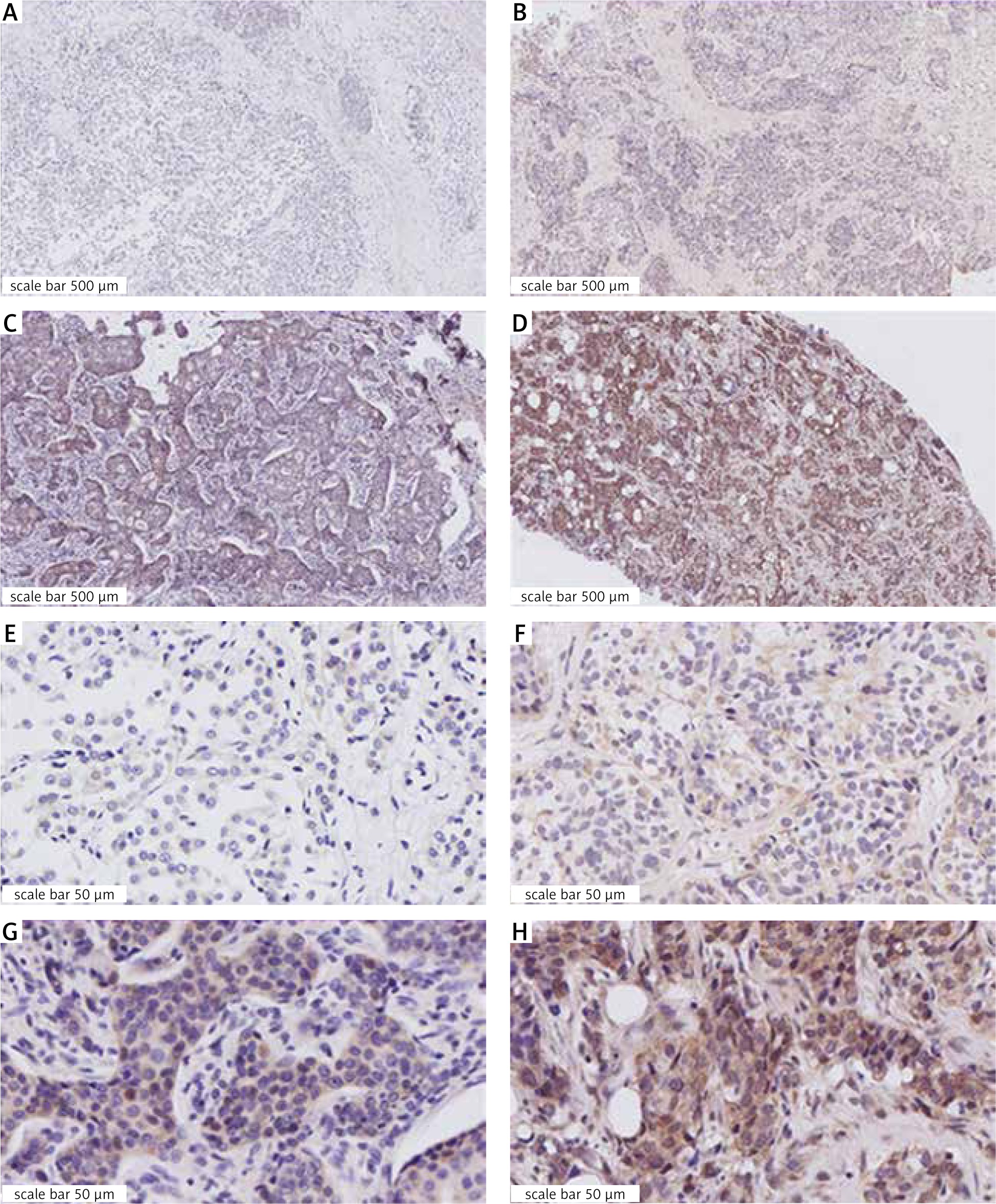
Table 1
Characterization of patients
Table 2
Clinical and pathological characteristics and Fisher test p-values for G protein-coupled oestrogen receptor-1 positive (≥ 4) vs. G protein-coupled oestrogen receptor 1 positive negative male breast cancer specimens (and ordinal by ordinal correlation)
The G protein-coupled oestrogen receptor 1 and clinicopathological parameters
We then tested for correlation between GPER-1 status and major clinicopathological parameters (Table 2). There was no significant difference in any of these clinical and pathological parameters, but we found a trend concerning tumour size (p = 0.093). Because most of the cases were ER and PR positive, we also correlated the ER and PR scores with the GPER-1score, but again, no significant result was obtained.
Survival analysis and G protein-coupled oestrogen receptor status
Data from 150 patients were available for the analysis of OS and RFS (Table 3). For relapse-free survival, 26 events were observed, whereas for overall survival, death was confirmed to be due to cancer in 5 cases, 11 deaths were attributed to other reasons, and for the remaining 11 the cause of death was not documented.
Table 3
Observation period stratified for G protein-coupled oestrogen receptor-1 status (≥ 4)
| Observation period [weeks] | All | GPER-1 positive | GPER-1 negative | p-value |
|---|---|---|---|---|
| Median | 208.9 | 215.6 | 206.3 | |
| Mean ± SD | 197.5 ± 72.0 | 204.8 ± 69.6 | 192.4 ± 73.5 | 0.555 |
| Range | 15–319 | 36–300 | 15–319 | |
| Total | 150 | 61 | 89 | |
| Missing | 11 | 4 | 7 |
Among the clinical and pathological parameters, obesity, tumour size, lymph node status, and type of axillary lymph node surgery were associated with OS. Lymph node status, as well as breast and axillary lymph node surgery, had a significant influence on RFS (Table 4, 5).
Table 4
Kaplan-Meier analysis of overall survival
Table 5
Kaplan-Meier analysis of relapse-free survival
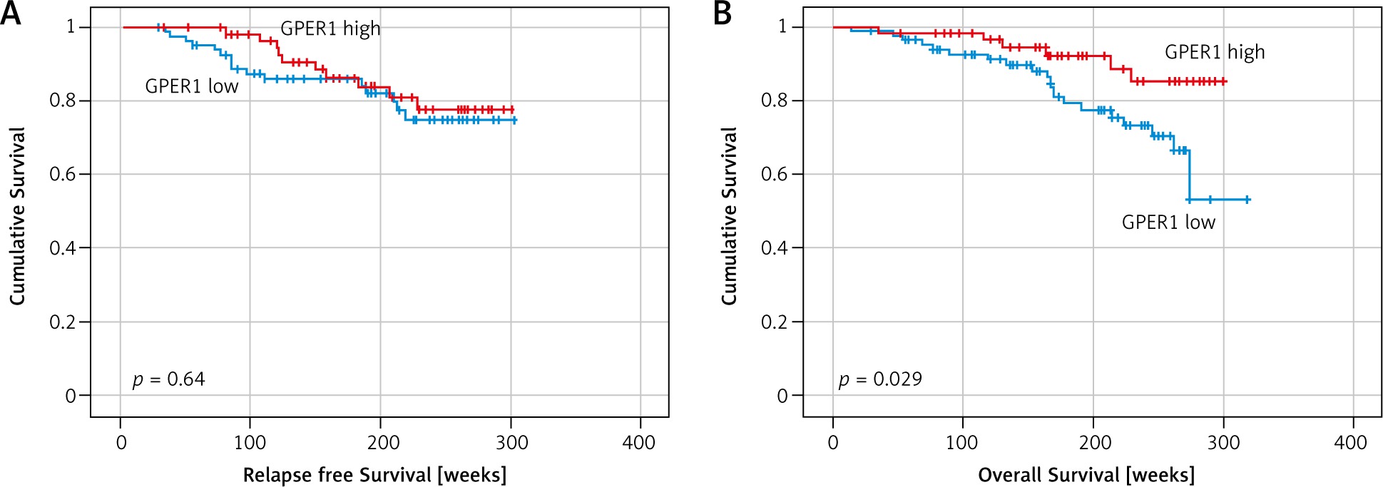
Kaplan-Meier analysis revealed that the GPER-1 status (≥ 4) was associated with better/improved overall survival, but not with RFS (Fig. 2, log-rank p = 0.029 and p = 0.637, respectively). However, after correction using a permutation test with 10,000 samples, the p-value for OS rose to 0.109 (this p-value was calculated using a pseudo random seed of 42).
Fig. 2
Kaplan-Meier survival analysis of the G protein-coupled oestrogen receptor-1 status (≥ 4) for relapse-free survival and overall survival. Log-rank p not corrected for age is given
As there was a significant difference in age at diagnosis depending on GPER-1 status (Table 1), we applied multivariable Cox regression analysis to correct the parameters effect for age (Table 6). For this purpose, age at diagnosis was used without further modification. Here, GPER-1 lost its significance for OS (p = 0.065, HR = 0.44, 95% CI: 0.18–1.11). Age at diagnosis itself was also a significant factor for OS (p = 0.043, HR = 1.034, 95% CI: 1.001–1.068). However, in multivariable analysis together with GPER-1 this significance decreased (p = 0.075, HR = 1.029, 95% CI: 0.996–1.06). In a multivariable Cox regression with algorithmic variable selection including GPER-1-status, age, tumour size, and N, only the lymph-node status remained a significant factor for RFS (p = 0.001. HR = 5.733, 95% CI: 2.208–14.884) and OS (p = 0.002, HR = 4,73, 95% CI: 1.796–12.477). There was also no significant interaction between tamoxifen therapy and GPER-1 status (data not shown).
Table 6
Cox regressions analysis for survival of male breast cancer patients. All data shown were adjusted for age
Discussion
This is the first study investigating GPER-1 in primary MBC cases. A total of 161 patients were studied, and an IRS value ≥ 4 was defined as a positive result. This cut-off point was based on Kaplan-Meier survival analysis log-rank p-value optimization. We then correlated OS and RFS, as well as clinical and pathological parameters, with this GPER-1 status.
GPER-1 status was positive in 40.4% of cases. In previous studies, the rate of GPER-1 positivity ranged from 56 to 80% in FBC [14, 19, 24] using a lower cut-off value (IRS ≥ 3) although the same staining protocol was applied. Using this cut-off value (≥ 3), 50.3% of MBC cases would be GPER-1 positive in our study. These results possibly indicate that expression of GPER is weaker in men, which would justify the higher cut-off value. Nevertheless, this requires further analysis. Another explanation for this might lie in technical differences such as different lots of the antibody, different fixation methods of the study centres, or storage conditions of the samples.
Also applying a cut-off value of IRS ≥ 3, the median age remained significantly higher in the GPER-1-negative cases (p = 0.027, difference: 4.8 years). Significant correlations to other clinicopathologic parameters were not detected. Nevertheless, significant differences in OS are lost with this cut-off (p = 0.061 Kaplan-Meier analysis, log-rank p). Therefore, splitting the collective at IRS ≥ 4 seemed most reasonable.
At first glance, GPER-1 expression was significantly related to favourable OS. This confirms the assumption of a prognostically favourable effect of GPER-1 in studies of women with ER-positive BC [19, 25]. GPER-1 has also been described as a favourable prognostic factor in early-stage cervical carcinoma, as well as in-vitro in ovarian cancer and in granulosa cell tumours [26–28]. Nevertheless, we did not find any association between GPER-1 status and the likelihood of relapse, but it should be taken into account that the statistical power of our study is lower than in most studies for FBC. Such an association of GPER-1 with metastasis has been described previously [29]. However, that study used a different system for the IRS evaluation. Nevertheless, another study on FBC [14] found a stronger effect of GPER-IRS on RFS than on OS. While the observation period was comparable for both groups (Table 4), we found a significant difference in both age at initial diagnosis and age at death (Table 1) depending on GPER-1 status. Previous studies examining women found no significant difference in age at initial diagnosis [19, 30]. This is a newly found association and possibly a distinct characteristic of MBC. However, this complicates the interpretation of the survival analysis. In our study, the cause of death is ill-defined. Additionally, the multivariable Cox regression analysis demonstrated a reduction of significance for GPER-1 status to a trend, when adjusted to age. This drop in significance also applies for age itself; thus, both factors contribute to OS. The effect of GPER-IRS on OS is uncertain, especially when we consider that the optimization of the cut-off value is known to cause an overestimation of significance in survival analysis [23, 31]. Indeed, when we corrected the log-rank p-value for this effect by permutation testing [32], significance was lost (p = 0.109). Altogether, as there was no significant correlation with RFS and an uncertainty of the cause of death in this study, we cannot conclusively show an effect of GPER-status on survival of our MBC patients. Based on these assumptions, GPER-1 might play a different role in MBC than in FBC.
We also found no association between tamoxifen therapy and GPER-1 status and its effect on RFS and OS. Men displayed a significantly prolonged OS for tamoxifen-treatment when compared with aromatase inhibitors (AIs), which was also shown in a larger sample of patients from this registry study [22, 33]. Presumably due to the small number of patients not given tamoxifen (n = 20) and patients with unknown endocrine therapy (n = 17), we could not demonstrate any effect. Also, a significant number of patients stopped using tamoxifen early. As a result, the administration time was often short. Tamoxifen resistance has been reported to occur especially in long-term use [20]. It is possible that the observed length of time the drug was administered in our study was too short for resistance to develop. However, GPER-1 may not be a factor determining tamoxifen resistance in men. Although a positive GPER-1 status was not associated with an improved probability of recurrence, the possibility of a therapy specifically tailored for GPER-1 status should be considered further. Prospective studies should evaluate whether this could have a significant impact on relapse-free time.
Investigations of MBC have revealed that compared to women, ER and PR are expressed more frequently – in approximately 95% and 80% of cases, respectively [5, 6]. Although our study supports this result, the observed positive ER and PR status of 98.1% and 95.0%, respectively, is higher than in some previous studies. These results are largely consistent with data from recent publications (ER: 96–97% PR: 91–92%) [34, 35]. A correlation between GPER-1 and ER or PR status was shown in female patients [19]. Our study does not confirm this observation, even when we correlated the respective scores to compensate for the low frequency of ER cases.
As in women [19], HER-2/neu expression did not correlate with GPER-1 status. Nevertheless, Sjöström et al. reported opposite results [36]; however, GPER-1 status was evaluated differently and only low-risk FBC patients were included, resulting in only 13.1% positive HER-2/neu cases. A review on this still controversial topic reported that the rate of HER-2/neu positivity is probably lower in men than in women because some of the older studies on HER-2/neu status may have been biased [37]. Comparable rates of 14% and 16% positives were reported in studies conducted in 2015 [34, 35].
We also found no significant association between GPER-1 status and tumour grading. Compared with other studies, these findings provide room for speculation. While one study was able to find an association between GPER-status and low tumour grading, others failed to do so [19, 29]. These 2 sources again considered FBC, which might allow the suggestion of the existence of another tumour entity in men.
We also did not determine the exact subcellular localization of GPER-1, which seems to be linked to other histopathological and prognostic parameters depending on its localization in the nucleus or cytoplasm [38, 39]. In particular, localization in the plasma membrane has been identified as a poor prognostic marker in one study [36].


