
Introduction
The term vulvar leukoplakia encompasses various non-inflammatory diseases that lead to skin discoloration of the external genitalia and white coloration. It includes various atrophic and hypertrophic diseases of the vulva, classified as vulvar dystrophies [1]. Mainly, these are vulvar lichen sclerosus (VLS) and squamous cell hyperplasia of the vulva (SCHV). These 2 conditions have different anatomical and pathological features but similar clinical manifestations and drug treatment. The frequency is 1 in 300-1000 [2].
Both diseases are easily clinically diagnosed, which is sufficient for clinical practice. However, their exact differentiation is histologically made after biopsy [3, 4]. Genital itching is the main clinical manifestation of both diseases and is more pronounced in SCHV [5]. Topical corticosteroids are the gold standard for their treatment [6, 7]. They present a very good result but have some side effects as well, such as atrophy, telangiectasia, and striae, and long-term use is not recommended. In addition, some cases are resistant to this therapy. For these reasons, other methods of VLS and SCHV treatment should be sought and used in practice.
We present 15 patients clinically diagnosed with VLS and SCHV, who were treated with Theresienöl herbal oil and monitored for 1 year.
Material and methods
This prospective study included 17 patients (15 of whom were in menopausal status) who were treated at a consulting-diagnostic centre at the Department of Gynecologic Oncology, D-r Georgi Stranski University Hospital – Pleven, for a period of 8 months (Jan. 1st, 2018 – Aug. 31st, 2018). All of them were clinically diagnosed with vulvar leukoplakia (VLS and SCHV). The prescribed treatment was topical administration of Theresienöl twice a day for 3 months.
One of the essential ingredients of this ointment is Butyrum bovis, which is a carrier of all active ingredients of Theresienöl – tocopherol and tocopherol acetate. They have effect on the repair of cells and on the epithelisation of the injured skin. This protects against scar and blister formation. The other ingredient is a fruit extract from Pyrus malus, which is rich in polyphenols – this is the reason for the antioxidant effect of the ointment. Theresienöl contains tannic and salicylic acids, which are responsible for the skin pH. The product contained the leaf extract from Stellarioides longibracteata, which has anti-fungal and antibacterial effects. It improves blood circulation and oxygenation and plays an important role in wound healing. The saponins contained in the ointment have anti-inflammatory, coagulation, and hormone stimulating effects and help treat swelling and haematomas
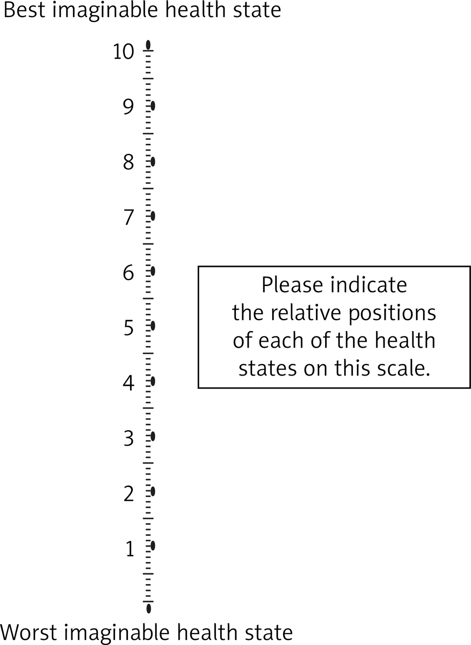
The 1-year follow-up reported the results obtained from visual analogue scale (VAS) assessment for vulvar itching (Fig. 1). Follow-up visits were at the beginning and at the end of the Theresienöl treatment, and 1 year after its initiation.
Results
The median age of the patients enrolled was 60.6 years (range 42–74). Seven of these cases were patients with recurrent disease (treated for vulvar leukoplakia at least 1 year ago); 5 of them were classified as VLS and 2 as SCHV. In 4 of the cases we had a previous histological examination – 1 case with SCHV and 3 with VLS. Two of the patients failed to attend the follow-up visits after the end of treatment. For this reason, they were omitted from the study. Some of the clinical and pathological characteristics of the patients are presented in Table 1.
Table 1
Clinical and pathoanatomical characteristics of patients
It is noteworthy that all patients considered vulvar itching as a very serious problem. The average score that we reported initially using VAS was 1.65 (0–5). There was a significant improvement in itching in all patients, as early as the 10th day of treatment, with a median score of 9 (8–10) at the 3rd month. The score remained at almost the same level at the 12th month – 8.67 (7–10). No side effects and recurrence of symptoms after the end of treatment were observed during the follow-up period.
Discussion
The occurrence of VLS and SCHV is determined by many factors such as immunity, sexual hormones, injuries, environment, enzymes, free radicals, and apoptosis. It is assumed that VLS and SCHV are genetic immune diseases [5]. Both are diseases of autoimmune genesis, with genetic predisposition for VLS [1, 5, 8]. Both diseases are more common in postmenopausal women [1, 9–11]; with VLS there is also a peak in pre-pubertal girls [12–14]. In VLS the vulva is shiny, dry, with no creases. Lesions are often symmetrical, and the skin appears thin. With SCHV, the skin is rough and thick. Thickened plaque-like lesions containing maturing squamous proliferation with hyperkeratosis and/or parakeratosis is observed.
The clinical picture in both diseases is similar – pruritus, pain when having sex, dysuria, or dyspareunia – with itching being the leading symptom. Both conditions have malignant potential – 3 to 6% for VLS and 2 to 4% for SCHV [5]. Long-term follow-up for these patients is required.
Treatment with topical corticosteroids in different doses and duration is the first-line therapy for these conditions. Topical corticosteroids relieve the symptoms in almost 100% of cases. In about 70% of cases, symptoms disappear completely, and in 20% – complete recovery of the skin is observed [7]. The side effects of these drugs are known; however, it should be noted that they are not well documented when administered to the vulva skin [15]. Nevertheless, their use should be limited to achieving the necessary effect.
In patients refractory to this treatment, alternative methods in different operating modes are used: topical hormonal products [16], topical and systemic retinoids [17], topical calcineurin inhibitors [18], Platelet-rich plasma [19], ablative lasers (carbon dioxide laser) [20–23], and non-ablative neodymium: yttrium aluminium garnet (Nd: YAG) [24]. Surgical treatment of both diseases should be avoided because of recurrence risk and should only be used in certain cases, such as patients with malignancy, or to correct irreversible scarring, adhesions, and micturition difficulties or sexual dysfunction caused by the subsequent anatomical changes [7].
We decided to use Theresienöl herbal oil for topical treatment due to its mechanism of action: it hydrates the tissues, reduces bacterial invasion and inflammation, stimulates fibroblast and endothelial cell migration and proliferation, and stimulates epithelialisation. Thereby, it has an analgesic, antiseptic, moisturising, and itchy skin reducing effect. The other reason for our decision was our previous experience with this product – we used it for the treatment of postpartum perineal tears, chronic wounds, ulcus cruris, postoperative wounds, perianal fissures, burns grade 2 a and 2 b, etc. [25]. The results obtained are promising. However, this study bears some weaknesses: a small number of patients, a relatively short follow-up period, a lack of control group, and a lack of histological verification. Our previous experience with topical steroids leads us to believe that the Theresienöl effect is commensurable with them, as with response time and duration of action. Furthermore, there were no adverse events reported.
Conclusions
Vulvar leukoplakia comprises a set of diseases impairing patients’ quality of life. Standard forms of treatment – conservative and operative – have their limitations, side effects, and complications. This calls for alternative methods to influence them, such as the use of Theresienöl, which is a safe and effective option. Monitoring of these patients should continue, especially given the malignant potential of the disease.


