
Transcatheter aortic valve replacement (TAVR) is recommended for high- and intermediate-risk patients with severe aortic stenosis and a suitable option for degenerated surgical bioprosthetic valves. The feasibility of a complete percutaneous approach in transfemoral TAVR was demonstrated, even in complex scenarios, such as valve-in-valve (ViV) procedures [1]. ViV TAVR is associated both with a higher stroke risk due to biodebris released from the degenerated bioprosthesis and increased risk of coronary occlusion, especially in patients with a short distance between the bioprosthesis and coronary ostia [2]. In such patients, the “chimney technique” or BASILICA technique (Bioprosthetic or Native Aortic Scallop Intentional Laceration to Prevent Iatrogenic Coronary Artery Obstruction) can be used to prevent coronary occlusion [2]. BASILICA is a novel transcatheter electrosurgical technique which increases the safety of ViV TAVR, but also enhances the risk of stroke [3]. Thus, the use of a cerebral protection device (CPD) has been advocated when performing BASILICA prior to TAVR [4].
We present a case of a 66-year-old man after the mini-Bentall procedure with severe dysfunction of the aortic bioprosthesis (Trifecta GT 25 mm), admitted to the cardiology department due to exacerbation of chronic heart failure to NYHA class III. His medical history included percutaneous coronary intervention with stent implantation in the left anterior descending artery and intermediate branch, peripheral artery disease and hypercholesterolemia. Transthoracic echocardiography (TTE) revealed global hypokinesis of the left ventricle (ejection fraction, EF 35%), severe stenosis (Vmax 5.6 m/s, max/mean gradient 125/77 mm Hg, indexed aortic valve area 0.24 cm2/m2) and moderate regurgitation of the aortic bioprosthesis. Computed tomography revealed a virtual transcatheter heart valve-coronary (VTC) distance to the left coronary artery of only 4.6 mm (Figures 1 A–C). Considering the high peri-operative risk (EuroSCORE II 13.2%) and high risk of coronary occlusion [5], the patient was qualified for ViV TAVR using BASILICA and SENTINEL CPD (Boston Scientific, US).
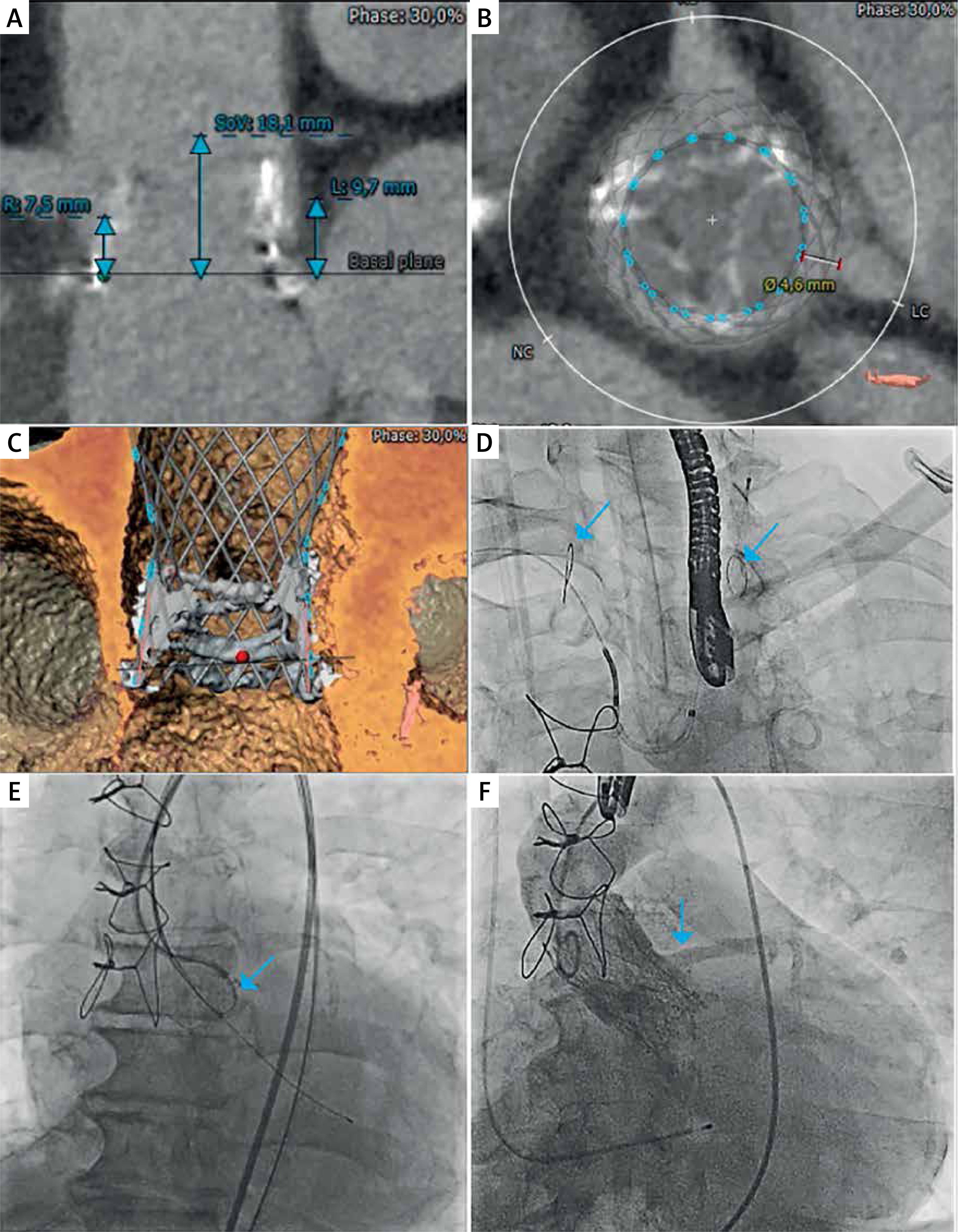
Figure 1
A – Aortic valve long axis showing the distance between the bioprosthesis and ostium of the right coronary artery (R), left coronary artery (L) and sinus of Valsalva (SoV). B – Aortic valve short axis with a virtual ring at the level of the coronaries revealing virtual transcatheter heart valve-coronary (VTC) distance to the left coronary artery of only 4.6 mm. C – Three-dimensional reconstruction showing the stent of the surgical bioprosthesis above the left main ostium. Red dot: hinge point of the bioprosthetic leaflet at left coronary cusp. Green dot: hinge point of the bioprosthetic leaflet at right coronary cusp. D – Deployment of the SENTINEL cerebral protection device – filters in the brachiocephalic trunk and left common carotid artery are indicated with blue arrows. E – Laceration of the left coronary cusp using the BASILICA technique – electrocautery wire is indicated with a blue arrow. F – Patent left coronary artery after valve-in-valve Evolut R 26 mm bioprosthesis implantation (blue arrow)
The SENTINEL was delivered via the right radial artery (6-Fr sheath) and the filters were deployed in the brachiocephalic trunk and the left common carotid artery (Figure 1 D). TAVR and BASILICA were performed via the right and left femoral accesses (14-Fr and 7-Fr sheath) under transesophageal echocardiography (TOE) guidance. For BASILICA, multipurpose and Amplatz left 2.0 catheters were inserted through the femoral arteries and placed either side of the aortic valve. A vascular snare and an electrified cutting wire were inserted into the left ventricular outflow tract to create an electric cutting knife below the left coronary leaflet (30 W). Next, the leaflet was lacerated (70 W), ensuring its splay around the left coronary ostium (Figure 1 E), and the Evolut R 26 mm bioprosthesis was implanted with no blood flow obstruction in the coronary arteries (Figure 1 F). Aortography and TOE showed no evidence of paravalvular leakage. The SENTINEL was removed at the end of the procedure. Small embolic debris (0.5–1 mm) were found in the filters. The femoral artery was closed using the ProGlide system. There were no conduction problems after the procedure.
At the 2-month follow-up, the patient’s condition improved to NYHA class I/II. TTE showed an improvement of left ventricle systolic function (EF 48%) and appropriate function of the bioprosthetic valve (Vmax 2.8 m/s, max/mean gradient 31/13 mm Hg, indexed aortic valve area 0.78 cm2/m2). Patient selection for CPD remains challenging and the experience with BASILICA for TAVR is limited. Our report supports the efficacy and safety of ViV TAVR in patients with low coronary ostia, if both cerebral protection and BASILICA technique are applied.


