
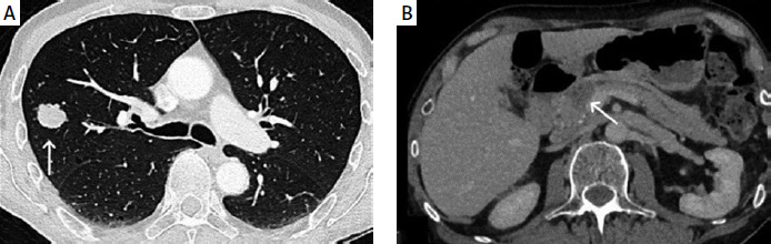
A 56-year-old male presented in the emergency department with symptoms of headache and ataxia. He had previous history of skin melanoma 7 years prior, treated with surgical excision. Given the patient’s symptoms, a head magnetic resonance imaging (MRI) was performed showing a few intraparenchymal brain lesions, consistent with metastasis (Figures 1 A, B). Contrast-enhanced computed tomography (CT) of the chest and abdomen revealed a solitary nodule at the periphery of the right lung (Figure 2 A), as well as a pancreatic lesion associated with mild pancreatic duct dilatation (Figure 2 B).
Figure 1
A, B – MRI axial images on T1-weighted image and FLAIR demonstrate a few scattered intraparenchymal lesions with surrounding oedema of metastatic origin. There is focal T1 hyperintensity in the intraparenchymal lesion, highly suggestive of a melanin component
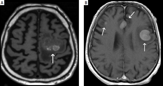
Figure 2
A – Axial CT of the chest demonstrates a slightly lobulated pulmonary nodule in the right upper lobe (arrow). B – Axial CT image of the abdomen shows a hypodense lesion at the head of the pancreas (arrow) with associated pancreatic ductal dilatation
The patient was discussed in our multidisciplinary team for the appropriate management because there was evidence of metastatic disease of unknown primary. The different biopsy options were discussed. The solitary pancreatic lesion thought to represent a metastasis and the radiological features of the brain lesions raised the possibility of metastasis from a previous melanoma. A lung biopsy was performed based on the location of the lesions, the safety risks of the procedure, and the urgency of therapy commencement. The lung nodule biopsy confirmed a melanoma origin of the lesion. The patient subsequently proceeded to an endoscopic ultrasound (EUS)-guided biopsy, which confirmed the initial diagnostic orientation and approach. Metastasis in the pancreas is an uncommon finding, only found in a small percentage (3–12%) of patients with widespread metastatic disease [1]. It accounts for only 2% of all pancreatic malignancies. The most common primary tumours metastasising to the pancreas are renal cell carcinoma, lung cancer, breast cancer, carcinoma of gastrointestinal origin, malignant melanoma, and prostate cancer [2–7]. Metastases from osteosarcoma, leiomyosarcoma, chondrosarcoma, and Merkel cell carcinoma are rare sites [8]. The most common radiological feature of a pancreatic metastasis is a well-circumscribed lesion (76%) with smooth borders, round or ovoid, with heterogeneous enhancement. It is usually not associated with significant ductal obstruction or vascular invasion [9]. Three patterns of pancreatic involvement can be identified: solitary lesion, multiple pancreatic lesions, and diffuse metastatic infiltration of the pancreas. The solitary pattern is the most common, accounting for 78.8% of cases in the Klein et al. study, followed by multifocal type, which accounted for 16.7% of the cases. The diffuse pattern is the most infrequent type, representing only 4.5% of cases [9, 10]. Pancreatic metastases are usually present as part of a late-stage cancer and are often found alongside metastases in other parts of the body. Although the well-circumscribed lesion and the absence of significant duct dilatation favours metastasis radiologically, these lesions mimic primary pancreatic neoplasms, and so radiological differential diagnosis is challenging and a definite diagnosis relies on histology. Treatment options are limited, but surgical resection offers the best chance for long-term survival; however, other treatments like immunotherapy, interferon therapy, and chemotherapy are used, often with a focus on palliative care.














