
Introduction
Metabolic dysfunction-associated steatotic liver disease (MASLD), the new term for non-alcoholic fatty liver disease (NAFLD), is characterised by steatotic liver disease along with cardiometabolic criteria, demonstrating a significant association with metabolic disorders [1]. Both terms can be used interchangeably; therefore, the term MASLD will be used throughout this paper. There is a two-way relationship between heart failure and liver diseases: liver complications can arise as a result of heart failure (e.g. congestive hepatopathy), and cardiac complications can develop in the course of liver diseases (e.g. cirrhotic cardiomyopathy) [2]. Patients with heart failure (HF) and MASLD have higher New York Heart Association (NYHA) classification, increased levels of natriuretic peptides, and greater risk of cardiovascular events [3]. A retrospective cohort study indicated that among patients with MASLD there was an approximately 20% higher risk of developing HF when compared with patients without MASLD. What is more, MASLD was associated with greater risk of heart failure with preserved ejection fraction (HFpEF) than heart failure with reduced ejection fraction (HFrEF) [4]. These results were also confirmed in meta-analysis of 11 observational cohort studies with aggregate data on 11,242,231 middle-aged individuals from different countries over a median of 10 years. MASLD was associated with a 1.5-fold increased risk of new-onset HF, independently of sex, age, diabetes, hypertension, and other cardiovascular risk factors [5].
Despite having a left ventricular ejection fraction (LVEF) over 50%, HFpEF is multisystem disease with diastolic and systolic dysfunctions, atrial and endothelial anomalies, vessel stiffness, pulmonary hypertension, and insufficient chronotropic reserve, which remains a prevalent chronic cardiac condition globally [2, 6]. Notably, HFpEF is linked to a significantly higher risk of adverse cardiovascular events and all-cause mortality. All the evidence suggests that chronic low-grade inflammation associated with metabolic syndrome and excess visceral adipose tissue correlates closely with the pathogenesis of HFpEF, as well as MASLD. Salah et al. described a bidirectional relationship between HF and liver disease, and proposed a “metabolic HFpEF/MASLD phenotype” that is related to inflammation, cardiac remodeling, elevated epicardial adipose tissue, pericardial restraint, and higher pulmonary artery [6, 7].
Methods
A PubMed literature search was conducted for English-language publications in peer-reviewed journals. We searched for metanalyses, retrospective and prospective cohort studies, reviews, or cross-sectional studies. The authors performed the literature search using the keywords: “HFpEF and MASLD”, “heart failure with preserved ejection fraction and chronic liver disease”, “HFpEF and MASLD”, “metabolic dysfunction associated steatotic liver disease and heart failure”, “nonalcoholic fatty liver disease and heart failure with preserved ejection fraction”, “HFpEF and liver”, and “chronic liver disease and heart failure”. Reference lists of included articles and reviews were also checked for additional studies. For the other sections of the article, a manual literature search was performed.
MASLD epidemiology
MASLD has become the most common cause of chronic liver disease (CLD). The prevalence of MASLD in developed countries is high, and a constant increase in the number of cases is forecast due to the increasing percentage of diseases predisposing to the occurrence of MASLD, mainly obesity and type 2 diabetes mellitus (T2DM). The incidence of MASLD varies and depends both on regional factors and the occurrence of diseases involved in the pathogenesis of MASLD – mainly components of the metabolic syndrome. According to data from 2018, a quarter of the world’s population may be affected by this disease [8]. Recent data from a meta-analysis that included 92 studies around the world showed an overall global MASLD prevalence of 30.05% (95% CI: 27.88–32.33%) [9]. Furthermore, additional analysis in the same paper revealed that the global prevalence of MASLD increased by 50.4%; particularly, it grew from 25.26% (with a confidence interval of 21.59–29.33) during 1990–2006 to 38.2% (with a confidence interval of 33.72–42.89) in the period 2016–2019 (p < 0.001). The rising trend in the prevalence of MASLD is consistent with the growing epidemic of type 2 diabetes and obesity. The highest prevalence for MASLD was reported in Latin America at 44.4% followed by the Middle East and North Africa region at 36.5%, South and Southeast Asia at 33%, North America 31.2%, East Asia at 29.7%, Asia Pacific 28.02%, and Western Europe at 25.1% [9]. Additionally, the prevalence and severity of MASLD tend to rise with age, peaking between 45 and 64 years old [10]. A higher percentage of patients with MASLD than in the general population is observed in patients with overweight or obesity. Because the underlying cause of MASLD is mainly insulin resistance resulting from excessive body weight, this disease is observed in 51% of overweight or obese patients [11]. The correlation between MASLD and overweight/obesity is undeniable, but it should be emphasised that MASLD may also occur in patients with normal body weight or with underweight as so-called lean MASLD. Chen et al. [12] showed that metabolic-associated fatty liver disease (MAFLD) in underweight, normal weight, overweight, and obese patients was present in 0.1%, 4%, 27.4%, and 59.8% of the subjects, respectively.
HFpEF epidemiology
HF, described as a global epidemic, affected more than 64 million people worldwide in 2017 [13]. According to the forecast in the United States, by 2030 over 8 million patients will suffer from HF and over 2 million of these will be over 80 years old [14]. The prevalence of HF is anticipated to increase due to improved survival rates following diagnosis, better and earlier treatment, and the overall longer life expectancy of the general population [13].
Data from the European Society of Cardiology Heart Failure Long-Term (ESC-HF-LT) Registry showed that 60% of patients were classified as HFrEF, 24% as HFmrEF, and 16% as HFpEF. Patients with HFpEF were older and 48% were female. Moreover, this group of patients was characterised by higher body mass index (BMI). Atrial fibrillation and systemic hypertension were most common in patients with HFpEF [15, 16]. In the Swedish HF registry, the estimates for HF were as follows: 56% for HFrEF, 21% for HFmrEF, and 23% for HFpEF [17]. Other epidemiological data indicate that the prevalence of HFpEF relative to HFrEF is increasing at a rate of 1% per year. This trend suggests that HFpEF is becoming the most common type of heart failure and correlates with significant hospitalisation and mortality, reaching up to 80% and 50%, respectively, within 5 years [16, 18].
Possible relationship between MASLD and HFpEF
Of the several subtypes of HF, Fudim et al. [3] evaluated that the association between MASLD and HF risk is stronger in patients with HFpEF (adjusted HR = 1.24; 95% Cl [1.14–1.34] p < 0.001), than in patients with HFrEF (adjusted HR = 1.09; 95% Cl [0.98–1.2] p = 0.12).
Systemic inflammation, metabolic syndrome, insulin resistance, and disrupted cardiac energy metabolism are shared pathophysiological features between HFpEF and MASLD. Both MASLD and HFpEF have similar prevalence, clinical manifestation, and risk factors including male sex, age, obesity, and type 2 diabetes mellitus (T2DM). MASLD is frequently diagnosed in patients with HFpEF [19]. Miller et al. [20] assessed the prevalence and correlation of MASLD among 181 patients with HFpEF. MASLD was diagnosed in 50% of patients with HFpEF with prior abdominal imaging, which is twice as high as among the general population (27%). These patients were more likely to have diabetes mellitus and obesity, which were associated with MASLD among a cohort of HFpEF patients.
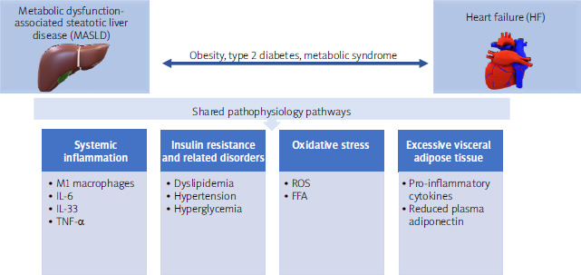
MASLD and HFpEF are systemic diseases with a significant interconnection. Emerging evidence highlights a link between them, primarily in the following aspects: common risk factors, systemic inflammation, insulin resistance, oxidative stress, and expended adipose tissue.
One of the most commonly documented pathomechanisms correlating with the development of HFpEF and MASLD is increased systemic inflammation. Similar pro-inflammatory cytokines are present in both MASLD and HFpEF [21]. For instance, M1 macrophages release cytokines like IL-6 and TNF-a, which lead to liver cell damage and the progression of MASLD. In turn, damaged hepatocytes release IL-33, which drives fibrogenesis through the IL-33 receptor (ST2) and galectin-3. Similarly, in the heart, IL-33 is released in response to myocardial fibre stretching. When it binds to its upregulated soluble ST2 receptor, this process, triggered by inflammation and fibrosis, promotes cardiomyocyte hypertrophy and heart fibrosis, mimicking the effect of galectin-3 [5, 21]. What is more, excessive and dysfunctional visceral adipose tissue releases increased levels of pro-inflammatory cytokines, along with reduced plasma adiponectin, which not only contribute to the development and progression of MASLD by affecting glucose and lipid metabolism or promoting insulin resistance but also negatively impact coronary arteries, increasing the risk of heart disease. Elevated serum IL-6 levels have been linked to subclinical atherosclerosis in population studies. Visceral fat produces significantly higher levels of IL-6, IL-1b, and TNF-a than the liver, and significant weight loss reduces the expression of these inflammatory cytokines [22, 23].
Another pathomechanism refers to oxidative stress, in which excessive free fatty acids (FFAs) lead to the production of reactive oxygen species (ROS) in the liver by inducing cytochrome 2E1 and endoplasmic reticulum stress, as well as impairing mitochondrial function. The created ROS promotes liver inflammation, apoptosis, and fibrosis, with ROS leaking into the bloodstream, triggering cardiac remodeling by damaging cardiomyocyte membranes and DNA. Additionally, ROS activates pathways such as NF-kB and ERK-1/2 in heart cells, driving cardiac remodeling and fibrosis. ROS also impairs calcium regulation in cardiomyocytes, worsening diastolic dysfunction and contributing to heart failure development [23, 24].
The pathophysiological overlap between HFpEF and MASLD is significant, particularly through the mechanisms of systemic inflammation, insulin resistance, and oxidative stress. These shared pathways not only contribute to the individual development of HFpEF and MASLD but also facilitate their co-occurrence (Figure 1) [25]. A multidisciplinary approach focusing on lifestyle changes, metabolic control, and anti-inflammatory treatments may be key in managing these intertwined disorders.
Clinical implications of MASLD and HFpEF diagnosis
The diagnosis of both HFpEF and MASLD significantly worsens a patient’s prognosis, so efforts should be made to identify groups of patients in whom testing for the aforementioned conditions is indicated. Considering that the aetiological factors of HFpEF and MASLD are similar, it is reasonable to look for MASLD in patients with HFpEF, and vice versa. Table I shows the prevalence of HFpEF and MASLD in patients with selected diseases [26–30].
Table I
| Conditions | HFpEF | MASLD |
|---|---|---|
| Overweight/obesity | 29–39% [26] | 50.7% [27] |
| Hypertension | 58–79% [26] | 50% [28] |
| Diabetes mellitus | 35–49% [26] | 55.5% [29] |
| Age | > 65 years – 59–70% [26] | > 60 years – 43.8% [30] |
Table II
Management of patients with heart failure with preserved ejection fraction and metabolic dysfunction-associated steatotic liver disease [35, 37]
The symptomatology of MASLD is non-specific, which may delay diagnosis. The guidelines of scientific societies recommend searching for MASLD among patients with fatty liver with a specific clinical profile, and not among people presenting specific symptoms [31, 32]. According to the definition of MASLD, patients require the presence of hepatic steatosis (histology or imaging or blood markers/scores) accompanied by one of the following clinical features: overweight or obesity or abdominal obesity, T2DM, prediabetes, hypertension, hypertriglyceridaemia, or low HDL-C level [33].
Unlike MASLD, which can be asymptomatic, clinical symptoms are part of the definition of HFpEF. Patients with HFpEF tend to be older and predominantly female compared to those with HFrEF and heart failure with mildly reduced ejection fraction (HFmrEF). Additionally, conditions like atrial fibrillation, chronic kidney disease, and non-cardiovascular comorbidities are more frequently observed in HFpEF patients than in those with HFrEF. According to the European Society of Cardiology (ESC) 2021 guidelines [34], an HFpEF diagnosis should include: a) signs and symptoms of HF, b) LVEF ≥ 50%, c) objective evidence of cardiac structural and/or functional abnormalities consistent with the presence of left ventricular (LV) diastolic dysfunction/raised LV filling pressures, including raised natriuretic peptides.
General principles of management in patients with HFpEF and MASLD
In patients with HFpEF and in those with MASLD, bidirectional management is necessary. First, drugs registered for these disease states should be used, but co-morbidities, such as obesity and type 2 diabetes, which are related to the aetiology of both disease entities, should be treated in parallel. When choosing substances for the treatment of type 2 diabetes and obesity, it is advisable to take into account their anti-steatosis effects.
In 2023, an update to the ESC 2021 guidelines on the management of patients with HFpEF was published [35]. The task force for the diagnosis and treatment of acute and chronic heart failure of the ESC proposed the management of patients with HFpEF based on three main pillars with class I recommendation: diuretics for fluid retention, dapagliflozin/empagliflozin, treatment for aetiology, cardiovascular (CV) and non-CV comorbidities.
Moreover, SGLT-2i (dapagliflozin or empagliflozin) is recommended in patients with HFpEF to reduce the risk of HF hospitalisation or CV death with level of evidence A [35]. Borlaug et al. [36] proposed a similar treatment plan for patients with HFpEF. After diagnosing HFpEF and ruling out other potential conditions, a comprehensive treatment should be initiated. Treatment begins with the inclusion of SGLT2i, if tolerated, followed by assessment of the congestion; if present, a diuretic should be included. The last stage concerns the evaluation of comorbidities and the use of targeted therapy. This plan should address both pharmacologic and nonpharmacologic therapies.
There is no specific drug for the treatment of MASLD. Instead, some non-specific, non-pharmacological, and pharmacological treatments are currently applied. Some of the treatments have been documented to play a main role in controlling common risk factors, reducing the impact of inflammatory and fibrotic mediators, or decreasing fat accumulation. Non-pharmacological treatment is the pivotal treatment of MASLD, which mainly includes dietary intervention and weight loss induced by exercise, caloric restriction, and/or bariatric surgery. These non-pharmacological approaches are effective in the treatment of MASLD, and they showed promising results in delaying the development of HFpEF. According to the current guidelines, in patients with MASLD, lifestyle modification including weight loss, dietary changes, physical exercise, and discouraging alcohol consumption, as well as optimal management of comorbidities and use of incretin-based therapies (e.g. semaglutide, tirzepatide) for T2DM or obesity, if indicated, are advised [37].
Among the drugs used for the treatment of HFpEF, SGLT2 inhibitors (SGLT-2i) may have potential anti-steatosis mechanisms relevant in the treatment of MASLD [38–40]. SGLT-2i treatment lowers glucose and insulin levels, particularly in diabetic patients, which significantly reduces liver fat production. These drugs also stimulate glucagon release from a-cells in the pancreas, increasing fat oxidation and shifting the body’s metabolism from carbohydrates to fatty acids, leading to a reduction in hepatic steatosis and liver triglycerides. Additionally, SGLT-2i has antioxidant properties, lowering oxidative stress, reducing free radicals, and enhancing antioxidant defences, such as glutathione peroxidases and superoxide dismutases (SODs) [40]. Androutsakos et al. [40], in their review of 25 clinical studies, demonstrated the benefits of SGLT-2i in patients with MASLD and T2DM. Most of them report improvements in liver enzyme levels and reductions in hepatic steatosis, assessed by methods such as ultrasound, magnetic resonance imaging (MRI), and noninvasive biomarkers like MASLD fibrosis score, FIB-4 score, and AST-to-platelet ratio (APRI) index. In some cases, improvements in liver fibrosis were observed through transient elastography or liver biopsy, but these findings were not consistently reported across all studies.
Another Korean cohort study compared oral antidiabetic drugs (OADs) such as SGLT-2i, thiazolidinediones, dipeptidyl peptidase-4 (DPP-4) inhibitors, or sulfonylureas to investigate which of them is associated with the best outcomes in patients with MASLD and T2D. The study involved 80,178 patients (mean [SD] age, 58.5 [11.9] years; 43,007 [53.6%] male). The rate of MASLD regression was significantly higher in patients treated with SGLT-2 inhibitors (ASHR = 1.99 [95% CI: 1.75–2.27]), thiazolidinediones (ASHR = 1.70 [95% CI: 1.41–2.05]), and DPP-4 inhibitors (ASHR = 1.45 [95% CI: 1.31–1.59]) compared to those using sulfonylureas. Among these, SGLT-2i showed the most favourable outcomes, with higher regression rates when directly compared to thiazolidinediones (ASHR = 1.40 [95% CI: 1.12–1.75]) and DPP-4 inhibitors (ASHR = 1.45 [95% CI: 1.30–1.62]), making them the most effective OAD class for MASLD regression. Compared with sulfonylureas, only SGLT-2i (ASHR = 0.37 [95% CI: 0.17–0.82]) was significantly associated with a lower incidence of adverse liver-related outcomes [41].
Despite the beneficial impact of SGLT-2i on MASLD in patients with T2DM, there are few data regarding the effect of SGLT-2i on MASLD in patients without T2DM. Taheri et al., in a prospective, randomised, double-blind, placebo-controlled trial, analysed the effect of empagliflozin on liver steatosis and fibrosis in patients with MASLD in the absence of T2DM. Participants were randomized for 24 weeks to empagliflozin 10 mg (n = 43) or placebo (n = 47). As a result, empagliflozin reduced ALT and AST levels, and improved hepatic steatosis and measures of liver fibrosis. Both groups had significantly reduced controlled attenuation parameter (CAP) scores; however, there was no significant difference between the empagliflozin and placebo groups (p = 0.396) [42]. In another prospective, single-centre, double-blind study, dapagliflozin 5 mg or teneligliptin 20 mg was administered for 12 weeks in patients with MASLD without T2DM. The study results showed decreased serum ALT levels in both groups [43].
In patients with MASLD and HFpEF, non-pharmacological and pharmacological treatment targeting comorbidities and HFpEF should be applied. Behavioural therapy includes the following: self-monitoring, enhancing patients’ self-efficacy and motivation, setting realistic negotiable goals, and overcoming barriers. Examples of unprocessed/minimally processed foods include vegetables, fruits (not juice), low-fat dairy, nuts, olive oil, legumes, unprocessed fish, and poultry. In adults with MASLD, improving diet quality (similar to the Mediterranean dietary pattern), limiting the consumption of ultra-processed food (rich in sugars and saturated fat), and avoiding sugar-sweetened beverages should be recommended to improve histologically or non-invasively assessed liver injury. In a meta-analysis of observational studies and clinical trials, the Mediterranean diet has repeatedly shown hepatic and cardiovascular health benefits, even without weight loss [44]. In adults with MASLD, physical activity and exercise should be recommended to reduce steatosis, tailored to the individual’s preference and ability (preferably > 150 min/week of moderate or 75 min/week of vigorous-intensity physical activity). Nutraceuticals cannot be recommended for adults with MASLD due to insufficient evidence of their effectiveness in reducing liver damage, fibrosis, or liver-related outcomes, whether assessed histologically or non-invasively, nor of their safety [37] (Table II).
Conclusions
MASLD and HFpEF are closely linked through shared mechanisms like inflammation, insulin resistance, and oxidative stress. This connection worsens patient outcomes and requires early diagnosis and a multidisciplinary treatment approach, including lifestyle changes, metabolic control, and targeted therapies like SGLT-2i. Understanding this relationship is crucial for improving patient care and managing these increasingly common conditions.














