
Purpose
Brachytherapy (BRT, interventional radiotherapy) is a vital component of modern oncological practice, and remains an indispensable resource in the treatment of tumors. This precise and localized radiation therapy technique allows for the delivery of high doses of radiation to the target area while sparing surrounding healthy tissue, offering significant advantages in terms of therapeutic outcomes and patient quality of life. However, the effective implementation of BRT is not without challenges.
One of the major obstacles to utilize the full potential of BRT is the need for clinicians to undergo prolonged and specialized training [1, 2]. Mastery of BRT intricate procedures demands not only a comprehensive understanding of radiation physics, but also a high degree of manual dexterity and procedural skills. As a result, many healthcare institutions face a challenging task of ensuring that their medical professionals acquire and maintain the required expertise. The demand for highly trained personnel often exceeds the available resources, and can hinder timely and widespread application of BRT.
In response to these challenges, several strategies have been proposed to enhance training and expertise in BRT. One approach involves intensifying educational programs and ongoing professional development to ensure that clinicians are well-prepared and up-to-date with the latest techniques and technologies [2]. Another avenue explores the use of medical simulators, which provide a safe and controlled environment for trainees to practice and refine their skills. These simulators offer a valuable bridge between didactic learning and real-world clinical experience, allowing clinicians to develop their proficiency in a risk-free setting [3-8].
Augmented reality (AR) has emerged as a transformative force within the field of medicine, reshaping the way healthcare professionals approach patient care and medical procedures. AR technology is leveraged across various domains within medicine, providing valuable support in critical aspects of healthcare delivery, education, and decision-making.
In the realm of surgical interventions, AR role is particularly pronounced [9]. Surgeons utilize AR to enhance precision and accuracy during procedures. By overlaying digital information onto the patient anatomy in real-time, surgeons can visualize critical structures, navigate complex anatomical regions, and precisely plan incisions and implant placements. AR assists in minimizing surgical invasiveness and reduces the risk of complications.
Additionally, AR contributes significantly to medical education and training [10]. It offers students and novice healthcare practitioners immersive learning experiences that bridge the gap between theoretical knowledge and practical skills. Through AR simulations, medical students can practice surgical techniques, diagnostic procedures, and even complex surgeries in a controlled and risk-free environment. This not only accelerates the learning curve, but also fosters a deep understanding of anatomical structures and pathologies.
Apart from the operating room and the classroom, AR facilitates remote consultations and telemedicine [11]. Physicians can remotely examine patients by accessing their medical data, and visualize diagnostic image or patient information in real-time. This is especially valuable in situations where physical presence is challenging, such as in rural or underserved areas, or during global health crises.
Furthermore, AR improves in-patient monitoring and rehabilitation [12]. Wearable AR devices can provide patients with real-time feedback and guidance during exercises, rehabilitation routines, or chronic diseases managements. It empowers patients to take an active role in their healthcare and enhances their overall quality of life. In general, the role of AR in medicine is multi-faceted, involving surgical assistance, medical education, remote healthcare delivery, and patient empowerment. Its capacity to improve visualization, decision-making, and training makes AR an valuable tool in advancing medical practices and ultimately enhancing patient outcomes. In the context of BRT, AR has shown promising benefits in enhancing the precision and safety of treatment delivery, simplifying complex procedures, and potentially mitigating challenges associated with training. Despite the potential of AR in BRT, the available literature has provided only glimpses of its application and impact. Existing experiences and studies are scattered, and a comprehensive synthesis of evidence is lacking. Therefore, the primary objective of this literature review was to collect, analyze, and synthesize the existing evidence on the use of AR in BRT. By adopting a question-answer framework, we aimed to explore the current state of knowledge, identify gaps and challenges, and encourage further research and innovation in this field. Ultimately, our goal was to provide valuable insights into the development and integration of AR solutions in BRT, thereby enhancing the quality of cancer care and improving patients outcomes.
Material and methods
Literature review design
This narrative review of the literature was conducted by a multi-disciplinary team, including radiation oncologists, medical physicists, and radiotherapy technicians, and was written based on scale for the assessment of narrative review articles (SANRA) [13]. The aim was to comprehensively explore the existing evidence on the use of AR in the context of BRT. The methodology followed a question-answer format, and to ensure comprehensive coverage of the topic, a narrative review checklist was applied (Supplementary Table 1).
Literature search
Literature search was performed using PubMed on September 29, 2023. The following search strategy was employed: (“Augmented Reality” OR “Augmentation Reality” OR “Mixed Reality” OR “Virtual Reality” OR “AR”) AND (“Brachytherapy”). The search was designed to acquire relevant articles on AR or related technologies in BRT, and included only full publications in English.
Study selection
Two authors (MF, BF) independently conducted the study selection process. Initial screening involved assessing the relevance of articles based on their titles and abstracts. Articles that appeared to be related to the use of AR in BRT were selected for full-text review.
Data extraction
Data extraction was performed independently by two authors (MF, LF). Information was systematically collected from each selected article, including details on study design, AR system or platform used, hardware and software components, integration into clinical workflow, outcomes, and conclusions. The extracted data were organized according to the question-answer framework.
Data analysis
Due to the limited and heterogeneous nature of the evidence available in terms of methods and evaluations, no statistical analysis was conducted. Instead, findings from the selected articles were qualitatively synthesized and presented in a narrative format to provide a comprehensive overview of the topic. The narrative review approach allowed for a synthesis of key insights, trends, challenges, and opportunities in the field of AR in BRT.
Results
Search results
Our search strategy identified a total of 84 publications. However, after a thorough analysis, only four of these papers met our criteria, and were considered relevant for inclusion in the literature review on AR in BRT. The summary of contents of this literature review is presented in Table 1.
Table 1
Summary of questions and answers on augmented reality (AR) in brachytherapy (BRT)
Questions and answers
1. What is AR?
Augmented reality is a cutting-edge technological advancement that smoothly integrates the virtual and physical realms, enriching the user’s perception of reality by overlaying digital information, such as images, videos, or three-dimensional models onto real-world environment in real-time. Unlike virtual reality (VR) that immerses users entirely into computer-generated environments, AR enhances the existing environment by superimposing contextually relevant data onto the user’s field of view [14]. This dynamic interplay between the physical and digital worlds opens up a wide range of possibilities in various domains, including medicine.
Augmented reality systems typically rely on devices, such as headsets, smartphones, or tablets equipped with cameras and sensors to capture the surroundings [15]. Advanced algorithms process these information, identify objects, surfaces, and spatial relationships, and then precisely position and display digital content within the user’s view [16]. The result is an interactive and immersive experience that can be harnessed for educational, training, entertainment, and professional purposes.
2. Why might AR be useful in BRT training?
Augmented reality has emerged as a groundbreaking tool in the realm of medical education, offering unique advantages that are particularly well-suited to the specialized field of BRT. Brachytherapy demands a high level of precision, spatial awareness, and hands-on skill development [17], making it an ideal candidate for AR-enhanced training methods.
Brachytherapy training traditionally involves a steep learning curve [18], with trainees having to master intricate techniques for precise placement of radioactive sources within the patient’s body. The introduction of AR into this training process could improve educational experience. In fact, AR may be useful in BRT training as follows:
Dynamic and interactive learning: AR can provide trainees with a dynamic and interactive learning environment. Rather than relying solely on static textbooks or lectures, learners can engage with 3D models, interactive simulations, and real-time feedback. This hands-on approach may allow trainees to explore the intricacies of BRT in an immersive and engaging manner.
Realistic simulation: AR can simulate realistic BRT scenarios. Trainees can practice procedures, such as needle placement and applicator positioning in a virtual environment that closely mimics the challenges of real clinical settings [19]. This practical experience enhances muscle memory and procedural skills.
Visualization of radiation dose: One of the critical aspects of BRT is understanding the radiation dose distribution within the patient’s body. AR allows trainees to visualize this distribution in real-time [20]. They can see how radiation sources interact with surrounding tissues and organs, helping them grasp the complexities of dose planning and optimization.
Confidence building: AR-based training can build trainees’ confidence. The ability to practice procedures repeatedly in a risk-free virtual space allows learners to refine their skills and reduce anxiety when performing BRT in clinical settings [21]. This enhanced confidence contributes to safer and more effective patient care.
Personalized learning: AR systems can adapt to individual learning needs. Trainees can progress at their own pace, with an option to return to specific modules or simulations as needed. This personalized approach ensures that each learner achieves a high level of competency before transitioning to clinical practice.
In conclusion, AR could offer a revolutionary approach to BRT training by providing dynamic, realistic, and interactive learning experiences. Trainees benefit from enhanced skills development, improved spatial awareness, and increased confidence, ultimately translating to safer and more effective BRT procedures in clinical practice.
3. What evidence is available on the use of AR in radiotherapy?
Radiotherapy is a crucial component of cancer treatment, aiming to deliver high doses of radiation precisely to cancerous tissues, while minimizing exposure to healthy surrounding tissues. The success of radiotherapy depends on accurate patient positioning and treatment planning. AR technology has emerged as a promising tool in the field of radiotherapy, offering potential benefits in the patient’s setup, treatment planning, and real-time monitoring. The following studies reported on possible applications of AR in the field of external-beam radiotherapy.
Zhang et al. focused on developing an AR-assisted radiotherapy positioning system using HoloLens. This system allows for enhanced accuracy and feasibility in clinical environment. The study reconstructed 3D models of phantoms and anthropomorphic phantoms, and used AR tracking to align virtual and real objects. The results indicated that the AR-assisted positioning system was feasible and positioning errors comparable with traditional laser-based methods. While setup time was longer, the system provided advantages, such as intuitive visual guidance and radiation-free position verification [22].
The same authors further reported on the integration of AR and optical surface imaging for precise patient positioning. In fact, traditional methods have limitations, including skin markers, additional doses, and lack of information integration. In their study, the authors proposed a non-invasive radiotherapy positioning system that combined AR and structured light-based surface imaging. The system used a two-step approach: AR-based coarse guidance and optical surface-based precise verification. The study findings demonstrated promising results, with maximum errors of 3.4 mm in coarse guidance and 1.6 mm in precise verification. Pearson’s correlation coefficients between the precise verification and cone-beam CT (CBCT) results were high, indicating the potential of AR in improving patient positioning accuracy [23].
Cardan et al. explored the accuracy of an AR holographic guidance system for patient alignment in radiotherapy applications. This system allowed users to see a holographic representation of scanned patient anatomy overlaid on the treatment vault. Therapists used HoloLens glasses to align a phantom with the hologram using couch controls. The study found that the holographic guidance system provided adequate accuracy for initial treatment alignment. However, it lacked a fine alignment accuracy of X-ray imaging systems [24].
Batista et al. observed that surface-guided radiation therapy (SGRT) is becoming a routine tool for patient positioning in many clinics. In their vision paper, the authors discussed the challenges in transitioning to SGRT, and explored its current and future role along with other imaging techniques. The paper highlighted the potential benefits of SGRT in improving patient setup accuracy and safety [25].
In conclusion, the integration of AR in radiotherapy has shown promising results in improving patient positioning accuracy, enhancing visualization, and reducing setup errors. Various studies have explored the feasibility and accuracy of AR-assisted systems, highlighting their potential benefits for both patients and healthcare providers. While challenges and opportunities remain, the evidence suggests that AR has a valuable role to play in the advancement of radiotherapy practices. Future research and developments in this field are likely to further refine and expand the applications of AR in radiotherapy, ultimately leading to improved treatment outcomes for cancer patients.
4. What evidence is available on the use of AR in BRT?
Brachytherapy is a medical procedure involving a precise placement of radioactive sources within or near the target tissue to treat various types of cancer. The use of AR technology could improve the accuracy, efficiency, and safety of BRT procedures. In this chapter, we reported the evidence available on the use of AR in BRT based on relevant studies.
The use of AR for intra-operative guidance in image-guided 3D interstitial BRT was explored in a study published by Krempien et al. Key findings were as follows: 1. The system utilized a video projector, cameras, and patient tracking for AR guidance; 2. Real-time visualization of planning data on the patient skin was achieved; 3. Dynamic adjustment of data to the patient position eliminated the need for rigid fixation; 4. Average deviation due to soft-part displacement was 1.1 mm. In conclusion, the low-cost AR system proved accurate and feasible for intra-operative guidance in BRT. This study, although conducted in 2008, laid the foundation for AR in BRT. It demonstrated that AR could provide real-time guidance, improving the precision of needle implantation, while allowing for dynamic adjustments during the procedure [26].
A study published by Stone et al. explored the application of AR in remote surgical education. The objective of the study was to investigate the use of an AR headset to remotely train clinicians on medical devices using anatomical models. The researchers developed disease-specific phantoms for training in various procedures, including multi-parametric magnetic resonance imaging-guided fusion of prostate biopsy and BRT. Key findings of the study were: 1. Disease-specific phantoms were developed for training in BRT; 2. Remote training was conducted using AR headsets; 3. Participants found the training realistic and valuable, with 70.9% requesting more training; 4. The remote training platform was successfully tested for trans-perineal prostate biopsy and rectal spacer insertion. The authors concluded that remote training using AR eliminates the need for traveling, reduces costs, and increases supervisor availability. Overall, the study demonstrates that AR can play a significant role in medical training, including training in BRT procedures. It provides a feasible solution for remote training, which is particularly relevant in the context of reducing traveling and exposure risks, ultimately benefiting medical education and patient care [27].
Another study published by Liu et al. investigated the use of intra-operative 3D holograms with mixed reality techniques based on CT-MRI fusion images for brain BRT. Key findings of the study were: 1. 3D holograms generated from CT-MRI fusion images were used to guide brain BRT; 2. Interventional surgeons could share and interact with the hologram in real-time; 3. The hologram provided visualization of the skull, tumor location, and nearby vessels; 4. The procedure resulted in improved clinical outcomes at 3- and 6-months post-implantation. In conclusion, intra-operative holograms with MR techniques offer potential benefits for guiding BRT procedures in the brain. This research highlights the use of AR in a highly sensitive and critical area of BRT, specifically for brain metastasis treatment. The ability to visualize and interact with 3D holograms during surgery can aid in precision and improve patient outcomes [28].
A study by Zhou et al. introduced a personalized mixed reality surgical assistance system for BRT. Key findings were: 1. The system incorporated virtual organ fusion and real-time surgical tool tracking; 2. A novel multi-information fusion method was applied for patient-specific planning; 3. The system’s accuracy was validated through phantom and animal experiments; 4. The needle location error in phantom experiments was 0.957 mm. In conclusion, the mixed reality surgical system demonstrates accuracy and efficiency, reducing the need for multiple CT scans, enabling real-time surgery. This study shows the potential of mixed reality in BRT by offering precise, patient-specific planning and real-time tool tracking. The results suggest that such systems could enhance the accuracy and efficiency of BRT procedures [29].
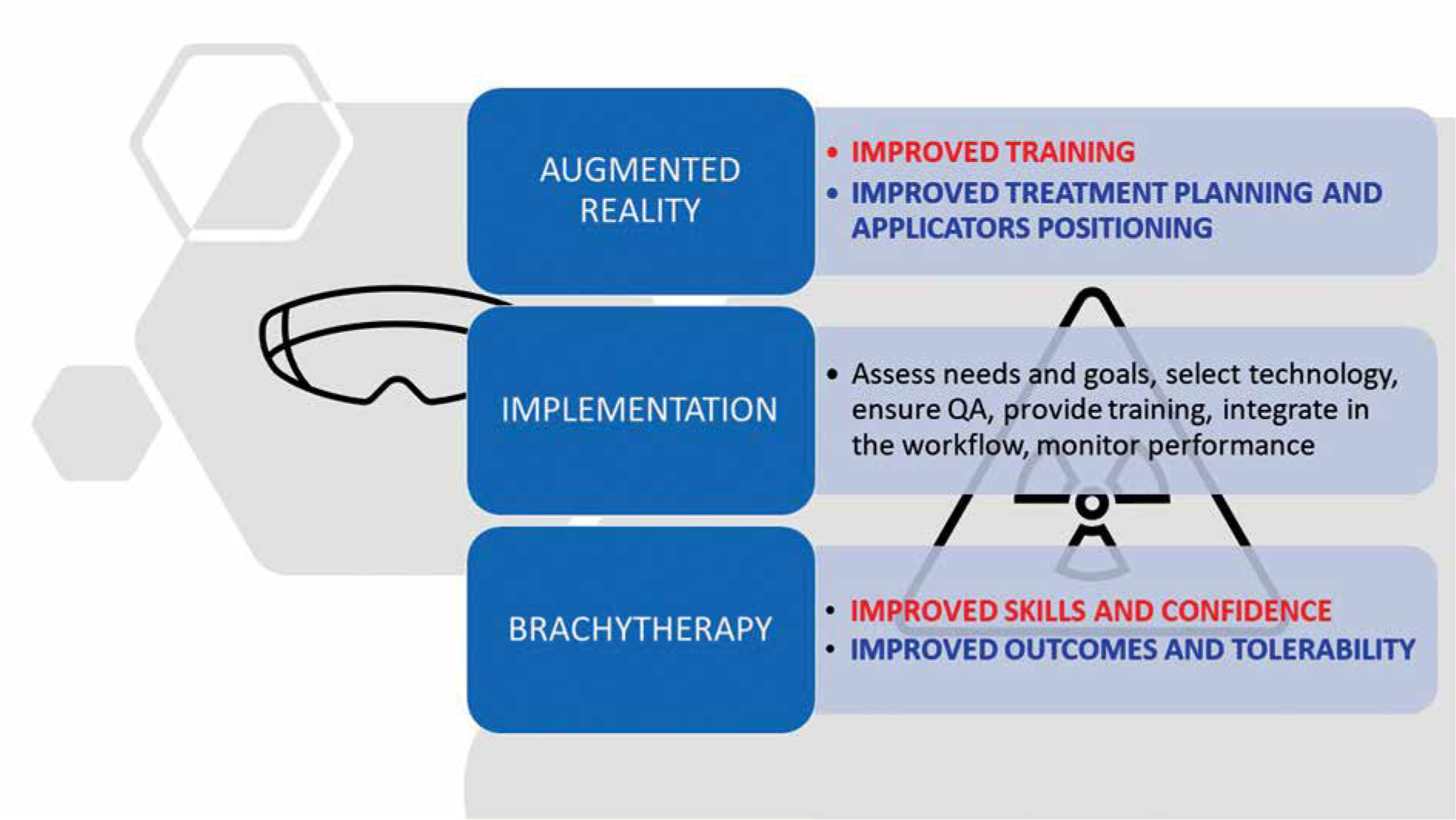
In summary, the above-mentioned studies collectively provide evidence on the potential benefits of AR in BRT. AR technology has the capacity to enhance training, improve precision, reduce the need for multiple imaging scans, and ultimately enhance patient outcomes in BRT procedures. Further research and clinical studies are likely to continue exploring the application of AR in this field, advancing the state of BRT practice (Figure 1).
5. What systems are potentially available to implement AR in a BRT department?
In recent years, the integration of AR technology into the field of medicine has gained momentum, offering the potential for enhanced precision and improved patient outcomes. Various AR systems and platforms have been developed, each with unique hardware and software components tailored to the specific needs of clinical departments [15]. In this chapter, we summarized information on AR systems available for future implementation in a BRT department, describing their features and integration into the clinical workflow.
Hardware components of AR systems
Head-mounted displays (HMDs): One of the core hardware components of AR systems is HMD. HMDs, such as Microsoft HoloLens or Magic Leap, provide clinicians with a wearable, see-through display that overlays virtual information onto the real-world environment. These devices enable surgeons to visualize anatomical structures and treatment plans during procedures [30].
Tracking devices: To achieve precise registration of virtual objects with the patient anatomy, tracking devices are crucial. These may include electro-magnetic trackers, optical trackers, or marker-based tracking systems. These devices continuously monitor the position and orientation of both the patient and surgical instruments [31].
Projectors: In some AR systems, projectors are used to project virtual information directly onto the patient body or onto surgical drapes [32]. This approach eliminates the need for HMDs, providing a hands-free visualization of critical data.
Stereoscopic cameras: High-resolution stereoscopic cameras capture real-time images of the patient and surgical field [33]. These cameras enable the system to track and superimpose virtual objects accurately.
Software components of AR systems
Image registration software: AR systems rely on sophisticated image registration algorithms to align pre-operative imaging data (CT or MRI scans) with the patient current anatomy [34]. These algorithms ensure that virtual objects match the real-world patient geometry.
Visualization software: Specialized software is used to render and display the augmented content [35]. This software may include 3D reconstruction tools, volume rendering, and segmentation algorithms to create a clear and informative AR overlay.
Navigation and guidance software: AR systems often feature navigation and guidance functionalities [36]. These tools help surgeons visualize critical structures, target locations, and trajectories for needle placement or seed implantation.
Interactive interfaces: User-friendly interfaces allow clinicians to interact with AR system, adjusting the display, moving between different views, or making real-time annotations [37].
Integration into clinical workflow
Integration of AR systems into the clinical workflow of a BRT department involves several key steps:
Patient data acquisition: Pre-operative imaging data (CT, MRI, etc.) are acquired and loaded into AR system. This data serves as the foundation for the AR overlay.
Patient registration: AR system uses tracking devices to register the patient current position and orientation with respect to the pre-operative imaging data. This step ensures accurate alignment.
Procedure planning: Clinicians use AR system to plan BRT procedure, visualizing the tumor target, critical structures, and treatment paths. This interactive planning phase enhances precision.
Intra-operative guidance: During the actual procedure, AR system provides real-time guidance, helping clinicians to accurately position needles or implants. This dynamic feedback improves the precision of intervention.
Outcome assessment: After the procedure, AR system can assist in assessing the placement of seeds or other treatment elements. This assessment can inform further treatment decisions and follow-up.
Data storage and documentation: AR systems often integrate with electronic health records (EHR) systems, allowing for continuous documentation and archiving of procedure data.
The integration of AR into BRT departments may significantly impact the field by potentially improving precision, decreasing the frequency of repeat imaging, and contributing to better patient outcomes. With ongoing technological progress, it is anticipated that both hardware and software aspects of AR will see enhancements, possibly making AR a more useful resource in BRT.
6. How could AR be introduced in a BRT department?
Introducing AR into a BRT department requires careful planning, quality assurance, and staff training. Table 2 summarize the practical aspects of integrating AR technology into a BRT department, outlining the steps involved, addressing quality assurance considerations, and discussing staff training. Additionally, Table 3 and Table 4 highlight the potential challenges and best practices to ensure a successful implementation, respectively.
Table 2
Steps for introducing augmented reality (AR) in brachytherapy (BRT) departments
Table 3
Challenges in introducing augmented reality (AR) in brachytherapy departments
Table 4
Best practices in introducing augmented reality (AR) in brachytherapy departments
Incorporating AR into a BRT department could offer benefits in terms of precision, patient outcomes, and overall efficiency. By considering these steps, acknowledging possible challenges and adhering to established best practices, healthcare facilities might be able to integrate AR technology into their clinical workflows, potentially improving care quality for patients undergoing BRT procedures (Figure 1).
7. What kind of clinical research could be done on the use of AR in BRT?
Exploring the use of AR in BRT opens numerous research opportunities to advance patient care and treatment outcomes. In Table 5, the potential avenues for clinical research in the field of AR-assisted BRT are summarized. As the integration of AR in BRT continues to evolve, these research opportunities will contribute to a deeper understanding of its clinical benefits, cost-effectiveness, and impact on patient care. Prospective studies and collaborative efforts will be essential in advancing the field and optimizing AR applications for the benefit of both patients and healthcare providers.
Table 5
Potential areas of research on the use of augmented reality (AR) in brachytherapy (BRT)
Discussion
Narrative
In this discussion section, the findings from the reviewed literature on the use of AR in BRT were summarized. We aimed to address three key aspects: the fundamental or key findings, limitations and quality of the research reviewed, and the need for future research.
Potential applications of AR in BRT
Augmented reality has the potential to significantly impact the clinical practice of BRT, presenting several benefits, which could influence the field in future. Given BRT requirement for high precision in radiation therapy, AR may offer enhancements in both precision and efficiency for this treatment approach. Key considerations suggesting AR utility in BRT clinical practice include:
Enhanced treatment planning: The success of BRT depends on detailed treatment planning. AR could assist in this aspect by offering healthcare professionals advanced visualization tools. These tools might help in the precise identification of treatment area, potentially leading to more accurate placement of radioactive sources. Such precision may possibly reduce the risks associated with underdosing or overdosing, aiming to ensure that radiation delivery is optimally concentrated on the tumor.
Real-time visualization of radiation sources: One of the unique advantages of AR is its ability to provide real-time visualization of radiation sources within the patient body. During BRT procedures, this feature is valuable, as it allows clinicians to monitor the exact positioning and distribution of radioactive sources throughout the treatment session. Any adjustments or corrections can be made promptly to optimize treatment outcomes.
Accurate applicator and needle positioning: In BRT, the precise positioning of applicators or needles is critical. AR technology can assist clinicians by superimposing virtual guides or markers onto the patient anatomy, ensuring that these devices are correctly placed. This reduces the margin of error and enhances the overall accuracy of the procedure.
Potential for improved treatment outcomes: The introduction of AR into treatment processes has the potential to refine precision and accuracy that may contribute to better treatment outcomes. AR capability to facilitate the delivery of more focused radiation doses directly to the tumor while minimizing exposure to adjacent healthy tissues, could potentially lead to more effective tumor control and a decreased number of adverse effects in patients.
Error reduction: BRT procedures are complex, and errors can have serious consequences. AR systems offer real-time guidance and feedback, reducing the likelihood of procedural errors. This is especially crucial in complex cases, or when treating tumors in challenging anatomical locations.
Streamlined procedures: AR can streamline BRT procedure, making it more efficient. Clinicians can work more confidently and quickly, ultimately reducing treatment time and improving patients’ overall experience.
In summary, AR holds the potential to significantly impact the clinical practice of BRT by potentially enhancing precision, enabling real-time visualization, and improving accuracy. These potential benefits may lead to better treatment outcomes, fewer errors, and more efficient procedures, aspects that are crucial given the targeted and complex nature of BRT.
Fundamental findings
The literature review showed several noteworthy findings regarding the integration of AR into BRT. Notably, studies have demonstrated the feasibility and potential benefits of AR in various aspects of BRT, such as education and training, intra-operative guidance, and treatment planning. For instance, Krempien et al. explored the utilization of a low-cost augmented reality system for intra-operative guidance in image-guided 3D interstitial BRT, achieving real-time visualization of planning data on the patient skin and dynamic adjustments to patient positioning [26]. Stone et al. reported that remote training using AR eliminated the need for traveling and was well-received by participants [27]. Additionally, Liu et al. explored the use of 3D holograms for intra-operative guidance, improving visualization and outcomes [28]. Zhou et al. presented a personalized MR surgical assistance system that achieved real-time tracking of surgical tools, and was validated with promising results [29].
Limitations and quality of research
Despite these promising findings, it is important to acknowledge the limitations and varying quality of the research reviewed. Firstly, the evidence on AR in BRT remains relatively scarce and heterogeneous, making it challenging to draw definitive conclusions. Secondly, most studies are in the experimental or early implementation phases, with limited long-term follow-up data. Additionally, there is a need for standardized assessment metrics and methodologies in evaluating the effectiveness and safety of AR in BRT. The research landscape would benefit from more robust clinical trials and comparative studies to establish the superiority of AR-assisted procedures over conventional methods.
Need for future research
The reviewed literature underscores the extreme need for future research in the field of AR in BRT. There are several avenues for exploration:
Prospective clinical trials: Well-designed prospective clinical trials comparing AR-assisted BRT with traditional methods are essential to establish the clinical efficacy and safety of AR technologies. These trials should consider patient outcomes, treatment accuracy, and long-term results.
Patient-centered outcomes: Investigating patient satisfaction, comfort, and outcomes with AR-assisted BRT is crucial. Understanding the impact on patient experiences and quality of life is vital for the acceptance and adoption of AR in clinical practice.
Cost-effectiveness analysis: Evaluating cost-effectiveness in implementing AR systems into BRT departments is pivotal for healthcare decision-makers. Cost-effectiveness studies can provide insights into the economic viability of AR technologies.
Standardization: Developing standardized protocols, guidelines, and assessment tools for AR in BRT will facilitate consistent evaluation and adoption. Collaboration among professional organizations and regulatory bodies is essential for achieving this standardization.
Conclusions
In summary, it is evident that AR has yet to fully penetrate the realm of BRT, despite its potential to revolutionize the field. This observation is striking, particularly when considering the expanding role of AR in other medical disciplines, such as surgery and interventional radiology. Our analysis not only highlights the current underutilization of AR in BRT, but also acts as a catalyst for further research and practical adoption of AR techniques in this area. The efficacy of BRT as a treatment method is undisputed. However, complexities associated with its training and execution are equally acknowledged. From advancements in adjacent medical fields, it is clear that AR can play a crucial role in streamlining BRT application and education processes.
For healthcare practitioners, policy-makers, and academicians, our findings emphasize the potential advantages AR offers in refining the accuracy and safety of BRT procedures. With the ongoing development and sophistication of AR technology, its incorporation into clinical settings is poised to not only elevate patient care, but also alleviate challenges associated with training and ensure more favorable treatment results. That said, the adoption of AR in BRT must be approached with a rigorous emphasis on evidence-based methods and continuous research to leverage AR capabilities fully. Developing standardized protocols and conducting comprehensive clinical trials are essential steps towards exploiting AR transformative potential to improve patient and provider experiences in BRT.


