
Purpose
Cervical cancer is the fourth most common cancer in women, with an estimated rate of 13.1 per 100,000 women. There is even higher rate of incidence in less-resourced countries due to lack of access to cervical cancer prevention programs, resulting patients having higher disease burden and advanced stages at diagnosis [1, 2]. Generally, cervical cancer patients with advanced stages indicate worse oncologic outcomes. The current standard treatment is external beam radiation therapy (EBRT) concurrent with weekly cisplatin, followed by brachytherapy (BT) [3]. Conventionally, BT was done with 2D technique, but it is gradually being replaced with 3D image-guided BT treatment, which allows optimization of the dose received by clinical target volume and organs at risk, leading to superior local control and less toxicity [4-7]. In addition to image guidance, improvement in applicators with the use of interstitial needles demonstrate better outcomes due to better target coverage [8], especially when bulky tumor at the time of BT tend to have inadequate coverage with conventional applicators [9]. The best modality for image guidance would be the use of magnetic resonance imaging (MRI) due to its superior soft tissue resolution [10], but in low- and middle-income countries, the access to MRI may be limited [11]. Therefore, the GYN Groupe Européen de Curiethérapie European Society for Therapeutic Radiology and Oncology American Brachytherapy Society (GEC-ESTRO-ABS) acknowledged the lack of access and medical resources, and published contouring guidelines considering the limitations of computed tomography (CT)-based planning [12]. The most commonly used fractionation schedules for intracavitary brachytherapy are once weekly or bi-weekly treatments consisted of four to six fractions [12, 14]. Interstitial BT is usually done as out-patient with multiple insertions, or in-patient with multiple insertions, and least commonly as in-patient with a single insertion [15]. A study comparing single application with multiple insertions showed that single insertion had lower costs while maintaining similar local control and toxicity [16]. Correspondingly, during the COVID-19 pandemic, some centers shifted to single application to ease anesthesia and in-patient strain [17]. For MRI-based interstitial BT, there have been reports of using only 3 fractions with favorable oncologic outcomes and toxicity, which are possible due to the superior soft tissue resolution of using MRI. Among studies with single application multi-fractionated CT-based interstitial BT, the minimum number of fractions used were 5 fractions only [15]. In the current study, we presented the oncologic outcomes and toxicity of patients treated using single application multi-fractionated CT-guided interstitial high-dose-rate (HDR) BT for locally advanced cervical cancer using 4 fractions only.
Material and methods
A retrospective review was done among all cervical cancer patients, who underwent single application multi-fractionated CT-guided interstitial HDR-BT from January 2018 till December 2022 at the Benavides Cancer Institute, University of Santo Tomas Hospital and Manila Doctors Hospital, Philippines. Included patients were at least 18 years old, histologically diagnosed with cervical cancer through cervical mass biopsy, and stage IIB-IVA according to International Federation of Gynecology and Obstetrics (FIGO) 2018 cervical cancer staging. Excluded were patients with metastatic disease, i.e., supraclavicular metastasis, previously diagnosed with a different malignancy, history of previous chemotherapy or radiation therapy, and patients with contraindication for radiation therapy. Patient characteristics, treatment parameters, and outcomes were recorded.
External beam radiation therapy
All patients received 50 Gy in 25 fractions EBRT in 3D conventional radiation therapy (3D-CRT), or intensity-modulated radiation therapy (IMRT) to the primary tumor and pelvic nodes, with or without concurrent weekly cisplatin chemotherapy. Pelvic nodal boost, para-aortic irradiation, or parametrial boost were allowed.
Brachytherapy
If the patient had a pre-BT pelvic MRI, it was used to guide and plan needle placement for optimum coverage. The applicators used included Mick®, Venezia®, and MUPIT® applicators. BT planning was CT-based with a slice thickness of 2.5 mm, and bladder filling of 50 cc PNSS with 1 cc of iodine contrast. HDR-BT plan was generated using Flexitron iridium-192 Oncentra, or Sagi-Nova Co-60, SagiPlan treatment units. Catheters/applicators were reconstructed manually. Dwell positions were activated every 3rd position. Dose was first prescribed to point A using activated positions from the tandem/intrauterine/applicator. Dwell positions were activated in the needles, and dwell times were adjusted to cover high-risk clinical target volume (HR-CTV) and reduce irradiated volumes of organs at risk (OARs). Manual/graphical optimization was applied. Treatment plan was guided by the IBS-GEC ESTRO-ABS recommendations for CT-based contouring in image-guided adaptive BT for cervical cancer, with a total planning target of HR-CTV D90 > 85 Gy EQD2, IR-CTV D98 > 60 Gy EQD2, 2 cc rectal dose ≤ 75 Gy, sigmoid 2 cc dose ≤ 75 Gy, and 2 cc bladder dose ≤ 90 Gy. To achieve the target prescribed dose, a dose of 6.5 Gy per fraction given in 4 fractions was prescribed with a total of 26 Gy, with EQD2 of 35.8 Gy.
Single application multi-fractionated CT-guided interstitial high-dose-rate brachytherapy
Preparations prior to admission consisted of cardiopulmonary clearance, clear diet, bowel preparation, and discontinuation of anticoagulants and contraindicated medications.
On day 1, pre-operative antibiotics were administered, and anti-embolic stockings were supplied to patients. Patients were then brought to the BT center for induction of spinal-epidural or intravenous anesthesia. An indwelling urinary catheter was inserted, and fixation of perineal template for Mick and MUPIT applicators was done. For Venezia applicator, it was fixed to the hip using four-point taping. Insertion of the interstitial needles was performed under abdominal ultrasound guidance, with trans-rectal guidance as indicated. CT simulation was done with the applicator in situ, and adjustment of needles as needed for optimal positioning or depth. The bladder was filled with 50 cc for each fraction. Fluoroscopic imaging port with orthogonal radiographs and documentation were taken prior to every treatment to check for alignment and positioning of the needles. Adjustment of depths was performed if shifts of 2-3 mm were identified, and repeated CT scan with re-planning of dosimetry was done if > 3 mm discrepancy was noted. Then, patients underwent the first fraction of radiation using HDR-BT in the afternoon of day 1. Patients were brought back to the room on the hospital bed, and maintained in supine position, with low back rest only to avoid movement of the applicator. Patients were maintained on the hospital bed for 3 days of treatment, and brought to the BT center for treatment only. On day 2, patients were allowed low-residue diet and underwent the second fraction of radiation in the morning. The third fraction was applied in the afternoon after at least 6 hours. On day 3, patients received the last fraction of treatment, and templates and interstitial needles were removed. Patients were then observed post-treatment and discharged in the afternoon.
Statistical analysis
The primary aim of the current study was to evaluate the safety and clinical outcomes. Oncologic outcomes were local control (LC), progression-free survival (PFS), and overall survival (OS). The analysis was done from the first day of EBRT to the date of last follow-up or any recordable event. LC event was defined as progression on the cervix or parametria. PFS event included local, regional, or distant progression. Adverse events documented were graded according to common terminology criteria for adverse events (CTCAE) version 5.0 [18]. Statistical analysis was performed using jamovi version 2.3.19.0 (jamovi project, Australia). Time-to-event analyses were done using Kaplan-Meier method, and Cox proportional hazard regression model analyzed univariate predictors for LC, PFS, and OS. P-value < 0.05 was considered statistically significant.
Results
Between January 2018 and December 2022, 22 patients with histologically proven cervical cancer FIGO stage IIB-IVA underwent definitive chemoradiation and interstitial BT. The median age was 50.5 years (range, 35-70 years). For histologic sub-type, 77.3% was squamous cell carcinoma (SCC), and 22.7% was adenocarcinoma. Clinical stage distribution was as follows: IIB – 13.6% (n = 3), IIIB – 27.3% (n = 6), IIIC – 22.7% (n = 5); and IVA – 36.4% (n = 8). Among stage IIIC patients, two were IIB and three were IIIB. The median T-score at diagnosis was 10 (range, 6-15); 20 of the 22 patients had IMRT, and 19 had chemotherapy. All patients underwent intracavitary + interstitial BT with needles (median, 12; range, 6-20 needles). The mean overall treatment time (OTT) was 66 ±21 days. Patient characteristics are shown in Table 1.
Table 1
Patient characteristics
For brachytherapy dosimetry outcomes, the mean ± standard deviation HR-CTV volume was 66.19 ±32.69 cm3, HR-CTV V200% was 18.89 ±10.3 cc, HR-CTV V150% was 35.47 ±16.8 cc, and HR-CTV V100% was 58.86 ±22.49 cc. The prescription dose of HR-CTV D90 was 86.8 ±1.7 Gy, and IR-CTV D98 was 63.5 Gy ±1.4 Gy. The 2 cc dose to the bladder, rectum, and sigmoid were 84.6 ±2.8 Gy, 71.5 ±2.4 Gy, and 65.6 ±4.0 Gy, respectively. The average coverage index was 1.70, and the average relative dose homogeneity index was 0.41 (Table 2).
Table 2
Brachytherapy parameters average
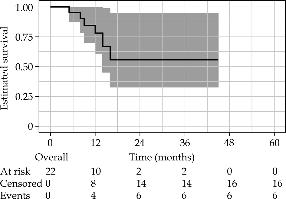
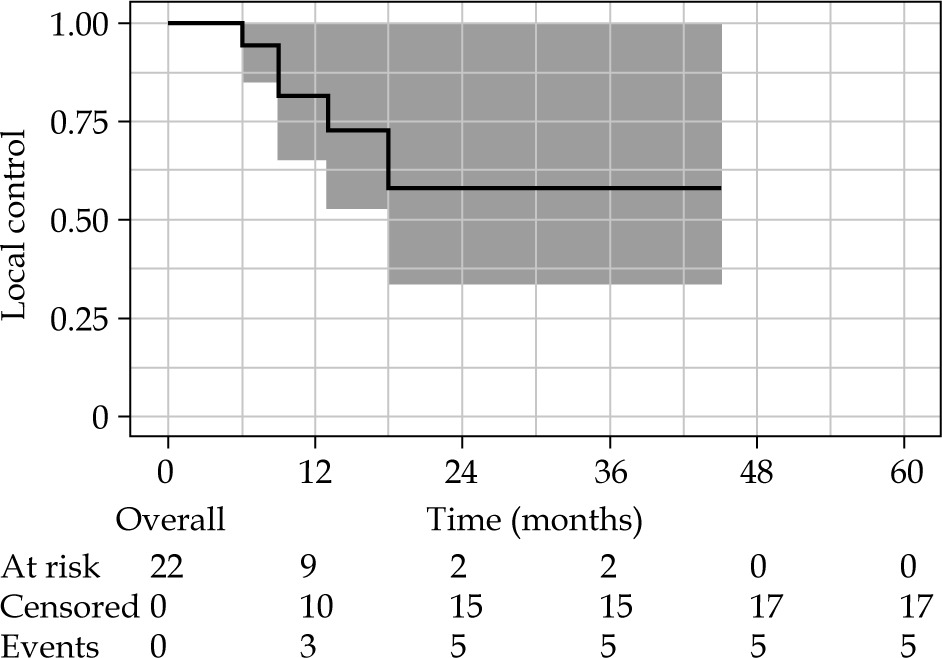
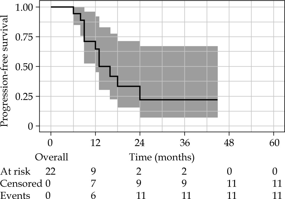
With a median follow-up of 11.5 months (range, 5-44 months), the median local control and median overall survival were not achieved, while median PFS was 16 months. The one-year LC, PFS, and OS rates were 82% (95% CI: 66-100%), 66% (95% CI: 46-93%), and 78% (95% CI: 61-100%), respectively. On univariate analysis, in analyzing factors related to LC, PFS, and OS, only OTT was consistently found to be significant in all outcomes (p < 0.05). The presence of lymph nodes at diagnosis was associated with worse PFS (Tables 3 and 4, Figs. 1-3).
Table 3
Oncologic outcomes
| Parameter | Median | 1-year |
|---|---|---|
| Overall survival | Not achieved | 78% (95% CI: 61-100) |
| Progression-free survival | 16 months | 66% (95% CI: 46-93) |
| Local control | Not achieved | 82% (95% CI: 66-100) |
Table 4
Univariate analysis for predictors of local control, progression-free survival, and overall survival
For acute toxicity, 11 patients had grade 1-2 toxicity, mainly gastrointestinal (GI) and genitourinary (GU) symptoms. Grade 3 toxicity was observed in 4 patients: 1 hematologic, 1 diarrhea, and 2 infections. The most severe was in a patient with grade 4 toxicity of urosepsis and pelvic abscess. For late toxicity, 5 patients had grade 1-2 toxicity, mainly GI, GU, and vaginal synechia. Grade 3 toxicity was observed in 2 patients: 2 proctitis needing intervention.
Discussion
In the current study, we presented our experience on the use of single application multi-fractionated CT-guided BT given in four fractions. The advantages of prescribing single application and less fractions include a smaller amount of trauma due to repeated implantations, lower resource utilization, less hospital stay, and reduced OTT. The patients were maintained on low-residue diet, and none required parenteral nutritional support. Disadvantages include issue toxicity and reproducibility. There is a theoretical toxicity due to less time for normal tissue repair between fractions. The use of MRI makes it possible to apply higher doses in less fractions, but with the use of CT scans, target delineation is not as accurate, which leads to higher toxicity [19]. Reproducibility is also an issue due to interfraction movement, such as movement of the needles and surrounding organs during succeeding fractions. The way of treatment is similar to low-dose-rate (LDR). Radiobiologically, LDR would have better therapeutic ratio, but with HDR, optimization of the prescribed treatment can be achieved as well as active nursing care during the treatment, since there is no issue with radiation protection for nursing care. Another disadvantage of single application is the inability to implement adaptive brachytherapy, as the same plan is delivered in all fractions. However, tumor shrinkage is not expected during short intervals between fractions, and interfraction movement is mitigated by uniform bladder filling.
The present study population consisted of patients with locally advanced disease and poor prognostic features. 36.4% of the patients had stage IVA disease, with a mean T-score of 10. The mean OTT was 66 days with 8 patients having OTT of more than 50 days. The mean HR-CTV volume was 66 cc, with 16 of the 22 patients having more than 45 cc. 91% of the patients had IMRT-EBRT, and all with enlarged lymph nodes received nodal boost.
Current literature on the use of interstitial BT have been summarized by a systematic review by Li et al., who reported a 2-year overall survival rate of 43-94%, 2-year local control of 61-95%, and grade 3-4 toxicity of 3-16.2% [14]. Consistent results was found in this study, with 1-year local control of 82%, 1-year PFS of 66%, and 1-year overall survival of 78%. This also shows the importance of distant and regional recurrence in these patients with a higher local control than PFS, which considers local, regional, and distant progression after treatment. A higher PFS indicate that distant metastases should be considered. A reported relapse by Kato et al. estimates a 20% local relapse rate, and 36.7% distant metastases rate [20]. Of those patients with tumor progression, three patients needed urinary diversion or dialysis for obstruction.
Toxicity reported was consistent with rates in the literature. Acute toxicity of urosepsis and pelvic abscess were seen in one patient with pyometra noted during brachytherapy procedure, which shows the importance of draining the pyometra prior to the procedure, especially in single application treatments. Late grade 3 toxicities included 2 gastrointestinal toxicity of proctitis. These patients received a brachytherapy rectum dose D0.1cc of 5.69 Gy per fraction, and D0.1cc of 4.71 Gy per fraction.
Univariate analysis in this study showed only OTT to be correlated with poorer LC, PFS, and OS. This is consistent with findings that increasing OTT of more than 50 days leads to loss of local control of 1% per day [21]. The presence of lymph nodes was also found to be correlated with decreased PFS. Similarly, histology of SCC showed a trend for worse PFS, which is the opposite from what is expected in the literature, and it could be explained by the population consisting of only 5 patients with adenocarcinoma histology and a wide confidence interval. Other factors were found not significant, probably due to the low sample size. In the literature, there are multiple studies showing a significant correlation between HR-CTV volume > 45 cc, HR-CTV D90, or T scoring with the probability of local control [22-24].
While this study is limited by its retrospective design, small sample size, short follow-up duration, and attrition, it adds to the current emerging evidence on the feasibility and safety of single-implant multi-fraction CT-based interstitial BT. Further, the cohort of the study reflects outcomes in a limited-resource setting combined with limitations of a global healthcare crisis.
Conclusions
This retrospective study suggests that single application multi-fractionated CT-guided interstitial HDR-BT given in four fractions in locally advanced cervical cancer seems to be feasible and safe, showing satisfactory response and acceptable toxicities. However, more evidence in the form of prospective studies and clinical trials are needed to generate more substantiated conclusions.


