
Dunbar syndrome, also known as median arcuate ligament syndrome (MALS), is a rare condition affecting approximately 2 per 100,000 individuals. It is most often diagnosed between 20 and 50 years of age, with a female-to-male ratio of 4 : 1. In this disorder, the celiac trunk is compressed by the median arcuate ligament (MAL). Typical symptoms include postprandial or exercise-induced epigastric pain, weight loss, nausea, and vomiting [1].
The MAL connects the crura of the diaphragm as they pass over the aorta, typically above the origin of the celiac trunk. However, in 10–24% of individuals the ligament runs lower and crosses the proximal segment of the celiac trunk [2]. The development of collateral circulation, primarily around the pancreas, plays a key role in symptom manifestation and significantly increases the risk of aneurysm formation in visceral vessels [3].
We report the case of a 19-year-old male with a history of Crohn’s disease (CD) diagnosed at the age of 8 years, who had been receiving upadacitinib since July 2024 with good clinical response and tolerance. The patient was admitted to the Gastroenterology Clinic with symptoms suggestive of disease exacerbation, including worsening abdominal pain, low-grade fever, unintentional weight loss (11 kg over 3 months), and nausea. The patient noted that, unlike his previous episodes of pain localised in the lower abdomen, the current pain was epigastric and correlated with meal volume.
Laboratory tests revealed elevated inflammatory markers (C-reactive protein [CRP], 86.3 mg/l; reference range < 5 mg/l). Computed tomography (CT) angiography was performed and compared with a scan obtained 6 months earlier. Inflammatory changes in the terminal ileum, ascending colon, and transverse colon had regressed. However, the new study revealed a 6-mm segment of the celiac trunk without contrast enhancement, caused by external compression and the presence of collateral circulation within the pancreatic head (Figures 1 A, B). The previous CT scan had shown only moderate celiac trunk stenosis with collateral circulation.
Figure 1
A – 3D Reconstructions from computer tomography (CT) angiography showing lack of contrast of the celiac trunk (white arrow); B – Computer tomography (CT) angiography showing external compression to the celiac trunk (yellow arrow); C – 3D Reconstructions from computer tomography (CT) angiography 1 month after surgery. The celiac trunk was patent, but its lumen narrowed by approximately 75% (white arrow). Peripancreatic collateral circulation (yellow arrow); D – Computer tomography (CT) angiography 1 month after surgery. Celiac trunk with luminal stenosis and post-stenotic dilatation (yellow arrow)
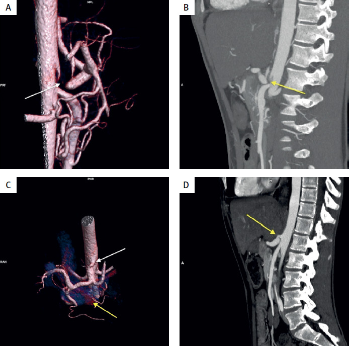
A laparotomy was performed, during which the MAL fibres were divided. Postoperatively, the patient’s pain improved, and CRP levels decreased. One month later, during follow-up, the pain recurred, accompanied by signs of CD exacerbation. Laboratory tests again revealed elevated CRP levels (80.9 mg/l). Repeat CT angiography demonstrated progression of inflammatory changes in the colon, while the celiac trunk remained patent but was narrowed by approximately 75% (Figures 1 C, D). Because of loss of response to upadacitinib, therapy was switched to adalimumab. In the following weeks, the patient’s abdominal pain subsided, and inflammatory markers gradually normalised.
The diagnosis of Dunbar syndrome is based on the exclusion of other gastrointestinal diseases. The most commonly used imaging modalities include CT angiography and magnetic resonance angiography. Many authors recommend Doppler ultrasonography with flow assessment of abdominal vessels as a screening tool due to its wide availability and lack of contrast media. However, its diagnostic utility is limited because anatomical variations may limit visualisation of the celiac trunk in some patients, and increased flow velocity observed during expiration may lead to false-positive findings suggestive of stenosis [4, 5]. Surgical treatment is indicated when symptoms persist, and MAL release is equally effective via open or laparoscopic approaches; the choice of method should be individualised. Previous studies have demonstrated symptom reduction following surgical intervention [6].
It is important to emphasise that MALS may lead to ischaemic changes in the jejunum, which can be mistaken for CD [7]. To date, only a few cases of Dunbar syndrome associated with CD have been reported [1, 8]. Due to its non-specific symptoms and relatively rare occurrence, Dunbar syndrome may mimic other gastrointestinal diseases, leading to incorrect diagnosis or suggesting an exacerbation of the disease. This case highlights the importance of considering MALS in patients with gastrointestinal disorders who develop meal-related epigastric pain that differs from their previous symptom patterns.














