
Introduction
Selective biliary cannulation is essential for the success of therapeutic endoscopic interventions. Conventional biliary cannulation success rates range between 70% and 90%, influenced by factors such as papillary anatomy, endoscopic technique, and the experience of the endoscopist [1–5]. Cannulation becomes particularly challenging in cases of anatomical variations, inflammation, periampullary diverticula, or small papillae [6].
Prolonged or repeated attempts at cannulation in such settings increase the risk of endoscopic retrograde cholangiopancreatography (ERCP)-related complications [7, 8]. To improve success rates in these difficult cases, several alternative techniques have been developed, including the double-guidewire technique (DGT), wire-guided cannulation over a pancreatic stent (WGC-PS), needle-knife precut sphincterotomy, and transpancreatic sphincterotomy [6, 9–11].
The DGT involves pre-insertion of a guidewire into the pancreatic duct (PD), facilitating biliary access. This technique is especially useful for patients with tortuous intraduodenal segments or prominent papillae within diverticula, as first described by Dumonceau et al. [12]. The WGC-PS approach, introduced by Coté et al. [11], involves placing a plastic pancreatic stent to protect the PD during further cannulation attempts, thereby potentially reducing the risk of post-ERCP pancreatitis (PEP) [13, 14]. Pancreatic stents have been shown to minimise both the incidence and severity of PEP, particularly in high-risk cases [15, 16].
Despite the theoretical benefits of each method, data directly comparing DGT and cannulation over a pancreatic stent (CPS) remain limited.
Aim
This study aims to evaluate and compare the clinical outcomes of these two techniques in patients experiencing unintended pancreatic duct cannulation during ERCP.
Material and methods
We conducted a prospective study involving 103 patients who underwent ERCP between June and December 2024 at Kafrelsheikh University Hospital, a tertiary referral centre. Patients were included if they experienced unintentional guidewire cannulation of the PD during attempts at selective biliary cannulation using the standard WGC technique. In these patients, either the DGT or CPS, with or without needle-knife sphincterotomy, was employed to facilitate biliary access.
Exclusion criteria included: 1) Prior sphincterotomy or endoscopic papillary balloon dilation; 2) ERCP performed for pancreatic rather than biliary intervention; 3) History of gastric bypass surgery; 4) Acute pancreatitis at the time of procedure; 5) Successful selective biliary cannulation using standard technique; 6) Use of needle-knife precut sphincterotomy without prior guidewire entry into the PD.
For included patients, the primary outcomes were time to successful deep biliary cannulation and the number of cannulation attempts. Data were collected prospectively in the ERCP database at Kafrelsheikh University Hospital.
Endoscopic procedures
All ERCP procedures were performed using a side-viewing duodenoscope (Olympus 190V, Japan) under conscious sedation with standard doses of midazolam, propofol, and meperidine. Selective biliary cannulation was initially attempted using a 0.035-inch guidewire (Jagwire™, Boston Scientific or Tracer Metro® Direct™).
Only patients with inadvertent guidewire insertion into the PD during WGC were included. An experienced ERCP assistant handled guidewire manipulation.
CPS technique: After successful PD guidewire insertion, a 5-F, 5-cm Zimmon™ polyethylene stent (Cook Medical) was placed into the main PD. Biliary cannulation was then attempted using a sphincterotome preloaded with a guidewire, directed toward the biliary orifice (11–12 o’clock position), using the PD stent as a reference.
DGT technique: A guidewire was inserted into the PD to at least the mid-portion (as confirmed fluoroscopically). A second guidewire was loaded into the sphincterotome, which was then advanced alongside the first wire. The sphincterotome tip was bent over the pancreatic guidewire and positioned at the 10–11 o’clock position to achieve bile duct cannulation.
In both techniques, needle-knife sphincterotomy (NKS) was used as needed to facilitate access. For NKS, a needle-knife was used to incise the ampullary mound at the junction of its upper and lower thirds to create a bilio-enteral fistula. Contrast was injected only after deep biliary cannulation was achieved.
The choice to leave or remove the PD wire and place a PD stent post-cannulation was at the discretion of the endoscopist. All procedures were performed by experienced endoscopists with consistent technical approaches across patients.
Statistical analysis
Statistical analyses were performed using SPSS version 14.0 (SPSS Inc., Chicago, IL, USA). Continuous variables were compared using Student’s t-test. Categorical variables were analysed using the c2 test or Fisher’s exact test, as appropriate. A p-value of < 0.05 was considered statistically significant.
Results
A total of 103 patients with native papillae who experienced unintentional pancreatic duct cannulation during ERCP were included during the study period. Patients were divided into two groups: those managed using the double-guidewire technique (DGT; n = 59) and those treated with cannulation over a pancreatic stent (CPS; n = 44) (Table I).
Table I
Patients’ characteristics, laboratory investigations, and findings in both groups
There was no significant difference in age between the two groups (p = 0.713). However, the CPS group had a significantly higher proportion of male patients (70.5%) compared to the DGT group (55%) (p = 0.0025).
Regarding liver function tests (e.g. liver enzymes, bilirubin, serum albumin), no significant differences were observed between the groups because most patients presented with obstructive jaundice. However, the total leukocyte count (TLC) was significantly higher in the CPS group (p < 0.001), probably reflecting the presence of associated cholangitis in some patients.
Indications for ERCP: The CPS group had a significantly higher proportion of patients with calculous obstructive jaundice (68.2%) compared to the DGT group (47%) (p = 0.005). There was no significant difference between groups for malignant obstructive jaundice (p = 0.601). Biliary strictures were significantly more common in the CPS group (42.9%) than in the DGT group (20%) (p = 0.034). Biliary colic was more frequent in the DGT group (11.7%) than in the CPS group (2.2%), but this was not statistically significant (p = 0.125). Biliary leak post-cholecystectomy was more common in the CPS group (22.2%) than in the DGT group (3.3%) (p = 0.045). Cholangitis was present only in the DGT group (11.6%) (p = 0.02) (Table II).
Table II
Procedural and anatomical characteristics
Papillary anatomy: Small papillae were slightly more common in the CPS group (52.9%) than in the DGT group (47.1%). No statistically significant differences were noted between groups regarding other papilla types, such as regular (type 1), pendulous (type 3), or creased/rigid (type 4).
Cannulation time and attempts: The DGT group achieved biliary cannulation in significantly less time (8.27 ±4.8 min) compared to the CPS group (11.84 ±3.2 min) (p = 0.0031). The number of cannulation attempts did not significantly differ between groups: 2.61 ±1.8 (DGT) vs. 2.48 ±1.24 (CPS) (p = 0.492).
Need for precut sphincterotomy (PS): A significantly higher proportion of patients in the CPS group required precut sphincterotomy (88.6%) compared to the DGT group (33.3%) (p < 0.0001).
Figure 1 demonstrates recorded complications (bleeding, perforation, pancreatitis) across both groups. Perforation occurred in 1 patient, representing approximately 0.9% of the total. Bleeding was observed in 9 patients, accounting for roughly 8.2%. Post-ERCP pancreatitis was noted in 6 patients, equivalent to approximately 5.5%.
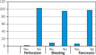
Figure 2 illustrates the need for precut sphincterotomy in two different ERCP cannulation techniques: the double guidewire technique (blue line) and the cannulation over pancreatic stent technique (orange line). The cannulation over pancreatic stent technique shows a higher count and percentage of patients requiring precut sphincterotomy compared to the double guidewire technique. Around 60% of cases using the pancreatic stent technique required precut sphincterotomy. In contrast, only about 20% of cases using the double guidewire technique needed it. Conversely, a higher percentage of patients did not require precut sphincterotomy in the double guidewire group compared to the stent group.
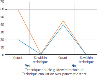
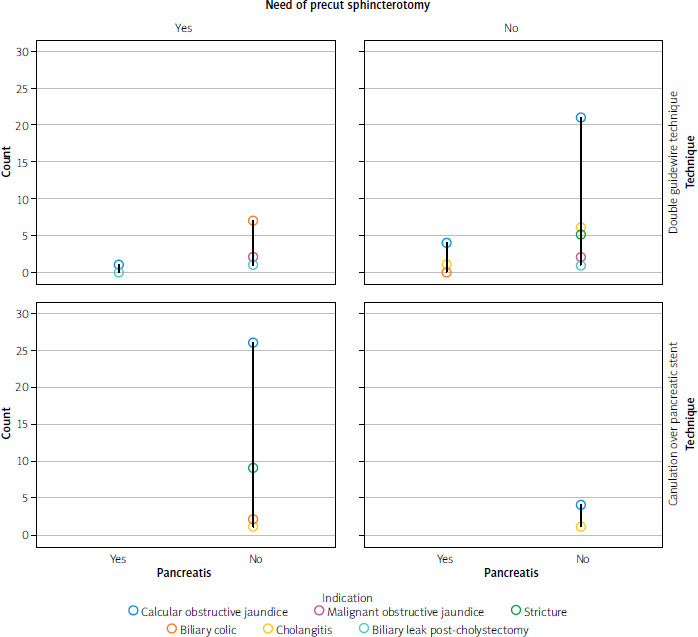
Figure 3 illustrates the incidence of post-ERCP pancreatitis across two different cannulation techniques – double guidewire technique and cannulation over pancreatic stent technique – in relation to the following: 1) the need for precut sphincterotomy, and 2) the clinical indications for ERCP, including calculous and malignant obstructive jaundice, strictures, biliary colic, cholangitis, and biliary leak post-cholecystectomy.
Discussion
Several techniques have been developed to facilitate selective deep biliary cannulation in difficult ERCP cases. Among these, the DGT and CPS have proven to be useful alternatives when standard methods fail.
In the DGT, placement of a guidewire into the pancreatic duct offers several mechanical advantages: it helps to stabilise and lift the papilla, straighten the common channel, reduce repeated PD injections, and allow for subsequent pancreatic stenting if needed [17]. Conversely, the CPS technique involves pancreatic duct stenting, which may facilitate biliary access by marking the PD orifice and minimising inadvertent pancreatic cannulation during repeated attempts [18].
Our findings indicate that CPS was associated with a significantly higher need for precut sphincterotomy (88.6%) compared to DGT (33.3%), consistent with previous studies such as the one by Bailey et al. [19]. They observed that the presence of a pancreatic stent, while protective against post-ERCP pancreatitis (PEP), can complicate biliary access, especially in anatomically challenging situations, thereby increasing the requirement for precut access.
Additionally, we found a higher proportion of small papillae in the CPS group (52.9%) compared to the DGT group (47.1%). Small papillae are anatomically more difficult to cannulate. In such cases, CPS may offer an advantage by stabilising the access point and guiding biliary entry. This is supported by Sangwan et al. [20], who emphasised that pancreatic stenting in small or atypically shaped papillae can improve cannulation success while reducing trauma to the PD.
Importantly, time to successful biliary cannulation was significantly shorter in the DGT group. This may be due to the enhanced manoeuvrability and directional control provided by the dual guidewires. In contrast, the CPS approach often necessitates additional steps such as stent deployment and, in many cases, precut sphincterotomy, both of which contribute to longer procedure time. This observation aligns with the findings of Uchida et al. [21], who demonstrated the efficiency of DGT in shortening cannulation time in difficult ERCP scenarios.
While the number of cannulation attempts did not differ significantly between the two techniques, the shorter cannulation time in DGT highlights its potential procedural efficiency. This supports an earlier report by Buxbaum et al. [22], who noted that advanced cannulation techniques like DGT can reduce procedural complexity without increasing the number of failed attempts.
Regarding ERCP complications in both techniques, there was 1 case of perforation, which was successfully controlled endoscopically using Hemoclips. There were 9 cases of bleeding – 7 primary and 2 secondary – all managed endoscopically. Additionally, 6 cases of post-ERCP pancreatitis were observed.
Interestingly, our results suggest that precut sphincterotomy may be associated with fewer complications such as bleeding, perforation, and pancreatitis, possibly due to controlled access and reduced mechanical trauma from multiple failed cannulation attempts. This notion is echoed by Cotton et al. [23], who proposed that a carefully executed precut can reduce cumulative trauma and bleeding risks when initial standard techniques fail.
Although DGT was more time-efficient, CPS may offer better protection against post-ERCP pancreatitis, particularly in high-risk patients. As supported by studies from Goldberg et al. [24] and Fan et al. [25], prophylactic PD stenting significantly reduces the incidence of PEP and facilitates safer cannulation in anatomically challenging situations.
This study has several limitations. First, the sample size was relatively small. Second, operator experience, although consistent across procedures, may still have introduced subtle bias. Third, selection bias cannot be entirely ruled out despite efforts to randomise technique usage. Finally, complication rates – especially bleeding and pancreatitis – were not statistically powered to detect subtle differences due to low event counts.
Conclusions
In patients with unintended pancreatic duct cannulation during ERCP, the DGT demonstrated several advantages over CPS. DGT was associated with a significantly reduced need for precut sphincterotomy and shorter time to biliary cannulation, indicating greater procedural efficiency.
However, CPS may still play a critical role in reducing the risk of post-ERCP pancreatitis, particularly in cases with challenging papillary anatomy, such as small or fibrotic papillae. The number of cannulation attempts was similar between the two groups, suggesting that the complexity of the anatomy probably dictated overall difficulty regardless of technique.
Ultimately, both DGT and CPS are valuable tools in the endoscopist’s armamentarium. The choice of technique should be tailored to individual patient anatomy, clinical indication, and endoscopist expertise. Further large-scale, multicentre trials are warranted to refine technique selection algorithms and optimise patient outcomes.














