
Gallstone ileus, a rare but clinically relevant complication of cholelithiasis, constitutes 1–4% of all mechanical small bowel obstructions and is particularly prevalent in the elderly, affecting up to 25% of this population [1]. The pathogenesis of gallstone ileus involves the spontaneous formation of a biliary-enteric fistula, predominantly with the duodenum, followed by the colon, with fistulas to the stomach being exceedingly rare [2]. The clinical presentation of gallstone ileus is site dependent. The diagnosis should be suspected in elderly patients presenting with a history of gallstones, acute cholecystitis, and abrupt onset of intestinal obstruction, particularly if supported by radiographic findings consistent with Rigler’s triad [3]. Treatment of gallstone ileus necessitates a multifaceted approach encompassing antibiotic therapy, fluid resuscitation, and surgical intervention. Surgical options include emergency enterolithotomy for immediate relief of obstruction, a one-stage procedure combining enterolithotomy, cholecystectomy, and fistula repair, or a two-stage approach involving initial emergency enterolithotomy followed by cholecystectomy and fistula closure 4–6 weeks later [4]. A 78-year-old male presented to the emergency department with a history of intermittent abdominal pain, vomiting, dehydration, and altered mental status that began 5 days prior. The patient’s medical history includes dyslipidaemia, hypertension, prostate cancer, aortic aneurysm, and a previous hospitalisation for acute cholecystitis. Upon arrival, the patient was afebrile and tachypneic (RR 24). Vital signs were as follows: heart rate 105 beats per minute, blood pressure 140/85 mm Hg, and oxygen saturation 96%. Physical examination revealed abdominal distension, absent bowel sounds, and mild right upper quadrant tenderness. Laboratory findings included leukocytosis (14,670 cells/mm3), elevated C-reactive protein (3.45 mg/dl; reference range < 0.5 mg/dl), elevated serum urea (90 mg/dl), and elevated serum creatinine (1.9 mg/dl), while haemoglobin (13.7 g/dl) and other laboratory parameters remained within normal limits. Nasogastric tube placement resulted in the immediate drainage of 1500 ml of intestinal fluid. Intravenous fluid resuscitation was initiated. Based on the clinical presentation, small bowel obstruction was considered the primary diagnostic hypothesis. Upon admission to our hospital, a contrast-enhanced computed tomography (CT) of the abdomen and pelvis was performed to further clarify the diagnosis. The CT scan revealed findings consistent with small bowel obstruction, including dilated small bowel loops, with air-fluid levels, proximal to the obstruction site, with the transition point located at the distal ileum (Figures 1, 2 A). Within the terminal ileum, hyperenhanced, round, well-defined lesions with a rim-enhanced pattern were visible, suggesting cholesterol gallstones as the obstructive cause (Figures 1, 2 A, B). Distal small and large bowel loops seemed to collapse (Figure 3). Additional findings included mild biliary ductal dilatation (Figure 4) and a thickened gallbladder wall with post-contrast enhancement (Fig- ure 3). Importantly, gas was detected within the gallbladder, indicating abnormal communication between the gallbladder and adjacent gastrointestinal structures. The presence of gas, in combination with the gallbladder findings, strongly supported the presence of a cholecystogastric or cholecystoduodenal fistula. Based on the anatomical location and the extension of the findings from a CT scan and the patient’s prior history of acute cholecystitis, gallstone ileus was determined to be the most likely diagnosis. The fistulous communication was hypothesised to involve the gallbladder and the gastric antrum (Figure 3), a finding that was subsequently confirmed by a gastroscopy which revealed the presence of a fistulous communication between the gastric antrum and the gallbladder. The patient was subsequently transported to the operating room. A midline incision was performed to access the peritoneal cavity, which revealed 150 ml of free fluid. The dilated small bowel loops were subsequently mobilised. The site of obstruction was identified within the ileum, approximately 100 cm proximal to the ileocecal valve. Following a thorough inspection, no evidence of bowel strangulation was observed within the obstructed loop. An enterolithotomy was subsequently performed. A large gallstone measuring 4 cm in diameter was extracted (Figure 5). The enterotomy was then closed in two layers using absorbable sutures, and one vacuum drain was placed. The patient demonstrated gradual clinical and laboratory improvement. The nasogastric tube was removed on the second postoperative day, followed by the removal of surgical drains on the fifth postoperative day. His postoperative period was uneventful, and the patient was discharged home on day 6 postoperatively. At the 1-month follow-up appointment, the patient reported satisfactory overall health with normal bowel function and no evidence of recurrence.
Figure 1
Axial post-contrast (venous phase) CT image showing the transition point at the distal ileum (yellow arrow), a hyperdense gallstone within the terminal ileum (red arrow), and dilated small bowel loops with air-fluid levels (yellow arrowheads)
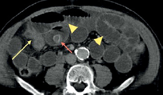
Figure 2
A – Coronal contrast-enhanced CT image highlighting cholesterol gallstones with a rim-enhanced pattern (red arrows) located in the distal ileum. The transition point is clearly visualised (yellow arrow). B – Multiple rim-enhanced cholesterol gallstones (red arrows) within the terminal ileum, consistent with the obstruction site
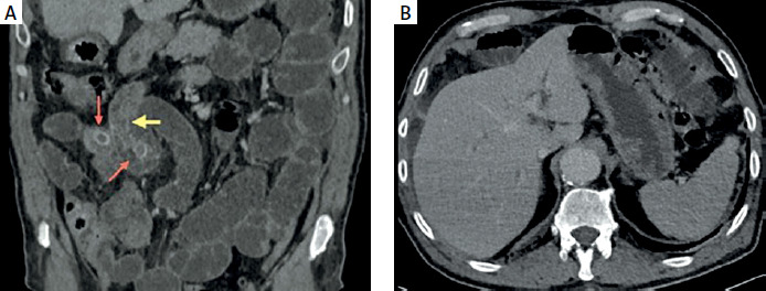
Figure 3
Coronal CT image demonstrating gallbladder wall thickening with post-contrast enhancement, along with gas within the gallbladder lumen (orange arrow), suggesting a fistulous communication (orange arrowhead is the potential site of fistulisation). Dilated small bowel loops (yellow arrowhead) and a collapsed distal terminal ileum (yellow arrow) are also noted
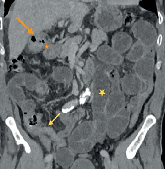
Figure 4
Axial post-contrast CT image showing mild biliary ductal dilatation, a secondary finding indicative of chronic cholelithiasis
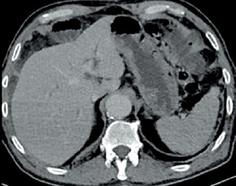
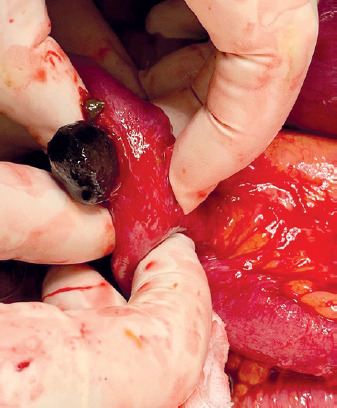
Figure 5
Intraoperative image depicting a 4 cm diameter spherical gallstone retrieved from an ileal intestinal loop
Biliary ileus, also known as gallstone ileus, exhibits a predilection for elderly female patients (> 70 years) with a history of cholelithiasis and multiple comorbidities [5]. The pathogenesis of biliary ileus involves the migration of a gallstone through an enteric fistula. Cholecystoenteric fistulas are the most prevalent, constituting approximately 60% of cases, followed by cholecystogastric and cholecystocolonic fistulas [6]. Cholecysto-intestinal fistula formation is frequently a consequence of acute cholecystitis. The associated inflammation within the gallbladder and surrounding tissues can result in the development of adhesions, which may subsequently evolve into a fistulous tract [7]. The proximal duodenum is the most frequent site of involvement, followed by the stomach, transverse colon, and other segments of the small intestine [8]. Alternative pathways for gallstone entry into the small bowel include migration through the common bile duct, a dilated ampulla of Vater, or as a potential complication following endoscopic retrograde cholangiopancreatography (ERCP) [9]. The size and morphology of the gallstone are crucial factors. Most reported obstruction cases involve gallstones exceeding 2 cm in diameter, while smaller stones may pass through the intestinal tract and be expelled naturally [10]. The most common gastrointestinal (GI) symptoms of gallstone ileus include nausea, vomiting, colicky abdominal pain, and varying degrees of abdominal distension [11]. Concomitant acute cholecystitis and jaundice may be present in 10–30% and < 15% of patients, respectively, at the time of bowel obstruction [12]. Plain abdominal radiographs may diagnose gallstone ileus, with the identification of Rigler’s triad (radiopaque gallstones, pneumobilia, and distended intestinal loops) contributing to diagnostic accuracy [13]. However, the sensitivity of plain radiographs is limited, ranging from 40% to 70%, and radiopaque gallstones are only observed in approximately 10% of cases. Conversely, CT with contrast is considered the gold standard for diagnosis, demonstrating a sensitivity exceeding 90% [4]. The therapeutic strategy for gallstone ileus (GI) conditions encompasses gastric decompression, administration of antibiotic therapy, and fluid and electrolyte resuscitation, particularly given the dehydrated state often observed in these patients, alongside necessary surgical intervention [7]. The management of gallstone ileus is primarily surgical and is defined by the classic triad of cholelithiasis, the presence of a biliary-enteric fistula, and subsequent bowel obstruction. The selection of the surgical procedure is largely contingent upon the patient’s clinical status. In the emergency setting, the performance of fistula closure during the initial procedure was independently associated with a higher risk of mortality compared to enterotomy and stone extraction alone [14]. Surgical interventions for this condition may include emergency enterolithotomy performed as a standalone procedure, one-stage enterolithotomy accompanied by cholecystectomy or fistula closure, and a two-stage approach in which enterolithotomy is conducted initially, followed by cholecystectomy or fistula closure after a period of 4 to 6 weeks [4]. One investigation reported that the mortality rates for patients undergoing enterotomy alone and those undergoing the one-stage procedure were 11.7% and 16.9%, respectively [15]. Furthermore, it was observed that 10% of patients who underwent enterotomy alone experienced recurrent biliary symptoms [16]. The persistence of a cholecysto-intestinal fistula constitutes a potential aetiological factor for the development of retrograde cholecystitis and gallbladder carcinoma [17]. A large-scale study demonstrated that urgent, one-stage fistula repair is associated with a significantly higher incidence of postoperative complications, including prolonged hospitalisation and increased mortality rates [6]. There are also different therapeutic strategies like endoscopic removal [18], as well as less commonly employed approaches such as shockwave lithotripsy and Nd:YAG laser lithotripsy, described in the literature for achieving obstruction relief [19]. Based on these findings, two-stage surgical intervention is considered the gold standard approach for managing cases of gallstone ileus and those involving impaction within the small intestine [6]. In contrast, while one-stage surgical procedures may be considered appropriate for duodenal and colonic impactions, a less invasive approach such as enterotomy with stone extraction alone appears more suitable for many GI patients. This is due to their typical profile as elderly individuals with multiple comorbidities, which makes them less tolerant of more extensive surgical techniques [17].
Gallstone ileus presents as a rare and diagnostically challenging clinical entity. The management of these patients necessitates an individualised approach, given the diverse range of therapeutic options, each with inherent advantages and drawbacks. Recognising the advanced age and comorbidities frequently observed in this patient population, initial management should prioritise the relief of intestinal obstruction through enterolithotomy. One-stage procedures may be considered for younger patients with favourable overall health and a clear indication for concomitant biliary intervention.














