
Background and methodology of the guidelines
Introduction
This document addresses the need to update and standardise the clinical care of patients with coeliac disease. Based on the latest scientific evidence and expert consensus, this text systematises diagnostic and therapeutic protocols. It incorporates the principles of coeliac disease monitoring and highlights the necessity of an interdisciplinary approach involving physicians (particularly gastroenterologists and general practitioners), while recognising the vital roles of histopathologists, clinical dietitians, and psychologists. It serves as a practical decision-making tool in daily clinical practice.
According to survey data published in 2023 regarding coeliac disease patients in the Polish population, only 55% of respondents rated the knowledge of gastroenterologists as good, and a mere 16.6% rated the knowledge of general practitioners as good [1]. The estimated mean duration from symptom onset to the definitive diagnosis of coeliac disease was 7.3 years [2]. It is anticipated that the development of these Polish clinical guidelines will significantly improve the quality of care for patients with suspected or confirmed coeliac disease.
Objectives of the guidelines
The primary objective of these guidelines was to establish and disseminate evidence-based, optimal protocols for diagnosis, treatment, and long-term monitoring in coeliac disease. These guidelines are designed to support clinical decision-making processes associated with the care of patients with suspected or confirmed coeliac disease, primarily by physicians, but also by other healthcare professionals and stakeholders involved in the organization and financing of healthcare services. Serving an educational purpose, this document summarises the current state of knowledge on coeliac disease and can serve as a foundation for future clinical research. However, it must be emphasised that the continuous evolution of medical knowledge regarding coeliac disease will necessitate subsequent updates to this document.
Clinical conditions addressed in the guidelines
This document focuses comprehensively on coeliac disease – its aetiology, epidemiology, diagnostic modalities, therapeutic interventions, and long-term surveillance. It features an extensive discussion on differential diagnosis, clinical conditions associated with coeliac disease, and other gluten-related disorders.
Target patient population
These guidelines apply specifically to adult patients with suspected and/or confirmed coeliac disease and, to a lesser extent, individuals presenting with other gluten-related disorders. They also provide key information regarding distinct management strategies for coeliac disease in the paediatric population.
Epidemiology and general characteristics of coeliac disease
Coeliac disease is a chronic, immune-mediated enteropathy of the small intestine triggered by the ingestion of dietary gluten in genetically predisposed individuals. It is characterised by the presence of disease-specific autoantibodies, including anti-tissue transglutaminase (anti-TG2) antibodies, and by histopathological changes in the small intestinal mucosa of varying severity (villous atrophy, crypt hyperplasia, and an increased number of intraepithelial lymphocytes) [3, 4].
The prevalence of coeliac disease has increased substantially over the past few decades. The disease can manifest at any age. In Western populations, serological screenings indicate a coeliac disease prevalence of approximately 1–1.6%, whereas the prevalence of histologically confirmed cases is estimated at approximately 0.7%. In Europe, notable geographical variations exist: the highest prevalence rates are documented in Northern Europe (1.6%), followed by Eastern Europe (0.98%), Southern Europe (0.69%), and Western Europe (0.6%) [5–8].
Coeliac disease occurs 1.5 to 2.0 times more frequently in females than in males. Familial aggregation is highly clinically relevant – the risk of developing coeliac disease among first-degree relatives of affected individuals ranges from 5% to 10% [5–8]. Furthermore, coeliac disease is strongly associated with various comorbidities, particularly type 1 diabetes mellitus, autoimmune thyroid diseases (Hashimoto’s thyroiditis, Graves’ disease), Down syndrome, Turner syndrome, Williams syndrome, selective IgA deficiency, and other autoimmune disorders [9].
The key genetic risk factors in the immunopathogenesis of coeliac disease are the HLA-DQ2.5 alleles (present in 90–95% of patients; associated with the classical clinical presentation of coeliac disease and a 30% lifetime risk of disease manifestation among carriers) and HLA-DQ8 alleles (present in 5–10% of cases). The HLA-DQ2 and/or HLA-DQ8 molecules consist of an alpha and a beta chain. These molecules are encoded by two alleles: most frequently A1*05 and B1*02 (for the HLA-DQ2.5 haplotype), A1*02 and B1*02 (for the HLA-DQ2.2 haplotype), and A1*03 and B1*03:02 (for the HLA-DQ8 haplotype). Individuals carrying two alleles encoding the HLA molecules are susceptible to coeliac disease, as are individuals having a single HLA-DQ2 chain encoded by a single allele [10–13]. The presence of these haplotypes is necessary but not sufficient for disease development; non-HLA genes also play a substantial role in pathogenesis [14]. Dietary exposure to gluten represents the indispensable environmental trigger. Other potential triggers include gastrointestinal infections, specific pharmacological agents (e.g., interferon alfa, immune checkpoint inhibitors), and surgical interventions [15–20].
Gluten is a collective term for the storage proteins found in cereal grains: wheat (gliadin and glutenin), barley (hordein), and rye (secalin). These proteins feature a high content of proline and glutamine residues, which imparts distinct viscoelastic properties but renders them highly resistant to complete proteolytic degradation in the human gastrointestinal tract, leading to the generation of immunogenic peptides [21, 22].
In individuals with coeliac disease, gluten ingestion triggers an aberrant immune response resulting in intestinal mucosal injury, malabsorption syndrome, and multiorgan manifestations. Dietary gluten can originate from various sources [23–26].
Natural sources:
– Wheat (including varieties such as spelt, kamut, and emmer), barley, and rye.
– Oats – although naturally lacking gluten sequences, commercial oats are frequently cross-contaminated with gluten during harvesting and processing. Some studies also suggest potential immunogenicity of avenin (the storage protein in oats) in a minor subset of coeliac patients.
Added/processed sources (gluten is a protein of great technological importance in the food industry):
Cross-contamination:
Coeliac disease presents with highly variable clinical phenotypes. The classical clinical presentation is dominated by symptoms of malabsorption: chronic diarrhoea, steatorrhea, weight loss, and iron deficiency anemia. However, in some patients, non-classical or extraintestinal manifestations predominate, such as chronic fatigue, neurological disorders (e.g., gluten ataxia, peripheral neuropathy), dermatological conditions (e.g., dermatitis herpetiformis), or laboratory abnormalities including anemia, elevated liver enzymes, or premature osteoporosis. Coeliac disease may also remain completely asymptomatic [4, 9, 27].
Management guidelines published over the last decade by scientific societies and expert groups show a definitive trend toward simplifying and accelerating the diagnostic pathway through high-quality serological testing. This includes considering a no-biopsy diagnostic approach in children and in a well-defined cohort of adult patients [28–30].
Methodology of the guidelines
These guidelines were developed on the initiative of the Intestinal Diseases Section of the Polish Society of Gastroenterology under the patronage of the National Consultant in Gastroenterology. The working group initiated the development of recommendations based on the PICO (Patients, Intervention, Comparator, Outcome) framework [31].
All stages of the systematic review and recommendation formulation utilised source data retrieved from electronic databases: PubMed, Cochrane Library, and Embase, alongside clinical practice guidelines published by international scientific societies. Particular emphasis was placed on documents utilising the GRADE (Grading of Recommendations Assessment, Development, and Evaluation) methodology, including those from the American College of Gastroenterology (ACG) and the European Society for the Study of Coeliac Disease (ESsCD) [28–30].
To evaluate the quality of evidence and the strength of the recommendations, a system adapted from the GRADE methodology was used. For each recommendation, experts determined the quality of evidence (high, moderate, low, or very low) and the strength of the recommendation (strong or weak; Table I).
Table I
Criteria for assessing the strength of recommendations
Following the formulation of recommendations and the assessment of evidence quality and strength, the final wording of each statement was subjected to detailed analysis. The degree of expert consensus regarding the final wording, quality of evidence, and strength of recommendation was evaluated using a 6-point Likert scale with 1 corresponding to complete disagreement/lack of support, 2 corresponding to disagreement/lack of support, 3 corresponding to partial disagreement/lack of support, 4 corresponding to partial agreement/support, 5 corresponding to agreement/support, and 6 corresponding to complete agreement/support (Table II).
Table II
The Likert scale
| Approval rating according to the Likert scale | |
|---|---|
| 1 | Complete disagreement |
| 2 | Disagreement |
| 3 | Partial disagreement |
| 4 | Partial agreement |
| 5 | Agreement |
| 6 | Complete agreement |
Following the formal voting process, revisions to the recommendations were performed as needed. A recommendation was considered permanently accepted if > 75% of voters scored their agreement between 4 and 6 points (indicating a high level of consensus). If the agreement rate was ≤ 75%, the consensus level was defined as low [32].
Subsequently, the quality of the guidelines was rigorously evaluated using the AGREE II instrument, in compliance with the protocols of the Agency for Health Technology Assessment and Tariff System (available at www.aotm.gov.pl). All reviewer comments were fully incorporated into the final version [33].
Additionally, the draft document was consulted with the Polish Coeliac Society – the biggest nation-wide organisation supporting people on a gluten-free diet, ensuring that the perspective of patients living with coeliac disease and gluten-related disorders was fully integrated.
Diagnosis of coeliac disease
Recommendation 1. We recommend diagnostic testing for coeliac disease in patients presenting with clinical symptoms and/or laboratory abnormalities suggestive of the disease, as well as in individuals with clinical conditions associated with an increased risk of developing coeliac disease.
| Strength of recommendation: Strong |
| Quality of evidence: High |
| Expert consensus vote: 100% total agreement (6 points) |
The diagnosis of coeliac disease remains clinically challenging, as symptoms may present at any age, including in older adults, and frequently involve extraintestinal manifestations affecting multiple organ systems [34, 35]. Furthermore, some individuals may be entirely asymptomatic despite belonging to a high-risk group, defined by a coeliac disease prevalence exceeding 2–2.5%; such individuals should undergo targeted screening [28].
Suspected coeliac disease and subsequent diagnostic workup should be considered in the presence of the following:
1) Clinical symptoms: Chronic diarrhoea (with or without abdominal pain), steatorrhea, constipation, postprandial abdominal distension and flatulence, recurrent aphthous stomatitis, unexplained weight loss, recurrent abdominal pain, recurrent headaches, psychomotor developmental delay, “brain fog”, chronic fatigue, mood disorders, muscle weakness, chronic arthralgia, or unexplained high-output ileostomy/colostomy fluid loss.
2) Laboratory abnormalities: Anemia and/or decreased mean corpuscular volume (MCV) and/or mean corpuscular haemoglobin (MCH), deficiencies in iron, ferritin, vitamin B12, folic acid, or vitamin D3, and unexplained increased activity of aminotransferases.
3) Associated clinical conditions: Dermatitis herpetiformis (Duhring’s disease), psoriasis, premature osteoporosis or osteopenia, incidental endoscopically or histopathologically detected villous atrophy, irritable bowel syndrome (IBS), microscopic colitis, hyposplenism, peripheral neuropathy, ataxia, epilepsy, depressive disorders, migraine headaches, dental enamel defects, Hashimoto’s thyroiditis, Graves’ disease, type 1 diabetes mellitus, autoimmune hepatitis, Sjögren’s syndrome, selective IgA deficiency, recurrent pancreatitis of unknown aetiology, unexplained female or male infertility, recurrent miscarriages, delayed menarche or secondary amenorrhea, premature menopause, and enteropathy-associated malignancies (lymphoma).
4) High-risk factors (screening indicated in asymptomatic individuals): First-degree relative diagnosed with coeliac disease, and specific genetic disorders including Down syndrome, Turner syndrome, and Williams syndrome.
Among the listed manifestations, chronic diarrhoea (with or without abdominal flatulence) is considered the most typical gastrointestinal symptom in adults, whereas iron deficiency anemia and unexplained weight loss represent the most common extraintestinal features [28, 30, 36–38].
Recommendation 2. The diagnosis of coeliac disease in adult patients is based on serological testing and gastroduodenoscopy with histopathological evaluation of duodenal biopsy specimens. We recommend measurement of serum IgA anti–tissue transglutaminase antibodies (anti-TG2 IgA) together with total serum IgA concentration as the initial diagnostic step. Regardless of the macroscopic endoscopic appearance of the duodenum, it is mandatory to obtain biopsy specimens from both the duodenal bulb and the descending duodenum. A positive serological test combined with histopathological evidence of intraepithelial lymphocytosis and villous atrophy confirms the diagnosis of coeliac disease.
| Strength of recommendation: Strong |
| Quality of evidence: High |
| Expert consensus vote: 89% Total Agreement (6 points), 11% Agreement (5 points) |
Serological testing in coeliac disease (first diagnostic step)
The routine first-line screening approach for coeliac disease involves serological testing via the determination of IgA anti-tissue transglutaminase antibodies (anti-TG2 IgA) concurrently with total serum IgA level measurement to rule out selective IgA deficiency, which could generate false-negative anti-TG2 IgA results [39, 40].
Testing must be performed while the patient is maintaining a gluten-containing diet. Initiation of a gluten-free diet prior to diagnostic testing confounds or prevents accurate serological interpretation.
The determination of other coeliac disease-associated antibodies (anti-endomysium [anti-EMA], anti-deamidated gliadin peptides [anti-DGP], anti-gliadin [AGA] of IgA or IgG classes, or anti-TG2 IgG) is indicated in specific clinical scenarios and diagnostic dilemmas. It must be noted that no single serological marker has 100% diagnostic sensitivity and specificity (Table III) [39–43].
Table III
Summary of serological tests in coeliac disease diagnostics and their clinical utility
In patients with a positive anti-TG2 IgA concentration, the traditional second diagnostic step in adults consists of upper endoscopy with macroscopic visualization of the duodenal bulb and descending duodenum accompanied by tissue sampling. A positive serological panel combined with intraepithelial lymphocytosis and villous atrophy confirms coeliac disease [38].
Endoscopic evaluation in coeliac disease (second diagnostic step)
Gastroduodenoscopy with macroscopic evaluation and multiple biopsies from the duodenal bulb and descending part of the duodenum is recognised by most international scientific societies as the standard tool for the diagnosis and differential diagnosis of coeliac disease in adults. However, emerging guidelines indicate that a no-biopsy approach may be considered in adult patients under highly specific clinical conditions:
1) High-concentration serology in young adults: In patients < 45 years of age presenting with a high anti-TG2 IgA (≥ 10 times ULN) on the initial test, confirmed by a positive serological result from a second, independent blood sample while on a gluten-containing diet, provided that alarm symptoms (“red flags” such as gastrointestinal bleeding, dysphagia, or signs of bowel obstruction) have been excluded. This approach was proposed for selected adults by the ESsCD in 2025 for the first time [28].
2) Endoscopy contraindications: In patients with contraindications to upper gastrointestinal (GI) tract endoscopy, a diagnosis may be considered based on two independent anti-TG2 IgA measurements as detailed above or, optionally, supported by a positive anti-EMA IgA confirmatory test. This non-invasive pathway is well established in paediatric population.
3) Premature dietary changes: If a symptomatic patient initiated a strict gluten-free diet due to severe clinical symptoms prior to undergoing endoscopy, provided serology remains high-positive.
Consequently, a no-biopsy diagnostic strategy may be considered in selected adult cases and must be thoroughly discussed with the patient [28, 30].
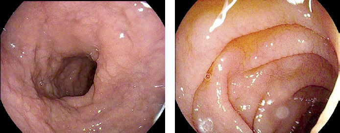
During upper GI endoscopy, specific macroscopic features indicative of coeliac disease should be actively sought in the duodenal bulb and the descending duodenum. These include loss or flattening of the duodenal mucosal folds, scalloping, mucosal smoothing, mucosal mosaic pattern, prominent submucosal vascular pattern, and mucosal erosions (Figure 1). While their presence strongly favours coeliac disease, approximately 22–50% of patients exhibit a macroscopically normal mucosa under standard white-light endoscopy [44]. This underlines the necessity of routine biopsy sampling.
Figure 1
Endoscopic presentation of duodenal changes in coeliac disease: flattening of intestinal folds and scalloping of circular folds (Images courtesy of Dr. Paweł Samborski, Central Endoscopy Unit, University Clinical Hospital, Poznan, Poland)
Microscopic mucosal changes in coeliac disease are frequently patchy and vary in intensity [45]. It is recommended to obtain ≥ 4 biopsy specimens from the descending duodenum and 1–2 specimens from the duodenal bulb at the 9 o’clock and/or 12 o’clock positions, as ultra-short coeliac disease may be confined entirely to the bulb. Each specimen should be obtained using a single-biopsy technique (one bite per pass of the forceps). Biopsies from the bulb and descending duodenum should preferably be submitted in separate containers. Tissue orientation on filter paper prior to fixation may be useful to optimise perpendicular cross-sections during subsequent histopathological processing [28, 46–50].
Histopathological evaluation in coeliac disease (third diagnostic step)
Microscopic evaluation of the duodenal architecture is the gold standard for confirming coeliac disease, differentiating other malabsorption syndromes, and monitoring mucosal healing in response to a gluten-free diet [51].
Staining
Routine evaluation requires standard hematoxylin and eosin (H&E) staining. Immunohistochemical (IHC) staining for CD3 may be performed to precisely quantify and evaluate the phenotype of intraepithelial lymphocytes.
Core histopathological parameters assessed [52]:
1) Intraepithelial lymphocytes (IELs): The normal threshold is < 25 IELs per 100 enterocytes.
2) Intestinal villi: The normal villus-to-crypt height ratio should be ≥ 3 : 1.
3) Intestinal crypts: Crypt epithelium contains enterocytes, neuroendocrine cells, goblet cells, and Paneth cells; mitotic figures normally do not exceed 1 per single crypt.
4) Lamina propria: Contains plasma cells, lymphocytes, eosinophils, histiocytes, and mast cells. Neutrophils are absent in a normal lamina propria.
Histopathology of coeliac disease
The histopathological diagnosis of coeliac disease requires characteristic alterations across all evaluated mucosal structures [30, 51–54].
1) The IELs count increases above 25–30 per 100 enterocytes, with a characteristic redistribution of lymphocytes toward the villous tips (tip intraepithelial lymphocytosis; a count of IELs of 6 per 20 apical enterocytes is considered pathological). The normal immunophenotype consists of CD3+ and CD8+ cells. Determining the percentage of TCR gamma/delta+ lymphocytes can be helpful in difficult cases, with a coeliac diagnostic cutoff set at 14% of total IELs.
2) The villus-to-crypt height ratio decreases below 3 : 1. Villi may appear broadened, stunted, or completely absent (atrophy). Accurate assessment requires properly oriented, perpendicular tissue sections demonstrating at least 5 continuous villi.
3) The crypt layer typically exhibits significant hyperplasia, branching, and increased mitotic activity.
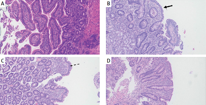
These features vary in severity and distribution, categorising the enteropathy into atrophic and non-atrophic forms. Table IV outlines the histological classifications utilised in coeliac disease, with the modified Marsh-Oberhuber grading scale being the most widely accepted. Figure 2 illustrates the distinct microscopic features of coeliac enteropathy.
Table IV
Histological classifications of coeliac disease enteropathy
Figure 2
Histopathological presentation of coeliac disease according to the Marsh-Oberhuber classification. A – Normal duodenal architecture. B, C – Partial villous atrophy (B, black arrow) and prominent intraepithelial lymphocytosis (C, dashed arrow), corresponding to Marsh 3a and 3b. D – Total villous atrophy corresponding to Marsh 3c.
Differential diagnosis of coeliac histopathology
The microscopic features of coeliac disease are non-specific; intraepithelial lymphocytosis and villous atrophy occur in various non-coeliac enteropathies and malabsorption syndromes. The core differential diagnosis includes autoimmune enteropathy, drug-induced enteropathy, common variable immunodeficiency (CVID), tropical sprue, collagenous sprue, infectious enteritis, and Crohn’s disease. Histological features that might be helpful in distinguishing these conditions are detailed in Table V [52–55].
Table V
Differential diagnosis of small intestinal enteropathies based on microscopic features
Definitive confirmation of coeliac disease and clinical phenotypes
A definitive diagnosis of coeliac disease in adults requires a positive anti-TG2 IgA serological result accompanied by intraepithelial lymphocytosis and villous atrophy (Marsh 3 architecture). In cases presenting with Marsh 2 histological changes, a high-positive anti-TG2 IgA confirms the diagnosis. If the serological concentration is low-positive with Marsh 2 architecture, clinicians must verify dietary gluten consumption, consider repeat serology, and perform a comprehensive differential diagnosis. Isolated Marsh 1 lesions (intraepithelial lymphocytosis with intact villi) are non-specific and typically do not represent coeliac disease, especially if serological screens are negative. However, if Marsh 1 coexists with high-positive anti-TG2 IgA, a diagnosis of coeliac disease is possible due to the potential patchiness (mosaicism) of the mucosal lesions, requiring further clinical evaluation.
Coeliac disease can be excluded in individuals maintaining a gluten-containing diet who present with negative serology and Marsh 0 histology, provided that mucosal biopsy sampling and processing adhered strictly to standard protocols.
In the non-invasive (no-biopsy) diagnostic pathway, coeliac disease is confirmed by a high-positive anti-TG2 IgA (≥ 10 times ULN) on the initial blood sample, backed by a positive serological result on a second independent sample in adults < 45 years old without alarm signs. In adults with an initial positive anti-TG2 IgA result that is < 10 times ULN, upper gastrointestinal endoscopy with duodenal biopsy remains mandatory to confirm the diagnosis [28].
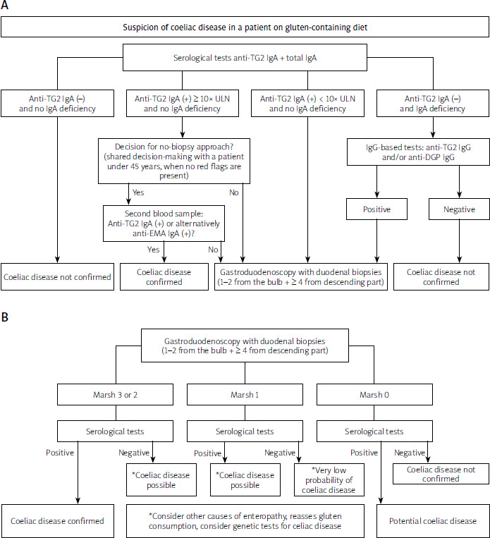
The standardised diagnostic algorithm for suspected coeliac disease in adults, alongside the integrated interpretation of histopathological and serological findings, is summarised in Figure 3.
Figure 3
A – Proposed diagnostic algorithm for suspected coeliac disease in adult patients. B – Suggested clinical interpretation of duodenal histopathological findings in relation to coeliac serology results
anti-TG2 – anti-tissue transglutaminase antibodies, anti-EMA – anti-endomysial antibodies, anti-DGP – anti-deamidated gliadin peptide antibodies, ULN – upper limit of normal.
Following clinical, serological, and histopathological correlation, several distinct clinical phenotypes of coeliac disease are recognised:
– Classical: Dominated by gastrointestinal symptoms of malabsorption, positive anti-TG2 IgA, and histopathological villous atrophy.
– Non-classical: Dominated by extraintestinal or systemic manifestations, positive anti-TG2 IgA, and histopathological villous atrophy.
– Potential: Intact, morphologically normal small-intestinal mucosa (Marsh 0) in individuals on a gluten-containing diet who have positive anti-TG2 IgA titres, with or without clinical symptoms.
– Silent: Completely asymptomatic individuals presenting with positive coeliac serology and histopathological villous atrophy.
– Seronegative: Histopathological evidence of villous atrophy (Marsh 3) in the presence of negative coeliac serology, with or without clinical symptoms, in which the enteropathy resolves following a strict gluten-free diet and all alternative causes of villous atrophy have been excluded [4, 38].
Coeliac crisis
A coeliac crisis is a rare, acute, and life-threatening manifestation of coeliac disease characterised by severe, rapid-onset malabsorption. Affected patients require hospitalisation, intravenous fluid resuscitation, electrolyte repletion, and the introduction of a strict gluten-free diet. Recognisable triggers include recent (in a few months) surgical interventions, severe infections, or pregnancy [56, 57].
Diagnostic modifications in patients on a gluten-free diet
The standard diagnosis of coeliac disease relies on serological testing and duodenal histopathology performed while the patient is actively consuming a gluten-containing diet. Clinicians should not recommend empirical implementation of a gluten-free diet prior to diagnostic testing. Symptomatic improvement on a self-prescribed gluten-free diet followed by symptom recurrence upon gluten reintroduction is insufficient to confirm a diagnosis of coeliac disease [30, 38, 58].
In patients on a gluten-free diet, coeliac-specific serological markers, endoscopic findings, and histopathological lesions usually normalise over time. However, they may persist if the diet has been maintained for a short duration (1–3 months). Prior to initiating a coeliac diagnostic workup in patients already adhering to a self-prescribed gluten-free diet, a formal gluten challenge must be performed. The protocol requires the daily consumption of 10 g per day of gluten for a minimum of 6–8 weeks. If a patient experiences severe symptom exacerbation, a modified short-term challenge consisting of 3 g of gluten daily for 2 weeks may be sufficient (one standard slice of wheat bread typically contains approximately 2.5–3.5 g of gluten) [59–61]. In this scenario, serological testing should be performed after 2 weeks of gluten reintroduction. If negative, it must be repeated at week 8. If serology is positive, upper GI endoscopy with duodenal biopsy is indicated. If serological testing remains negative at week 8, upper GI endoscopy should still be considered to investigate alternative causes of the patient’s symptoms. If a standard gluten challenge is unfeasible due to severe intolerance, HLA genetic testing represents a valuable diagnostic tool (Recommendation 3) [61, 62].
Recommendation 3. We do not recommend routine HLA-DQ2/8 genetic testing for the primary diagnosis of coeliac disease.
| Strength of recommendation: Strong |
| Quality of evidence: Moderate |
| Expert consensus vote: 72% Total Agreement (6 points), 22% Agreement (5 points), 6% Partial Agreement (4 points) |
Routine HLA-DQ2/HLA-DQ8 genetic testing is not recommended due to its low positive predictive value (approximately 35–40% of the general population carries these haplotypes without developing the disease). The diagnosis must rely on clinical, serological, and histopathological criteria. HLA testing is indicated when primary diagnostic results are discordant, borderline, or ambiguous (e.g., suspected seronegative coeliac disease, potential or silent coeliac disease, or evaluation of patients already on a gluten-free diet who refuse a formal gluten challenge).
Approximately 90–95% of coeliac patients express the HLA-DQ2.5 heterodimer. Individuals homozygous for HLA-DQ2.5 carry the highest relative risk of developing classical coeliac disease and severe clinical courses. The remaining 5–10% of coeliac patients carry either HLA-DQ2.2 or HLA-DQ8 [11, 13, 63].
The primary clinical utility of HLA testing lies in its negative predictive value (> 99%). A negative HLA-DQ2 and HLA-DQ8 test almost undoubtedly excludes coeliac disease, eliminating the need for periodic serological screening in asymptomatic first-degree relatives or individuals with associated risk conditions [63–65].
In a patient on a gluten-free diet, a positive HLA test necessitates a formal gluten challenge to establish the diagnosis, whereas a negative test result allows for avoidance of an unnecessary gluten challenge.
Recommendation 4. We do not recommend the routine use of radiological imaging or video capsule endoscopy for the primary diagnosis of coeliac disease.
| Strength of recommendation: Strong |
| Quality of evidence: Low |
| Expert consensus vote: 89% Total Agreement (6 points), 11% Agreement (5 points) |
Radiological imaging modalities are not indicated for routine coeliac screening or monitoring. However, cross-sectional imaging (CT or MR enterography) can detect structural abnormalities associated with advanced coeliac disease, including small bowel dilation, mural thickening, fold intussusception, mesenteric lymphadenopathy, vascular changes, or splenic atrophy. These imaging modalities are useful in differential diagnosis, evaluating mechanical complications, or resolving complex diagnostic queries [66].
Although video capsule endoscopy (VCE) demonstrates a pooled sensitivity of 89% and a specificity of 95% for detecting severe villous atrophy, neither VCE nor device-assisted enteroscopy is recommended for routine diagnostic screening or monitoring of mucosal healing. This is primarily due to VCE’s inability to obtain tissue biopsies for histopathological confirmation [67]. VCE and enteroscopy are reserved for specific clinical indications, particularly the evaluation of non-responsive or suspected refractory coeliac disease. They allow clinicians to assess the extent of mucosal disease, detect complications such as ulcerative jejunoileitis or enteropathy-associated T-cell lymphoma (EATL), and are useful in differential diagnosis [68]. Device-assisted enteroscopy is preferred when histopathological evaluation of small-bowel lesions is required [66].
Recommendation 5. We recommend a comprehensive assessment of nutritional status, including the evaluation of macro- and micronutrient deficiencies at the time of coeliac disease diagnosis.
| Strength of recommendation: Strong |
| Quality of evidence: Low |
| Expert consensus vote: 78% Total Agreement (6 points), 22% Agreement (5 points) |
Small intestinal mucosal injury in active coeliac disease frequently causes malabsorption and macro- and micronutrient deficiencies. Identifying these deficiencies at baseline guides appropriate nutritional supplementation, supports management of metabolic complications, and accelerates symptom resolution.
Baseline evaluation should include a complete blood count (as 2–5% of patients presenting with unexplained iron-deficiency anemia have underlying coeliac disease), iron biochemistry (serum iron, ferritin, total iron-binding capacity), serum vitamin B12, total calcium, vitamin 25(OH)D3, and serum folate (where accessible). These parameters represent the most common deficiencies in coeliac disease (iron deficiency affects up to 80% of patients, and vitamin B12 deficiency affects up to 41%). Given the shared autoimmune background, baseline screening must also include serum thyroid-stimulating hormone (TSH), fasting plasma glucose, and liver enzymes (serum aspartate and alanine transaminases – AST and ALT) [28, 30, 38, 69, 70]. Measurement of serum vitamin B6, zinc, and copper concentrations should be considered if clinical signs of deficiency are present.
Nutritional assessment must include a thorough physical examination and evaluation of the body mass index (BMI). Although protein-energy malnutrition is common in untreated classical coeliac disease, modern epidemiological data indicate that an increasing proportion of newly diagnosed adult patients present with an elevated BMI – approximately 28% are overweight and 11% are obese at baseline [71].
Anthropometric measurements and BMI calculation must be performed during the initial clinical consultation. Due to the wide availability of bioelectrical impedance analysis (BIA) devices, an advanced body composition analysis (quantifying fat mass, skeletal muscle mass, and total body water) should be integrated into the specialised dietary consultation [72]. Dual-energy X-ray absorptiometry (DXA) can also provide precise body composition metrics, but the availability of this technique is lower.
Treatment of coeliac disease
Recommendation 6. A strict, lifelong gluten-free diet is currently the only established and effective treatment for coeliac disease.
| Strength of recommendation: Strong |
| Quality of evidence: High |
| Expert consensus vote: 100% Total Agreement (6 points) |
The complete, permanent elimination of dietary gluten remains the sole effective therapeutic intervention for coeliac disease. Adherence to a strict gluten-free diet induces clinical symptom remission, normalises coeliac-specific serological changes, and promotes complete histological resolution of the small intestinal mucosa in most patients. The diet requires the total exclusion of all gluten-containing grains: wheat, rye, barley, and their strains (e.g., spelt, emmer) [28, 73].
The status of oats remains clinically nuanced. Pure oats do not inherently contain gluten. However, commercial oat supplies are heavily cross-contaminated by wheat, rye, or barley during standard agricultural cultivation, harvesting, transport, and milling. Clinical trials confirm that pure, certified gluten-free oats are well tolerated by most coeliac patients. Nevertheless, a small subset of individuals exhibits a distinct intraepithelial T-cell-mediated immune response triggered by avenin (the storage protein in oats), which shares structural homology with gluten, leading to symptom exacerbation and mucosal inflammation [74–79].
According to European and international legislative standards, foods labelled as “gluten-free” must contain less than 20 parts per million (ppm) of gluten (equivalent to 20 mg of gluten per kilogram of product). While an absolute safe lower threshold of gluten ingestion has not been defined, clinical evidence demonstrates that a daily intake of less than 10 mg of gluten is unlikely to induce mucosal damage in the majority of coeliac patients. Chronic ingestion of gluten above this threshold can trigger the immunopathogenic cascade and cause histological abnormalities, even in the complete absence of overt clinical symptoms [80, 81].
Strict dietary elimination resolves gastrointestinal symptoms in most patients. Amelioration or total cessation of diarrhoea is reported in up to 80% of classical coeliac patients within 60 days of initiating the diet [82].
Achieving adequate long-term dietary compliance requires a direct involvement of a specialised clinical dietitian [83]. Comprehensive nutritional education should be initiated immediately following diagnosis and must incorporate:
– identification of safe gluten-free grains and nutritional alternatives,
– recognition of hidden sources of gluten in processed products (e.g., spice blends, sauces, processed meats),
– mastering food label literacy,
– systematic management of cross-contamination risks in domestic, occupational, and social settings (e.g., dedicating separate cooking appliances, using separate preparation surfaces, toasters, and distinct storage areas),
– appropriate balancing the macro- and micronutrient composition of the diet [84–86].
Follow-up dietary consultations must be scheduled at regular intervals to monitor nutritional parameter evolution and maintain compliance with diet.
Pharmacological agents manufactured and registered within the European Union are generally considered safe for coeliac patients. When wheat starch is used as an excipient, its presence must be disclosed in the product leaflet, and its total gluten content remains well below the statutory threshold for gluten-free foods. Medications and dietary supplements registered outside standard pharmaceutical frameworks require individualised safety verification [87].
Although mucosal healing enhances nutrient absorption, a strict gluten-free diet can introduce unique nutritional deficiencies due to its restrictive nature. Patients frequently demonstrate insufficient intakes of iron, folate, vitamin B12, vitamin D, copper, and zinc [88–93]. Furthermore, commercially available ultra-processed gluten-free products are often low in dietary fibre and high in saturated fats and simple sugars. This structural profile can increase the risk of developing lifestyle diseases, e.g., metabolic syndrome, and hypercholesterolemia in coeliac patients [94, 95].
Adherence to a gluten-free diet is associated with significant financial burdens and risks of social isolation. Access to professional psychological support and active participation in patient advocacy groups enhance treatment compliance and improve health-related quality of life [84, 96, 97].
Investigational therapies currently in clinical development include use of proteolytic enzymes [98], immunotherapy [99, 100], and targeted microbiome modulations [101]. None of these therapeutic modalities have received regulatory approval, and they cannot be utilised outside formal clinical trials [102].
Dietary management across clinical phenotypes
A gluten-free diet is mandatory for all patients diagnosed with classical, non-classical, or silent coeliac disease. In potential coeliac disease, a gluten-free diet is indicated if the patient presents with persistent symptoms. In asymptomatic potential coeliac disease, initiating a gluten-free diet can be considered if high-positive anti-TG2 IgA or anti-EMA IgA concentrations persist. In confirmed seronegative coeliac disease, particularly when symptomatic, a strict gluten-free diet is recommended after excluding alternative enteropathies and verifying HLA susceptibility. In these cases, complete mucosal healing demonstrated via follow-up biopsy after 1–3 years of dietary elimination confirms the diagnosis [38, 103].
Recommendation 7. In patients with persistent symptoms or laboratory abnormalities despite 6–12 months of gluten-free diet, we recommend a re-evaluation of the primary diagnosis and a formal assessment of dietary adherence.
| Strength of recommendation: Strong |
| Quality of evidence: High |
| Expert consensus vote: 89% Total Agreement (6 points), 11% Agreement (5 points) |
Clinical data indicate that 7–36% of adult coeliac patients experience persistent symptoms or unresolving laboratory abnormalities despite 6–12 months of dietary restriction. This clinical scenario is defined as non-responsive coeliac disease (NRCD) [82]. The initial step in managing suspected NRCD requires verifying the primary diagnosis. This includes a re-evaluation of the baseline serology, histopathology, and genetic profiles.
Once the primary diagnosis of coeliac disease is confirmed, serological testing must be performed to assess dietary compliance [29, 30, 70]. Assays quantifying gluten immunogenic peptides (GIP) in urine or stool specimens provide direct evidence of recent gluten exposure; however, their clinical use is currently constrained by variable commercial availability.
Persistent positive serological concentration and/or failure to achieve clinical improvement after 12 months of dietary restriction strongly indicate ongoing, inadvertent gluten ingestion [29, 30, 70, 104]. Inadvertent or intentional dietary contamination accounts for 35–50% of all NRCD cases, representing the single most common cause of non-responsiveness [61, 105]. In these scenarios, a meticulous evaluation of dietary habits, food label comprehension, and nutritional status using targeted laboratory and anthropometric profiling is indicated. Self-reported adherence obtained via a structured clinical history remains a cost-effective, non-invasive indicator that correlates well with mucosal injury markers [106].
Barriers to strict dietary compliance include insufficient patient education, hidden cross-contamination in the food supply, and inadequate or ambiguous food labelling. Psychological distress, feelings of social exclusion during dining out, and financial constraints due to the high cost and limited availability of specialised gluten-free foods further reduce long-term compliance [107–110].
During clinical follow-up, providers must identify these dietary vulnerabilities [109]. Ensuring that patients understand cross-contamination dynamics and have adequate food label literacy is critical to maintaining long-term dietary adherence [110]. Domestic hygiene principles – such as separating gluten-free flour storage, utilising dedicated preparation surfaces, and thoroughly cleaning shared kitchen equipment – must be verified [111]. Misinterpretations regarding the safety of ingredients can lead to unrecognised, chronic gluten exposure [29, 30, 70, 104].
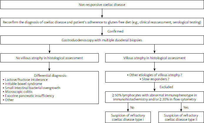
If gastrointestinal symptoms persist despite verified, strict dietary compliance, clinicians must systematically investigate alternative, overlapping gastrointestinal comorbidities (Figure 4) [112].
Figure 4
Schematic representation of the standardised diagnostic pathway for non-responsive coeliac disease (NRCD), including differentiation of refractory coeliac disease (RCD) subtypes
Recommendation 8. Refractory coeliac disease is defined by persistent clinical symptoms and villous atrophy despite verified, strict adherence to a complete gluten-free diet for a minimum of 12 months. The accurate differentiation and diagnosis of refractory coeliac disease subtypes require specialised histopathological evaluation, including immunohistochemical staining, flow cytometry, and/or molecular clonality analysis.
| Strength of recommendation: Weak |
| Quality of evidence: Low |
| Expert consensus vote: 83% Total Agreement (6 points), 11% Agreement (5 points), 6% Partial Agreement (4 points) |
Refractory coeliac disease (RCD) is a rare clinical entity affecting less than 1% of all patients diagnosed with gluten-sensitive enteropathy [28, 30, 113]. RCD should be diagnosed cautiously. The condition is suspected in patients with a verified primary diagnosis of coeliac disease who demonstrate persistent, severe symptoms and unresolving villous atrophy despite documented adherence to a strict gluten-free diet for at least 12 months [30, 113, 114]. Affected patients commonly present with marked quantitative and qualitative malnutrition, with peripheral oedema in severe cases and occasional ascites due to hypoalbuminemia.
Laboratory findings in RCD include persistent anemia, elevated liver transaminases, multi-mineral deficiencies, or thrombocytosis. Thrombocytosis may indicate underlying hyposplenism in severe clinical courses.
Prior to establishing a diagnosis of RCD, the accuracy of the baseline coeliac diagnosis must be verified [114]. A thorough evaluation of dietary compliance by a specialised dietitian is mandatory, as persistent inadvertent gluten ingestion remains the leading cause of ongoing mucosal injury [30, 66]. Elevated or rising coeliac serological concentrations point toward continuous gluten exposure.
The next diagnostic step requires gastroduodenoscopy with extensive tissue sampling. Clinicians should obtain 1–2 biopsies from the duodenal bulb, at least 4 specimens from the descending duodenum, and target any macroscopically abnormal mucosal areas. For cases with a high clinical suspicion of RCD, tissue specimens should be processed for standard histopathology and immunohistochemistry. Additionally, parallel fresh biopsy specimens should preferably be preserved – ideally frozen at –80°C or placed in nucleic acid–stabilising buffers for molecular assays, or submitted in saline or other dedicated transport media for flow cytometric analysis. Confirmatory diagnostics require advanced immunomorphological, molecular profiling.
If a patient presents with persistent symptoms but demontrates complete mucosal healing (intact villi and no significant intraepithelial lymphocytosis), coeliac disease does not seem to be the cause of non-responsiveness. Clinicians must initiate a diagnostic workup for non-coeliac gastrointestinal comorbidities (Figure 4) [28, 30, 113, 114].
A diagnosis of RCD can be considered only when persistent severe enteropathy (villous atrophy) is histopathologically documented in a symptomatic patient with verified strict dietary adherence for several months. In some adult patients, complete mucosal recovery may require more than 12–24 months. However, such slow-healing individuals typically demonstrate clinical improvement, and their overall condition is not severe. Additionally, in cases of persistent villous atrophy, alternative causes must be excluded (Table V).
Refractory coeliac disease is classified into two distinct immunophenotypic subtypes: Type I (RCD-I) and Type II (RCD-II). This differentiation is prognostically critical, as RCD-II is considered a low-grade intraepithelial lymphoproliferative disorder. It is associated with a severe clinical course and a high risk of progression to enteropathy-associated T-cell lymphoma (EATL), development of ulcerative jejunoileitis, or functional hyposplenism. Consequently, confirmation of RCD, particularly type II, necessitates advanced staging with magnetic resonance (MR) enterography or computed tomography (CT) enterography, as well as device-assisted enteroscopy and/or VCE, provided gastrointestinal patency has been confirmed [28, 30, 113, 114].
The standard light microscopic appearance of RCD resembles untreated coeliac disease, featuring severe villous atrophy, crypt hyperplasia, and intraepithelial lymphocytosis (> 25–30 IELs/100 enterocytes). In RCD-I, the intraepithelial lymphocytes maintain a normal surface T-cell immunophenotype (CD3+, CD8+) and demonstrate polyclonal T-cell receptor gene rearrangements (TCRαβ). Conversely, RCD-II is characterised by a clonal expansion of phenotypically aberrant intraepithelial lymphocytes. These neoplastic cells can infiltrate deeper mucosal layers, the gastric or colonic mucosa (causing lymphocytic gastritis or colitis), mesenteric lymph nodes, peripheral blood, and extraintestinal organs.
On immunohistochemical evaluation, the aberrant IELs in RCD-II demonstrate intracellular (cytoplasmic) expression of CD7+, CD103+ and CD3+ but characteristically lack surface expression of CD4, CD8, and surface CD3. They display monoclonal rearrangements of the TCR gene.
The diagnosis of RCD-II requires the detection of this aberrant immunophenotype in ≥ 50% of total intraepithelial lymphocytes via IHC, or ≥ 20% via multi-colour flow cytometry. Although flow cytometry on fresh tissue is highly sensitive, semi-quantitative IHC evaluation of CD3 and CD8 on standard formalin-fixed tissue is sufficient for routine clinical practice. Duodenal biopsy material in RCD-II typically features more severe, diffuse villous atrophy compared to the patchier changes seen in RCD-I (96% vs. 50% of cases). The primary differential diagnosis of RCD-II includes RCD-I and overt EATL. NKp46 has emerged as a novel biomarker to assist in identifying RCD-II and EATL clones.
Enteropathy-associated T-cell lymphoma (EATL) is a severe neoplastic complication of RCD-II, with a cumulative transformation rate of 50–60% within 4–6 years following the diagnosis of Type II disease. It typically presents in patients >60 years of age and affects both sexes equally. Histologically, EATL is characterised by a diffuse, neoplastic transmural infiltration of atypical, pleomorphic T-lymphocytes situated within a prominent inflammatory background containing histiocytes, plasma cells, and eosinophils. The malignant cells vary from medium-sized to large immunoblastic or anaplastic elements.
While initially confined to the mucosa, EATL typically causes deep transmural invasion of the small intestinal wall, forming tumour masses. This infiltration leads to intestinal strictures, deep ulcerations, acute mechanical obstruction, or perforation. Angioinvasion and associated ischaemic necrosis are common features. Neoplastic cells express cCD3, CD7, NKp46 and CD103 (mirroring the baseline RCD-II clone), alongside markers including TIA1, granzyme B, perforin, and CD30, with a high Ki-67 mitotic index (usually > 50%). They typically lack surface CD4, CD5, CD8, CD56, ALK, and EBER, as well as TCRαβ/TCRγδ (though up to 25% may express CD8 or TCR proteins). Molecular analysis confirms monoclonal TCR gene rearrangement. Given the characteristic clinical context, structural morphology, and immunophenotypic markers, EATL is distinct from other peripheral T-cell lymphomas. Management requires systemic multi-drug chemotherapy, occasionally combined with emergency surgical resection for obstruction or perforation; however, the long-term oncological prognosis remains poor [28, 52, 113].
Recommendation 9. There are no standardised, universally accepted guidelines for the treatment and long-term surveillance of refractory coeliac disease. Management must be customised according to the specific RCD subtype and clinical severity. Steroids represent the first-line pharmacological intervention in most cases. Long-term surveillance requires systematic evaluation for oncological complications and effective management of nutritional deficits.
| Strength of recommendation: Weak |
| Quality of evidence: Very Low |
| Expert consensus vote: 72% Total Agreement (6 points), 22% Agreement (5 points), 6% Partial Agreement (4 points) |
The primary objectives of RCD management are achieving clinical symptom remission, reversing nutritional deficiencies, and preventing transformation into EATL.
Steroids are the first-line pharmacological option. In RCD-I, open-capsule budesonide is the preferred agent [30, 113, 114]. The standard induction dose is 9 mg daily. The American Gastroenterological Association suggests a split-dose protocol (3 mg TID): opening the capsules and mixing of the microgranules in applesauce (to target the proximal duodenum) for the first dose and in water for the second daily dose, while the final dose is swallowed as an intact capsule [113]. No optimal maintenance duration or tapering schedules have been established. A prolonged induction phase (8–12 weeks) followed by a gradual taper is preferred. Tapering must be guided by clinical and laboratory parameters. Clinical relapse is common upon steroid discontinuation, often requiring long-term maintenance of the lowest effective dose. Systemic steroids (e.g., prednisone) are reserved for severe cases due to their adverse effect profile [30, 113, 114].
In steroid-refractory RCD-I, second-line immunomodulatory therapies may be considered, though clinical data are limited. Weak evidence supports the use of thiopurines (azathioprine at 2–2.5 mg/kg/day, 6-mercaptopurine at 1 mg/kg/day, or thioguanine at 0.3 mg/kg/day). Individual case reports note successful deployment of controlled-release mesalamine, elemental diets, or infliximab.
The treatment of RCD-II remains challenging. Induction is typically attempted with budesonide. In severe, steroid-resistant RCD-II, cladribine or infliximab may be considered. Limited evidence also supports the use of autologous stem cell transplantation in selected cases. Thiopurines are contraindicated in RCD-II because they may accelerate transformation into overt EATL. Targeted monoclonal antibodies directed against interleukin-15 (e.g., ordesekimab) are currently under clinical investigation. None of the available therapeutic options have been definitively proven to prevent transformation into EATL.
Functional hyposplenism is a serious complication of untreated RCD, particularly Type II. Diagnostic and prophylactic measures for hyposplenism are described in another section of these guidelines.
Parallel to pharmacotherapy, intensive nutritional support is critical in RCD. Patients must undergo systematic laboratory screens to detect iron, vitamin B12, folate, fat-soluble vitamin (A, D, E, K), zinc, selenium, and vitamin B6 deficiencies. Personalised dietary optimisation must be implemented immediately; severe cases may require enteral and/or total parenteral nutrition. Screening, prevention, and management of secondary osteopenia and osteoporosis are essential, as bone loss is exacerbated by chronic enteropathy and prolonged steroid use [30, 113, 114].
Surveillance protocols for RCD are empirical. The American Gastroenterological Association suggests clinical, laboratory, and nutritional reviews every 3 months until stable remission is achieved, followed by bi-annual evaluations. Follow-up gastroduodenoscopies with multiple duodenal biopsies should be performed 3– 6 months after initiating treatment, and annually thereafter in stable responders. If a patient fails to respond, immediate endoscopic re-evaluation within 3–6 months is indicated to guide therapy modification. Any clinical suspicion of lymphoproliferative transformation requires immediate cross-sectional imaging (MR or CT enterography), VCE if gastrointestinal patency has been confirmed, device-assisted enteroscopy, and/or positron emission tomography (PET-CT) [113].
Monitoring and follow-up
Recommendation 10. We recommend systematic clinical, laboratory (including serological), and endoscopic surveillance for all patients diagnosed with coeliac disease. Follow-up serological testing should be performed at 6 and 12 months post-diagnosis, and annually thereafter. A follow-up gastroduodenoscopy with duodenal biopsy for histopathological evaluation should be considered 1–2 years after initiating a gluten-free diet.
| Strength of recommendation: Strong |
| Quality of evidence: Moderate |
| Expert consensus vote: 72% Total Agreement (6 points), 28% Agreement (5 points) |
The goals of coeliac disease management are achieving sustained clinical symptom resolution, normalising coeliac-specific serological markers, and promoting complete small intestinal mucosal healing. Surveillance must encompass all three axes. Because a lifelong gluten-free diet is the only effective treatment, evaluating dietary compliance via clinical interviews and/or dietary logs is needed at every follow-up visit [82, 115–118].
The initial follow-up consultation should be scheduled 3–6 months after diagnosis. This evaluation tracks clinical symptom resolution, dietary adaptation, and initial serological response (anti-TG2 IgA, or anti-TG2/anti-DGP IgG in patients with confirmed selective IgA deficiency). Reassessment of baseline laboratory abnormalities (e.g., anemia, iron parameters, vitamin B12, folate, or vitamin D3) should be considered. Subsequent routine follow-up is scheduled at 12 months post-diagnosis and annually thereafter, incorporating clinical, serological, metabolic, and anthropometric profiling [29, 30, 70, 73].
Dietary gluten elimination leads to a predictable decline in concentration of antibodies. Low-baseline concentration may normalise within weeks. At 6–12 months post-diet initiation, serology is negative in approximately 80% of compliant patients, while remaining low-positive in 20%. After 5 years of strict dietary restriction, over 90% of patients achieve complete seronegativity [119, 120].
Clinical data indicate that after 12 months of dietary restriction, 75% of patients achieve clinical symptom remission and significant villous recovery, although persistent intraepithelial lymphocytosis remains present in 50–70% of cases [121, 122].
A follow-up gastroduodenoscopy with duodenal biopsy should be considered 1–2 years after diagnosis, customised to the patient’s individual risk profile. The time required to achieve complete small intestinal mucosal healing varies. Approximately 85% of compliant patients achieve complete histological resolution after 5 years of dietary restriction [58, 121, 122, 123].
Although complete mucosal healing is the optimal therapeutic target associated with reduced long-term metabolic and oncological complications, architectural abnormalities can persist even in highly compliant patients. Performing a follow-up endoscopy to evaluate mucosal healing is essential in patients with persistent or recurrent clinical symptoms, unexplained alarm signs, or persistently positive serological parameters despite verified dietary restriction [113]. Advanced monitoring protocols for suspected or confirmed RCD are detailed in Recommendation 9.
Recommendation 11. A confirmed diagnosis of coeliac disease represents an indication for the regular monitoring of the patient’s nutritional status.
| Strength of recommendation: Strong |
| Quality of evidence: Low |
| Expert consensus vote: 72% Total Agreement (6 points), 22% Agreement (5 points), 6% Partial Agreement (4 points) |
Following the implementation of a strict gluten-free diet, a patient’s nutritional status and micronutrient parameters improve gradually, though they might not return to baseline levels [124]. Persistent enteropathy is documented in patients diagnosed after 45 years of age, but it also affects approximately 20% of patients diagnosed under 21 years of age despite reported dietary compliance [125–127]. Patients adhering to a strict gluten-free diet remain at a higher risk for nutritional deficiencies than the healthy population due to the lower nutrient density of commercial gluten-free alternatives [128].
Given that the most common nutritional deficiencies in coeliac disease include iron, calcium, zinc, vitamin B12, vitamin D, and folate, these parameters should be considered for regular monitoring during follow-up. Clinicians should prioritise reassessing baseline abnormalities, tailored to the evolving clinical context (e.g., verifying serum vitamin B12 level if neurological symptoms develop, even if baseline levels were normal) [129–134].
Nutritional surveillance requires an annual anthropometric review, including weight tracking and BMI calculation [29].
Coeliac disease: major clinical challenges
Recommendation 12. In patients with coeliac disease presenting with selected extraintestinal manifestations and/or comorbidities, we recommend that diagnostic evaluation and clinical management be carried out by a multidisciplinary team of specialists.
| Strength of recommendation: Strong |
| Quality of evidence: Low |
| Expert consensus vote: 83% Total Agreement (6 points), 11% Agreement (5 points), 6% Partial Agreement (4 points) |
Coeliac disease frequently coexists with diverse extraintestinal manifestations and multi-organ comorbidities. These involve the hepatobiliary system, alongside oncological, neurological, psychiatric, dermatological, reproductive, and metabolic conditions. They are especially prevalent among patients with an established autoimmune background. Recognising these associations optimises diagnostic pathways, and managing these complex interactions requires structured collaboration across medical specialties.
Hepatobiliary diseases
The relationship between coeliac disease and hepatic dysfunction is bidirectional. Elevated serum aminotransferase activity can stem from concurrent primary liver diseases or direct coeliac-induced hepatic injury [135]. Hypertransaminasemia is observed in 13–60% of patients with undiagnosed or untreated coeliac disease, and higher aminotransferase levels correlate with the severity of intestinal barrier damage [136].
A defining feature of coeliac-associated liver injury (often termed coeliac hepatitis) is its resolution following adherence to a strict gluten-free diet. Unexplained, persistent elevation of serum transaminases requires screening for underlying coeliac disease. Conversely, if liver enzyme abnormalities persist in a coeliac patient despite verified dietary compliance, clinicians must suspect a concurrent chronic liver disease (CLD) or/and advanced major adverse liver outcomes (MALO), such as cirrhosis, warranting a comprehensive hepatology workup [137–139].
Coeliac disease patients carry an increased relative risk for developing primary autoimmune liver diseases. This is driven by shared genetic susceptibility, intestinal dysbiosis, and chronic enteropathy-mediated impairment of the gut-vascular barrier. This “leaky gut” allows for increased translocation of dietary antigens, bacterial components, and endotoxins into the portal circulation, activating immune system and triggering pro-inflammatory cytokine cascades.
Unexplained hypertransaminasemia requires evaluation for autoimmune hepatitis (AIH), whereas cholestatic laboratory or imaging profiles require evaluation for primary biliary cholangitis (PBC) or primary sclerosing cholangitis (PSC) [10, 140, 141]. Patients with confirmed AIH or PBC should be screened for coeliac disease. A meta-analysis of 8 clinical trials encompassing 567 AIH patients identified a biopsy-confirmed coeliac disease prevalence of 4.1% [142]. The pathogenetic link is supported by the identification of anti-TG2 IgA immune complexes co-localised in both duodenal and hepatic tissue biopsies in affected individuals [143].
In PBC, which exhibits bidirectional prevalence tracking with coeliac disease, molecular mimicry triggered by chronic portal exposure to bacterial antigens may drive the autoimmune attack against cholangiocytes once the immunological tolerance threshold is breached [144].
The interaction between coeliac disease, metabolic dysfunction-associated steatotic liver disease (MASLD), and alcohol-related liver injury is equally complex. Initiating a gluten-free diet can cause rapid weight gain if the diet relies on processed alternatives rich in simple sugars and saturated fats, predisposing patients to metabolic syndrome and secondary hepatic steatosis. Many patients are overweight or obese at diagnosis, and dietary elimination might not normalise body composition. An increasing trend in baseline BMI is also observed in paediatric coeliac cohorts [66, 145].
While structured data detailing alcohol misuse in coeliac disease are limited, the disease is associated with reduced health-related quality of life and higher rates of mood disorders, anxiety, panic disorder, social phobia, and major depression. These psychiatric vulnerabilities are established risk factors for alcohol abuse, indirectly increasing the risk for alcohol-related liver injury [146, 147].
A large 2025 population-based cohort study with a median follow-up of 16 years demonstrated that coeliac disease patients carry a two-fold increased risk for developing alcohol-related liver disease compared to matched controls. This risk peaked near the time of coeliac diagnosis and persisted for over 25 years. The risk for any aetiology-independent CLD and MALO was significantly elevated, with compensated or decompensated cirrhosis representing the most frequent presentation. Coeliac disease was associated with an increased risk for hepatocellular carcinoma (HCC) and liver-related mortality. Chronic liver disease was identified in 23.8% of coeliac patients prior to an index MALO event, driven primarily by autoimmune and alcohol-related aetiologies. Coeliac disease did not increase the risk for viral hepatitis [135].
Importantly, patients with persistent villous atrophy on follow-up biopsies at 6 months and 5 years post-diagnosis demonstrated a significantly higher risk for liver-related mortality [135].
In summary, coeliac disease increases the risk of chronic liver disease regardless of aetiology, including autoimmune, metabolic, and alcohol-related diseases. This highlights the need for clinical vigilance regarding hepatic parameters in coeliac patients. Baseline liver function tests should be performed in all patients, regardless of their primary clinical phenotype, to ensure early detection of transaminasemia or cholestasis, optimise interventions, and prevent progression to advanced CLD or MALO. High-risk sub-cohorts with concurrent metabolic or autoimmune conditions require close monitoring.
Autoimmune comorbidities
The prevalence of coeliac disease among patients with established autoimmune disorders is 10 to 30 times higher than in the general population, frequently presenting as atypical or silent phenotypes [148]. Early identification optimises control of both conditions. Family members of coeliac patients exhibit higher rates of rheumatoid arthritis, juvenile idiopathic arthritis, hypothyroidism, insulin-dependent diabetes mellitus, alopecia areata, and inflammatory bowel disease [148–152].
Malignancies
Coeliac disease is associated with an increased risk for specific malignancies, particularly certain types of intestinal adenocarcinomas [123, 153], lymphoproliferative disorders, and neoplasms of the oral cavity and pharynx. This risk is highest in patients diagnosed after 40 years of age [154–156]. Consequently, adhering to age-appropriate oncological screening is essential. While specific guidelines for accelerated screening are not established, early implementation of surveillance protocols is clinically justified.
The chronic mucosal inflammation, persistent pro-inflammatory cytokine release, continuous antigenic driving, and secondary genomic instability seen in untreated coeliac disease create a pro-oncogenic environment. Persistent mucosal damage and intraepithelial T-cell activation, particularly in RCD, predispose patients to malignant transformation. RCD-II is a recognised pre-neoplastic state for EATL [157, 158]. However, cancers develop in the course of coeliac disease regardless of its type.
Additionally, the risk for small bowel adenocarcinoma – a rare tumour representing less than 5% of all gastrointestinal cancers – is significantly elevated in coeliac patients compared to the general population [157, 159]. High-risk individuals include those diagnosed with coeliac disease after 40 years of age who experience persistent symptoms despite verified adherence to a strict gluten-free diet [157].
Reproductive disorders and infertility
Untreated coeliac disease can cause unexplained infertility, which may resolve following strict dietary elimination. Pathophysiological mechanisms include chronic malabsorption-induced nutrient depletion, systemic autoimmune activity, and secondary sexual dysfunction. Females with untreated coeliac disease experience higher rates of ovarian dysfunction, delayed menarche, severe dysmenorrhea, secondary amenorrhea, premature menopause, and recurrent spontaneous miscarriages [24, 160].
The increasing use of assisted reproductive technologies, such as intracytoplasmic sperm injection and in vitro fertilisation, has highlighted the need to manage modifiable factors influencing embryonic implantation and early pregnancy outcomes. Active, untreated coeliac disease can disrupt reproductive health and alter the intestinal microbiome. Secondary intestinal dysbiosis can impair systemic hormone metabolism and sustain a pro-inflammatory state that compromises implantation [161].
Accordingly, scientific bodies including the American College of Obstetricians and Gynecologists (ACOG) and the European Society of Human Reproduction and Embryology (ESHRE) recommend screening for coeliac disease in females presenting with unexplained infertility or recurrent miscarriages, and matching gastrointestinal complaints (chronic diarrhoea, malabsorption, iron or vitamin B12 deficiencies) [162–164]. Diagnosing coeliac disease during pregnancy requires immediate dietary elimination to protect foetal growth and mitigate maternal nutritional deficits [160, 161, 164].
In males, untreated coeliac disease can cause reproductive dysfunction through semen quality abnormalities (reduced sperm concentration, impaired motility, morphological defects) and erectile dysfunction. In addition to active malabsorption, deficiencies in vital trace elements, such as selenium and zinc, contribute to male infertility [160].
Down syndrome and Turner syndrome
Like other autoimmune diseases, coeliac disease is more common in people with Down syndrome than in the general population. Diagnosing coeliac disease in this cohort is challenging because baseline features of Down syndrome – including linear growth failure, chronic anemia, intermittent diarrhoea, epilepsy, neurodevelopmental delays, and severe constipation – overlap with coeliac symptoms [165]. Emotional and behavioural shifts caused by active coeliac enteropathy are often underestimated or attributed to the primary genetic syndrome. Similarly, females with Turner syndrome carry an increased risk for autoimmune disorders, including coeliac disease, with a prevalence twice that of the general female population. Routine screening facilitates early detection and intervention.
Dermatitis herpetiformis (Duhring’s disease)
Dermatitis herpetiformis is a cutaneous manifestation of gluten-sensitive enteropathy. It is an autoimmune, IgA-mediated blistering dermatosis directly triggered by dietary gluten ingestion. This condition is most common in Caucasian populations due to the high prevalence of predisposing HLA alleles and high dietary wheat consumption.
The pathogenesis involves genetic, environmental, and immunobiological factors. Diagnosis relies on clinical features (intensely pruritic, burning, symmetric polymorphic eruptions consisting of erythematous papules, urticarial plaques, and small vesicles concentrated on the extensor surfaces of the elbows, knees, buttocks, posterior scalp, and sacrum), skin biopsy of perilesional tissue for histopathology, and direct immunofluorescence microscopy. The detection of granular IgA deposition clustered within the dermo-epidermal junction is pathognomonic. Patients also present with positive serum anti-TG2 IgA concentration [166, 167].
While coeliac disease diagnoses have risen over recent decades, the incidence of overt dermatitis herpetiformis has decreased, likely due to earlier serological detection of coeliac disease. The epidemiological and immunobiological links between coeliac disease and dermatitis herpetiformis are well established. However, coeliac disease is also associated with higher rates of other dermatological conditions with less defined immunopathogenic links, including alopecia areata, atopic dermatitis, psoriasis, rosacea, vitiligo, and chronic urticaria [167–169].
Neurological and psychiatric manifestations
Gluten-related disorders, including coeliac disease and NCGS/non-coeliac gluten sensitivity, can cause extraintestinal neuropsychiatric manifestations. These include sensory peripheral neuropathy, cerebellar ataxia, and cortical myoclonus. Less common features include chronic headaches, cognitive impairment (“brain fog”), and seizures.
Neurological injury can result from autoimmune activity, where coeliac-induced autoantibodies cross-react with neural antigens due to structural homology with gluten peptides. Additionally, malabsorption-induced deficiencies in B-complex vitamins (particularly vitamin B12 and folate) can exacerbate neuropathy and cognitive issues, though they are rarely the primary cause. Diagnosing these conditions is challenging when gastrointestinal symptoms are minor or absent [9, 29, 170–172].
Coeliac disease is also linked to psychiatric conditions, including major depressive disorder, bipolar disorder, schizophrenia, autism spectrum disorders, attention deficit hyperactivity disorder, and eating disorders [173–175]. Psychological or psychiatric evaluation should be integrated into the care of selected coeliac patients, though the underlying pathophysiological mechanisms require further study.
Gluten ataxia (GA) is the most common neurological manifestation of gluten sensitivity and warrants distinct clinical focus. Ataxia involves impaired coordination due to structural damage to the cerebellum (cerebellar ataxia) or the posterior columns of the spinal cord responsible for proprioception (sensory ataxia). Symptoms include balance disorders, gait and limb movement coordination, abnormal body posture, dysarthria or nystagmus [176–178].
Gluten ataxia is a leading cause of sporadic idiopathic ataxia, accounting for 15% of all ataxias and up to 40% of sporadic idiopathic cohorts [179]. The prevalence of GA among all diagnosed coeliac patients is estimated at 0–6% [180]. A prospective study of 100 newly diagnosed coeliac patients identified subjective gait instability in 24%, persistent sensory neuropathic symptoms in 12%, and chronic headaches in 42% [181].
Gluten ataxia, originally defined as idiopathic, sporadic ataxia with serum AGA IgG or IgA antibodies, typically presents as a slowly progressive, insidious cerebellar ataxia with an average age of onset near 50 years. It is rare in children. Patients frequently present with concurrent autoimmune conditions. Importantly, less than 10% of GA patients report gastrointestinal symptoms, and approximately 40% demonstrate structural enteropathy on duodenal biopsy. The clinical presentation is usually mild, dominated by lower limb dysmetria and gait ataxia. Rare presentations include focal myoclonus, palatal tremor, or opsoclonus [9, 180, 182, 183].
Neuropathologically, GA causes a distinct pattern of permanent neurological injury characterised by the loss of cerebellar Purkinje cells. Post-mortem studies demonstrate Purkinje cell depletion, cerebellar cortical granular layer degeneration, astrocytic gliosis within the dentate nucleus and inferior olivary bodies, and variable inflammatory involvement of supratentorial tracks, the spinal cord, and peripheral nerves [184].
Gluten ataxia should be suspected in cases of late-onset sporadic ataxia, irrespective of enteropathy status. Diagnosis is difficult due to the lack of isolated, specific biomarkers. A subset of patients shows elevated IgG or IgA AGA, anti-TG2, or anti-tissue transglutaminase 6 (anti-TG6) antibodies. Anti-neural autoantibodies directed against glycine receptors, glutamic acid decarboxylase (GAD) or anti-DGP may also be detected. None of these markers are entirely specific, and false-positive results can occur in other neuro-inflammatory or autoimmune conditions [185].
A study evaluating serum anti-TG2 IgA, anti-TG6 IgA/IgG, and AGA IgA/IgG in 476 patients with idiopathic cerebellar ataxia versus 195 healthy controls found no significant differences between the cohorts using standard cutoffs. However, using lower threshold adjustments, patients with idiopathic ataxia displayed a higher prevalence of positive AGA titres than controls [186].
A 2025 study identified elevated anti-TG6 IgA titres in the cerebrospinal fluid (CSF) of GA patients compared to controls. CSF antibody levels did not correlate with serum titres. The presence of intrathecal plasma cells and anti-TG6 antibodies in GA is associated probably with prolonged disease duration and chronic gluten exposure [187].
Neurofunctional and structural imaging (CT, MRI) help confirm cerebellar disease and exclude alternative aetiologies. In the cohort of 100 newly diagnosed coeliac patients noted above, structural MRI abnormalities were detected in 60%, and 47% demonstrated metabolic deficits on cerebellar MR spectroscopy. Serum anti-TG6 autoantibodies were identified in 40% of these patients, correlating with significant subcortical brain atrophy compared to antibody-negative peers. Follow-up MR spectroscopy after 12 months on a strict gluten-free diet demonstrated partial metabolic regression of the cerebellar lesions [181].
Long-term follow-up of 30 patients from this cohort at 7 years identified a reduction in headache prevalence (from 47% to 20%) but an increase in objective coordination deficits (from 27% to 47%). Persistent gluten-specific autoantibodies were detected in 50% of the reviewed patients. Follow-up imaging demonstrated higher rates of cerebellar grey matter atrophy in patients with persistent positive serology compared to those who achieved seronegativity. This demonstrates that coeliac patients with suboptimal dietary compliance and persistent seropositivity are at a high risk for progressive cerebellar atrophy, highlighting the necessity of strict dietary monitoring.
Strict adherence to a gluten-free diet and subsequent serological normalisation can stabilise GA progression and promote partial neural recovery. However, clinical reversal may be incomplete because Purkinje cell loss and cerebellar atrophy are irreversible. In patients with verified dietary compliance for at least 1 year who experience progressive neurological decline, intravenous immunoglobulin therapy or specialised immunosuppressive protocols may be considered.
Current American guidelines recommend screening for underlying coeliac disease using standard serological panels in patients presenting with peripheral neuropathy, unexplained ataxia, refractory epilepsy, “brain fog”, or idiopathic recurrent migraines [29, 30, 138]. Similarly, consensus guidelines for progressive ataxias recommend screening all idiopathic sporadic cerebellar ataxia cases for gluten sensitivity, prioritising sensitive anti-TG6 assays where available. Patients demonstrating serological evidence of gluten sensitivity must be advised to initiate a strict gluten-free diet and receive structured serological follow-up, regardless of their enteropathy status [188].
In summary, coeliac disease is a systemic disorder that benefits from a multidisciplinary care model. To ensure comprehensive care, patients should be managed in centres that provide access to specialists in gastroenterology, dermatology, neurology, psychiatry, endocrinology, diabetology, gynaecology, oncology, and other relevant specialties as needed, as well as to dietetic and clinical psychological support.
Recommendation 13. We suggest diagnostic testing for osteoporosis followed by long-term surveillance in asymptomatic coeliac disease patients after 30–35 years of age, as well as in younger patients presenting with clinical signs of osteoporosis or features of intestinal malabsorption.
| Strength of recommendation: Weak |
| Quality of evidence: Low |
| Expert consensus vote: 61% Total Agreement (6 points), 33% Agreement (5 points), 6% Partial Agreement (4 points) |
Metabolic bone disease (osteopenia and osteoporosis) represents one of the most common and robustly documented extraintestinal manifestations of coeliac disease. Bone tissue undergoes lifelong remodelling balancing synthesis and resorption processes. The implementation of bone mineral density (BMD) measurement using dual-energy X-ray absorptiometry (DXA) has enabled objective quantification of BMD at various skeletal sites. Bone mass loss is a frequent finding in untreated individuals with coeliac disease [189, 190]. Concurrently, it has been demonstrated that patients with a classic clinical presentation of the coeliac disease are at a higher risk of BMD impairment compared to those with subclinical or asymptomatic courses. Although malabsorption syndrome is causally linked to adverse bone metabolism, additional contributing factors to skeletal degradation are substantial; these include genetic determinants, immunological mechanisms, physical inactivity, and alterations in the gut microbiota [66, 191].
In coeliac disease patients, the presence of osteoporosis evaluated via DXA is associated with sex, specific skeletal site measured, age at coeliac disease diagnosis, clinical phenotype, presence of menopause or andropause, and the level of adherence to a gluten-free diet. Due to the multifactorial nature of osteoporosis, its prevalence in coeliac disease patients ranges from 1.7% to 42%. Concurrently, within the population of patients with osteoporosis, a significantly higher prevalence of coeliac disease has been proven compared to individuals with normal BMD (3.4% vs. 0.2%) [189, 190].
An early-onset diagnosis of osteoporosis is considered an indication for active screening to exclude underlying coeliac disease. Routine screening for coeliac disease in all patients with postmenopausal or postandropausal-onset osteoporosis/osteopenia remains a subject of ongoing debate due to the low prevalence of undiagnosed coeliac disease in older populations and the costs associated with serological screening. Conversely, menopause and andropause can heavily impact mineralisation and BMD. Consequently, performing a DXA scan is suggested for any coeliac disease patient who has reached this stage of life [192–195].
The most common complication of osteoporosis observed prior to the diagnosis of coeliac disease is bone fractures, which occur with higher frequency in coeliac individuals compared to the general population [196, 197].
Adherence to a gluten-free diet contributes to an improvement in BMD. Within one year of initiating the diet, fracture risk and incidence become comparable to those of the general population, despite the fact that complete normalisation of BMD is not yet achieved in the majority of cases [198].
Other surrogate markers of bone metabolism are also recognised, such as serum levels of ionised calcium and vitamin D3, the activity of the bone-specific isoenzyme of alkaline phosphatase, parathyroid hormone levels, and markers of bone resorption, such as serum C-terminal telopeptide of type I collagen (CTX) or urinary N-terminal telopeptide of type I collagen (NTX). Measurement of these indices can provide valuable insights regarding the improvement or deterioration of bone tissue metabolism [189].
DXA scans and serum bone turnover markers may be useful for assessing baseline bone health at the time of coeliac disease diagnosis. Precise evaluation of bone tissue status enables timely clinical interventions aimed at preventing or delaying the complications of osteopenia and osteoporosis. Unfortunately, the evaluation of bone health via DXA in patients with subclinical or asymptomatic coeliac disease has not yet been thoroughly investigated [199].
Recommendation 14. We recommend pneumococcal vaccination for coeliac disease patients presenting with confirmed functional hyposplenism.
| Strength of recommendation: Strong |
| Quality of evidence: Moderate |
| Expert consensus vote: 67% Total Agreement (6 points), 33% Agreement (5 points) |
Prophylactic immunisation plays a vital role in mitigating the dissemination of invasive infections triggered by Streptococcus pneumoniae. This clinical intervention is of paramount importance in coeliac disease, as this enteropathy significantly increases the risk of severe septic complications associated with this pathogen [200–202]. This applies specifically to patients presenting with hyposplenism (functional asplenia), defined as impaired splenic function irrespective of the anatomical presence of the organ. Functional hyposplenism affects approximately 30% to 70% of individuals diagnosed with coeliac disease. Crucially, the introduction of a strict gluten-free diet frequently induces partial or complete restoration of splenic function, providing indirect evidence of its secondary nature in the course of coeliac enteropathy [203].
Hyposplenism in a coeliac disease patient may be suspected when the individual experiences frequent infections linked to an impaired immune response, most commonly directed against encapsulated bacteria such as Streptococcus pneumoniae, Neisseria meningitidis, Haemophilus influenzae, Streptococcus agalactiae (group B streptococcus), as well as Salmonella spp., Shigella spp., enterotoxigenic Escherichia coli (ETEC), and Yersinia enterocolitica [201, 202, 204].
In the context of hyposplenism, a complete blood count frequently reveals thrombocytosis and leukocytosis, and the identification of Howell-Jolly bodies on a peripheral blood smear is sufficient to establish the diagnosis in the majority of cases. In clinical scenarios demanding a more precise evaluation of splenic function, the quantification of pitted red cells (erythrocytes containing residual inclusions) can be utilised. However, this assay is performed exclusively in specialised laboratories. The diagnosis of hyposplenism also incorporates the quantitative assessment of memory B cells utilising their surface markers CD22 and CD27 [204–206].
In cases of hyposplenism/asplenia, it is advised not only to administer the pneumococcal vaccine but also to consider all vaccinations recommended for post-splenectomy patients, including available immunisations against infections caused by encapsulated bacteria (N. meningitidis, H. influenzae type b) [29, 172, 203, 206].
In Poland, 10-valent and 13-valent pneumococcal conjugate vaccines, as well as the 23-valent polysaccharide vaccine, are currently available. The immunisation schedule depends on the patient’s age, comorbidities, and prior vaccination record. Prior to immunisation, consultation with an infectious disease specialist or an immunologist is recommended to design an individualised vaccination calendar.
Other gluten-related disorders
Recommendation 15. In patients presenting with gluten-dependent gastrointestinal symptoms who do not fulfil the diagnostic criteria for coeliac disease, the presence of other gluten-related disorders, such as non-coeliac gluten sensitivity or wheat allergy, should be considered.
| Strength of recommendation: Strong |
| Quality of evidence: Moderate |
| Expert consensus vote: 59% Total Agreement (6 points), 29% Agreement (5 points), 12% Partial Agreement (4 points) |
Gluten-related disorders inducing gastrointestinal symptoms are classified immunopathogenetically into conditions with an autoimmune aetiology (coeliac disease), an allergic aetiology (wheat allergy), and a non-allergic, non-autoimmune aetiology (non-coeliac gluten sensitivity) [207].
The clinical presentation of non-coeliac gluten sensitivity and wheat allergy, particularly regarding gastrointestinal symptoms, can closely mimic coeliac disease. Given this phenotypic overlap, the initial diagnostic step must prioritise the exclusion of coeliac disease; once ruled out, a comprehensive diagnostic workup for wheat allergy or non-coeliac gluten sensitivity should be systematically pursued.
Non-coeliac gluten sensitivity
Non-coeliac gluten sensitivity (NCGS) is a clinical entity characterised by intestinal and extraintestinal symptoms mechanistically triggered by gluten ingestion. Establishing a definitive diagnosis requires the strict exclusion of coeliac disease and wheat allergy, alongside the documentation of an unambiguous clinical response to a gluten-free diet and a subsequent symptomatic relapse during a formal gluten challenge. Because growing evidence suggests that non-gluten components of wheat may also actively drive the pathogenesis of NCGS, an increasing consensus among experts advocates for the terminology “non-coeliac wheat sensitivity” (NCWS) rather than gluten sensitivity [208].
NCGS was first described in 1978 by Ellis et al. However, it was not until 2010 that definitive evidence emerged demonstrating a distinct immune response distinguishing coeliac disease from NCGS. In 2012, the definition of NCGS was formally standardised, establishing it as a distinct clinical entity within the global consensus nomenclature and classification of gluten-related disorders [209-212].
Aside from gluten, other wheat components implicated in the pathogenesis of non-coeliac gluten/wheat sensitivity (NCGS/NCWS) include fructans belonging to the FODMAPs group (fermentable oligosaccharides, disaccharides, monosaccharides, and polyols), amylase-trypsin inhibitors (ATIs), and wheat germ agglutinin (WGA). ATIs function as potent activators of the innate immune system, stimulating the release of pro-inflammatory cytokines responsible for triggering inflammation. WGA similarly induces pro-inflammatory cytokine cascades, compromised the structural integrity of the intestinal epithelial barrier. Fructans exhibit high osmotic activity, inducing fluid retention within the intestinal lumen, and undergo rapid bacterial fermentation in the colon, leading to excessive gas production and generating symptoms that overlap with irritable bowel syndrome (IBS) [212, 213].
The lack of isolated, specific biomarkers and associated diagnostic complexities prevent an accurate assessment of the true global prevalence of NCGS/NCWS. In a study published by Aziz, synthesising data across multiple international cohorts, the prevalence was estimated at approximately 10% (ranging from 4.3% to 14.9%) [214]. The exact pathogenesis of NCGS/NCWS remains partially elucidated; it is inherently multifactorial, involving intestinal mucosal inflammation, dysbiosis, impaired intestinal barrier function, bacterial translocation, and the activation of both innate and adaptive immune pathways [215]. This hypothesis is supported by clinical data demonstrating elevated serum concentrations of intestinal fatty acids and lipopolysaccharide-binding proteins in patients with NCGS/NCWS [216]. Consequently, the dynamic interaction of wheat components via the modulation of intestinal permeability and microbiota composition triggers a highly polymorphic spectrum of gastrointestinal and extraintestinal symptoms [212, 213].
The clinical features of NCGS/NCWS are non-specific and typically manifest within hours or days following gluten ingestion. Gastrointestinal symptoms include abdominal pain, diarrhoea, bloating, constipation, and flatulence, whereas systemic manifestations present as asthenia, chronic fatigue, or “brain fog”. Organ-specific features may involve peripheral neuropathy, ataxia, encephalopathy, chronic headaches, depressive or anxiety disorders, dermatological lesions (eczema), and musculoskeletal pain. Intestinal and extraintestinal symptoms may or may not occur together [217–219]. Due to this clinical heterogeneity, NCGS/NCWS can be easily misdiagnosed as coeliac disease, wheat allergy, IBS, or functional dyspepsia.
Diagnosis
A diagnosis of NCGS/NCWS is established following the exclusion of coeliac disease and wheat allergy, combined with a documented resolution of symptoms upon initiating a gluten-free diet and their recurrence during a gluten challenge. The absolute lack of highly specific diagnostic biomarkers remains a critical challenge, introducing substantial risks of diagnostic errors and the unvalidated empirical implementation of restrictive elimination diets [112, 220]. In daily practice, the diagnosis is frequently based on self-reported symptoms. However, a pooled analysis of 10 clinical trials comprising 1,312 patients with suspected NCGS/NCWS demonstrated that the diagnosis could not be objectively confirmed in over 80% of cases using a double-blind, placebo-controlled crossover gluten challenge protocol. Although the majority of participants reported significant symptom exacerbation, only 16% exhibited strictly gluten-dependent responses, and the calculated nocebo effect in this cohort reached 40% [221]. In a 2021 study evaluating 205 self-reported NCGS/NCWS patients and 74 individuals presenting with IBS symptoms, full gastroenterological workup revealed alternative diagnoses in 11.7% of the NCGS/NCWS cohort and 17.6% of the IBS cohort. Within the self-reported NCGS/NCWS group, coeliac disease was the single most common definitive diagnosis identified (8%) [222].
Currently, the most robust and widely accepted diagnostic standard is the Salerno Experts’ Criteria. Diagnostic protocols must be initiated while the patient is maintaining a gluten-containing diet, following the prior formal exclusion of coeliac disease and wheat allergy. In Step 1, the patient identifies 1 to 3 primary index symptoms linked to gluten consumption, quantifying their baseline severity using a numerical rating scale (NRS). Subsequently, a strict gluten-free diet is implemented for a minimum duration of 6 weeks, during which symptomatic questionnaires are completed weekly. A positive dietary response is defined as meeting the response criteria (more than 30% reduction in the severity of 1 to 3 core index symptoms or at least 1 symptom without worsening of others for at least 50% of the follow-up time). The lack of improvement after a 6-week gluten-free diet excludes the presence of NCGS/NCWS, whereas, in the case of a clinical response, a second diagnostic step should be undertaken, involving a gluten–placebo challenge protocol. In routine clinical settings, a single-blind challenge may be sufficient, whereas a double-blind design remains mandatory for research purposes. The recommended daily challenge dose is 8 g of purified gluten. The protocol requires 1 week of gluten/placebo administration, followed by a 1-week wash-out phase on a strict gluten-free diet, and a final 1-week period of placebo/gluten exposure. A minimum 30% difference in symptom severity scores between the gluten and placebo phases is required to confirm a positive diagnostic result. In patients who were already adhering to a gluten-free diet prior to evaluation, a modified, shortened protocol limited strictly to Step 2 can be considered [223].
The implementation of the Salerno criteria features distinct practical constraints, primarily its labour-intensive nature and the confounding influence of the nocebo effect. A 2017 systematic review demonstrated that the pooled percentage of patients experiencing symptom recurrence after a gluten challenge was 36% compared to 31% in the placebo arm. When challenges strictly conformed to the Salerno methodology, recurrence rates were 40% for gluten vs. 24% for placebo. This discrepancy underscores not only the nocebo effect but also the clinical heterogeneity of patient cohorts, symptom overlap with other functional gastrointestinal disorders, the fluctuating nature of NCGS/NCWS, and methodological variations during challenges [224].
To optimise the diagnostic pathway, recent research has investigated potential biomarkers evaluating enterocyte injury (e.g., FABP2, syndecan-1), innate mucosal immunity (pro-inflammatory cytokines), intestinal inflammation (fecal calprotectin), and barrier integrity (zonulin). To date, no single biomarker has sufficient diagnostic sensitivity and specificity. The most extensively evaluated serological marker is the presence of anti-gliadin antibodies of the IgG class, which are detected in over 50% of NCGS/NCWS patients. Notably, in NCGS/NCWS, these antibodies belong primarily to the IgG2 and IgG4 subclasses, whereas coeliac disease patients characteristically express IgG1 and IgG3. AGA IgG can also be found in other autoimmune conditions and in a subset of healthy controls. In NCGS/NCWS patients, serum AGA IgG concentration rapidly declines following gluten elimination [225].
Barbaro et al. evaluated the diagnostic accuracy of serum zonulin concentrations (the study included 86 patients with NCGS/NCWS, 59 patients with diarrhoea-predominant IBS, 15 patients with coeliac disease, and 25 controls), demonstrating an 81% accuracy in differentiating NCGS/NCWS from diarrhoea-predominant IBS (IBS-D), which increased to 89% after the formal exclusion of coeliac disease patients. Furthermore, a wheat-free diet reduced serum zonulin levels exclusively in NCGS/NCWS patients carrying the HLA-DQ2/DQ8 haplotypes [226]. Regarding fecal calprotectin, a diagnostic cutoff > 41 µg/g demonstrated a 58.6% sensitivity and a 98% specificity in distinguishing NCGS/NCWS from IBS-D [227].
The duodenal histopathology in NCGS/NCWS is typically characterised by a normal architecture or minimal alterations, including low-grade intraepithelial lymphocytosis or normal overall IELs counts featuring a distinct architectural clustering within the apical epithelium and a linear distribution in deeper mucosal layers, occasionally accompanied by a prominent infiltration of eosinophils within the lamina propria [228]. A multicentre trial by Rostami et al. revealed that the median villous height (VH) in NCGS/NCWS was significantly lower (600 µm) than in healthy controls (900 µm). NCGS/NCWS patients presenting with Marsh 1–2 lesions exhibited mucosal measurements (VH and villus-to-crypt ratio, VCR) that were highly comparable to those of coeliac disease patients (465 µm vs. 427 µm). To effectively differentiate Marsh 0 NCGS/NCWS from healthy controls, an IELs diagnostic cutoff value of 14 demonstrated a sensitivity of 79% and a specificity of 55% [229]. The diagnostic utility of confocal laser endomicroscopy is currently constrained by low overall accuracy and its invasive nature [230].
Treatment
A strict gluten-free diet is the primary therapeutic strategy for NCGS/NCWS, and longitudinal tracking of AGA concentration can serve as a valuable surrogate marker for dietary adherence. Due to the potentially transient nature of NCGS/NCWS, maintaining the diet for 1–2 years followed by a formal reassessment of gluten tolerance is suggested. The dietary restriction in NCGS/NCWS can be less stringent than in coeliac disease, as individual tolerance thresholds vary significantly. If symptoms persist despite gluten elimination, a trial of a low-FODMAP diet is clinically indicated [231, 232]. Clinical trials show that a low-FODMAP protocol effectively reduces residual gastrointestinal symptoms and enhances health-related quality of life and psychological metrics in NCGS/NCWS, though larger confirmatory trials are required [233–235]. Because both the gluten-free diet and the low-FODMAP diet have limitations, neither is recommended for healthy individuals. A long-term dietary management should be guided by an interdisciplinary team involving a gastroenterologist and a clinical dietitian [236].
Wheat allergy
While a broad array of foods can induce hypersensitivity, the Codex Alimentarius identifies wheat and other gluten-containing grains among the eight primary food groups responsible for over 90% of all documented food allergies globally [237]. Twenty-eight distinct allergenic proteins have been characterised within wheat [238]. Cereal proteins are traditionally segregated into four distinct chemical fractions: water-soluble (albumins), salt-soluble (globulins), 70% ethanol-soluble (prolamins, including gliadins, secalins, hordeins, and avenins), and acid/alkali-soluble (glutelins) [238, 239].
Wheat allergens are distributed across all four fractions, with the following proteins driving primary allergic reactions:
1) Amylase-trypsin inhibitors (ATIs: Tri a 15, Tri a 28, Tri a 29, Tri a 30, Tri a 40) – structurally linked to systemic anaphylaxis, wheat-dependent exercise-induced anaphylaxis (WDEIA), paediatric wheat allergy, and baker’s asthma.
2) Non-specific lipid transfer proteins (nsLTPs: Tri a 14) – implicated in systemic WDEIA phenotypes and baker’s asthma.
3) Gliadins – specifically omega-5 gliadin (Tri a 19), recognised as the dominant allergen driving WDEIA episodes.
4) Low-molecular-weight (Tri a 36) and high-molecular-weight (Tri a 26) glutenins [240–242].
The global incidence of allergic disorders is rising, though its exact prevalence in adult cohorts remains difficult to accurately define. Epidemiological data indicate that while self-reported, lifelong wheat sensitivity reaches 19.9%, the prevalence of sensitisation verified by allergen-specific IgE (sIgE) is 16.6%, by skin prick testing (SPT) is 5.7%, and by positive oral food challenges is 0.8% [243].
A 2023 meta-analysis calculated the worldwide prevalence of wheat allergy at 0.63% via self-reporting, 0.7% via physician-confirmed self-reports, 0.22% via positive SPT, 0.97% via positive sIgE screens, and 0.04% via formal oral food challenges [244]. Food allergies are more common in children, and IgE-mediated hypersensitivities to wheat, soy, cow’s milk, and hen’s eggs frequently resolve spontaneously as the child reaches adolescence [245].
Wheat allergy is broadly categorised into IgE-mediated and non-IgE-mediated immunological pathways triggered by ingestion, inhalation, or direct cutaneous contact. IgE-mediated conditions encompass classic food allergy, wheat-dependent exercise-induced anaphylaxis (WDEIA), and baker’s asthma. Classic food allergy manifests as urticaria, angioedema, asthma, allergic rhinitis, abdominal pain, vomiting, or acute systemic anaphylaxis. WDEIA is a distinct syndrome where severe anaphylactic episodes occur during or immediately following strenuous physical exertion preceded by wheat ingestion. Co-factors that amplify this anaphylactic response include the concurrent use of non-steroidal anti-inflammatory drugs (NSAIDs) or alcohol consumption – a clinical scenario designated as wheat anaphylaxis dependent on augmentation factors (WANDA). Baker’s asthma and allergic rhinitis represent occupational respiratory disorders induced by the chronic inhalation of wheat flour; crucially, these individuals can typically ingest cooked wheat products safely. Cutaneous contact dermatitis can rarely result from direct skin exposure to wheat proteins utilised as additives in cosmetic formulations [246].
The diagnostics of IgE-mediated disorders relies on a rigorous clinical history, SPT, and the quantification of serum total IgE and allergen-specific IgE (sIgE) using singleplex or multiplex molecular platforms. However, the overall diagnostic accuracy of these modalities for wheat is lower than for other major food allergens, rendering the oral food challenge (OFC) the definitive diagnostic gold standard despite its associated risk of triggering severe systemic reactions, especially in children. To minimise challenge-associated risks, modern research evaluates the utility of basophil activation tests (BAT), mast cell activation tests (MAT), and bead-based epitope assays (BBEA), though these require further standardisation for wheat. Currently, these tests are only recommended for patients with an equivocal diagnosis of IgE-dependent peanut or sesame allergy. A double-blind, placebo-controlled food challenge (DBPCFC) remains reserved for highly ambiguous open-challenge results and formal research designs, in accordance with updated consensus protocols. In 2023, updated guidelines were published regarding the qualifications, conditions of conducting, types and safety of food challenge tests in both IgE-dependent and non-IgE-dependent allergies [247–249].
The second major category comprises non-IgE-mediated allergic reactions, characteristically defined by dense eosinophilic infiltration across various segments of the gastrointestinal tract. Depending on the anatomical distribution, these are classified as eosinophilic esophagitis (EoE), eosinophilic gastritis (EoG), eosinophilic gastroenteritis, and eosinophilic colitis. Dietary wheat plays a well-documented role in driving the pathogenesis of EoE and EoG, whereas it does not appear to serve as a major trigger for eosinophilic conditions of the intestine [250].
Eosinophilic esophagitis is the most prevalent entity within this group (prevalence: 42.2 per 100,000 individuals). The clinical presentation varies by age: infants and toddlers present with vomiting, feeding refusal, and failure to thrive; school-aged children present with vomiting, epigastric pain and symptoms mimicking gastroesophageal reflux disease (GERD); adolescents and adults present primarily with acute or/and chronic dysphagia, food impaction episodes, esophageal strictures, luminal food stasis and underweight. Symptoms mimicking GERD are not typical in this group of patients. EoE displays a high rate of co-occurrence with other atopic disorders like asthma, allergic rhinitis, IgE-mediated food allergy, and atopic dermatitis. The incidence of EoE is 10-fold higher in patients with IgE-mediated food allergy compared to the general population. Identifying specific dietary triggers is clinically challenging because sIgE testing lacks predictive sensitivity and specificity for the two most common food triggers in EoE – milk and wheat. Peripheral blood eosinophilia is frequently observed. A definitive diagnosis of EoE requires upper GI endoscopy coupled with histopathological verification. Therapeutic management involves dietary elimination strategies, proton pump inhibitors (PPIs), steroids, targeted biologic therapy (dupilumab), and endoscopic dilation of symptomatic esophageal strictures or a combination of the above [30, 251].
In eosinophilic gastritis (EoG), symptoms are dominated by abdominal pain, distension, nausea, vomiting, and early satiety. Concurrent involvement of the duodenum can precipitate malabsorption syndromes, protein-losing enteropathy, or transmural mechanical obstruction and perforation if eosinophilic infiltrates extend into the submucosa and muscularis layers. Establishing a diagnosis of EoG necessitates formal histopathological confirmation, and standard allergy panels have insufficient diagnostic accuracy to effectively guide targeted dietary elimination protocols [251].
Table VI summarises the core parameters distinguishing coeliac disease, IgE- and non-IgE-mediated allergy, and non-coeliac gluten/wheat sensitivity [250, 252, 253].
Table VI
Differential diagnosis of coeliac disease, IgE- and non-IgE-mediated allergy, and non-coeliac gluten/wheat sensitivity. Table based on [250, 252, 253]
[i] AGA – anti-gliadin antibodies, anti-DGP – antibodies against deamidated gliadin peptides, anti-EMA – anti-endomysial antibodies, anti-TG2 – anti-tissue transglutaminase antibodies, FODMAP – fermentable oligo-, di-, monosaccharides, and polyols, HLA – human leukocyte antigens, IL – interleukin, NCGS/NCWS – non-coeliac gluten/wheat sensitivity.
Diagnosis, treatment, and surveillance of coeliac disease in children
Robust diagnostic testing, prompt therapeutic intervention, and systematic long-term surveillance are critically important in the paediatric population. The aim of this section is to present the principles of management in suspected and diagnosed coeliac disease in individuals under 18 years of age, with particular attention to differences compared with the adult population.
Recent epidemiological data indicate a clear trend toward diagnosing coeliac disease at an older age in children, accompanied by a declining proportion of patients presenting with severe, classical malabsorption syndromes. Monosymptomatic and atypical clinical presentations have become predominant, and an increasing number of cases are identified through active screening within established high-risk groups [254]. According to recently published data from the Polish paediatric registry, coeliac disease was diagnosed secondary to overt clinical signs in 71% of children, whereas the remaining cases were identified through active screening within high-risk cohorts. The most common manifestations in symptomatic children included recurrent abdominal pain, growth failure, diarrhoea, and weight loss [255].
A diagnosis of coeliac disease should be actively considered in children and adolescents presenting with:
1) Clinical symptoms: chronic or intermittent diarrhoea, persistent constipation non-responsive to conventional laxative therapy, chronic abdominal pain, abdominal distension, recurrent nausea or vomiting, weight loss or failure to thrive, linear growth deceleration (resulting in short stature), delayed puberty or primary/secondary amenorrhea, behavioural changes (marked irritability), chronic fatigue, peripheral neuropathy, arthralgia or arthritis, recurrent low-energy bone fractures, dermatitis herpetiformis (Duhring’s disease), recurrent aphthous stomatitis, or dental enamel defects.
2) Laboratory and diagnostic abnormalities: chronic iron-deficiency anemia, unexplained elevations in serum transaminases (hypertransaminasemia), or reduced bone mineral density (osteopenia/osteoporosis).
3) High-risk screening indications: type 1 diabetes mellitus, autoimmune thyroiditis, autoimmune hepatitis, selective IgA deficiency, Down syndrome, Turner syndrome, and Williams syndrome [73].
The initial diagnostic step for children with suspected coeliac disease must comprise the concurrent quantification of serum total IgA and anti-tissue transglutaminase 2 antibodies in the IgA class. The routine use of other antibody panels (anti-EMA IgA, AGA IgA, or anti-DGP IgA) is not recommended for initial screening purposes. Anti-TG2 IgA serology must be performed using stringently validated enzyme-linked or chemiluminescent assays featuring a broad calibration curve that encompasses the 10 times upper limit of normal threshold within its standard reading range, and testing should be conducted exclusively in accredited medical laboratories [73].
In children demonstrating an initial anti-TG2 IgA concentration ≥ 10× ULN, the second step in the non-invasive diagnostic pathway requires a separate, independent blood draw to test for anti-EMA IgA. A positive anti-EMA IgA result on this second independent sample confirms the diagnosis of coeliac disease, allowing the clinician to bypass endoscopy and initiate a gluten-free diet. The decision to pursue this non-invasive pathway (no-biopsy approach) must be thoroughly discussed with and mutually agreed upon by the patient and/or their parents or legal guardians. In Poland, the no-biopsy diagnostic algorithm is currently applied in over 50% of newly diagnosed paediatric coeliac cases [255].
For children with documented selective IgA deficiency or profoundly low total IgA levels (< 0.2 g/l in children > 3 years of age), diagnostic screening must utilise IgG-class serological markers (anti-TG2 IgG, anti-EMA IgG, or anti-DGP IgG). In this specific cohort, a positive IgG antibody test, irrespective of the absolute concentration, represents an indication for an upper gastrointestinal endoscopy with multiple duodenal biopsies to histopathologically confirm coeliac enteropathy. Endoscopic evaluation with tissue sampling is also mandatory for a child presenting with a normal level of total IgA and a low-positive anti-TG2 IgA (< 10 times ULN), or in scenarios where a positive anti-TG2 IgA screen is paired with a negative confirmatory anti-EMA IgA test [73].
During gastroduodenoscopy in children, standard biopsy protocols dictate obtaining multiple tissue specimens from the small intestinal mucosa, comprising ≥ 1 biopsy from the duodenal bulb and ≥ 4 biopsies from the descending part of the duodenum. The severity of mucosal alterations must be graded using the modified Marsh-Oberhuber classification system. Clinicians must remain aware that microscopic mucosal lesions can be patchy and display variable intensity across different duodenal segments [73].
Serological and histopathological evaluations can yield false-negative results if the child has experienced inadequate dietary gluten exposure prior to testing, such as an empirical restriction of gluten intake secondary to household dietary adjustments or an inappropriately short gluten challenge period [73].
There is an ongoing debate in the literature regarding the amount of gluten and duration of challenge sufficient to induce the development of coeliac disease-specific antibodies and typical changes in the small intestine. In paediatric practice, a formal gluten challenge requires the daily ingestion of 10–15 g of gluten for a minimum duration of 3 months before clinical and serological reassessment [86]. Clinical evidence indicates that coeliac-specific autoantibodies are detectable in 66% of coeliac children by month 3, and in 89.9% by month 6 of a steady, low-dose challenge comprising 5–10 g of gluten daily [256]. Formal gluten challenges are strictly contraindicated during periods of rapid pubertal growth spurts in children [73].
HLA genetic testing to confirm the presence of HLA-DQ2.5 and/or HLA-DQ8 haplotypes is not required for a standard diagnosis of coeliac disease in children. Its primary clinical utility is restricted to resolving diagnostic dilemmas (e.g., when high-positive autoantibodies are paired with a morphologically intact Marsh 0 mucosa), establishing long-term screening indications in asymptomatic, seronegative children belonging to high-risk groups, or evaluating patients already on a self-prescribed gluten-free diet. A negative HLA test effectively rules out coeliac disease. Formal laboratory reports should explicitly specify the analysed alleles and include a comprehensive clinical interpretation [73].
A strict, lifelong gluten-free diet is the cornerstone of coeliac disease management and must be initiated only after the completion of all diagnostic evaluations and establishing the diagnosis [257]. The definitive diagnosis and initial therapeutic planning should be conducted within a specialised paediatric gastroenterology framework (Figure 5) [73]. Secondary lactose restriction should be recommended exclusively for children demonstrating overt clinical signs of lactose intolerance despite documented mucosal recovery on a strict gluten-free diet. According to the position paper of the European Society for Paediatric Gastroenterology, Hepatology and Nutrition (ESPGHAN), pure, certified gluten-free oats can be safely consumed by paediatric coeliac patients in controlled amounts not exceeding 20–25 g/day [73].
Figure 5
A – The standardised algorithm for the initial approach to suspected paediatric coeliac disease. B – Proposed diagnostic algorithm for suspected coeliac disease in paediatric patients
anti-TG2 – anti-tissue transglutaminase antibodies, anti-EMA – anti-endomysial antibodies, anti-DGP – anti-deamidated gliadin peptide antibodies, ULN – upper limit of normal, HLA – human leukocyte antigen.
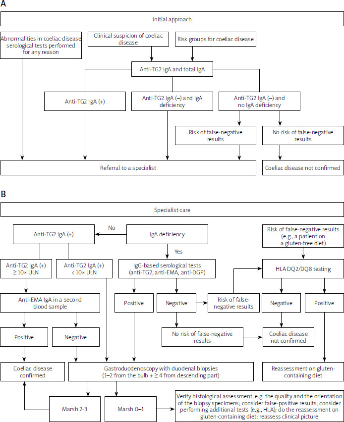
Children diagnosed with coeliac disease require regular clinical monitoring, ideally managed by a paediatric gastroenterologist and clinical dietitian experienced in coeliac care. ESPGHAN guidelines recommend scheduling the initial follow-up consultation 3–6 months post-diagnosis, followed by bi-annual reviews until complete normalisation of serum anti-TG2 IgA is achieved (anti-TG2 IgG in case of selective IgA deficiency), and every 12–24 months thereafter. Surveillance visits comprise a detailed clinical history regarding intestinal and extraintestinal signs, anthropometric mapping (weight, height, and growth velocity tracking), and targeted serological testing. Core laboratory parameters – including a complete blood count, iron biochemistry, vitamin D3, vitamin B12, and transaminase activities – should be reassessed if abnormalities were present at baseline. Screenings for autoimmune thyroid disorders (TSH, fT4, anti-TPO, thyroid autoantibodies) should be tailored individually. ESPGHAN does not recommend routine bone densitometry screening for all children with coeliac disease [86].
There is no single gold standard for monitoring dietary compliance in children and adolescents. Long-term surveillance requires a multimodal approach combining clinical tracking, structured dietary logs, and serial anti-TG2 measurements, which serve as a reliable surrogate marker for mucosal healing. Persistently elevated autoantibody concentrations after 6–12 months of therapy necessitate a comprehensive evaluation of dietary habits [86]. The sensitivity and specificity of serological tests as markers of significant damage to the small intestinal mucosa in children treated with a gluten-free diet for more than a year are estimated at 75% and 85%, with a positive predictive value of 22% and a negative predictive value of 98% [258]. Routine follow-up endoscopies to document mucosal healing are not recommended for asymptomatic children with a favourable clinical and serological response. Refractory coeliac disease is an exceedingly rare phenomenon in paediatrics; if a patient fails to respond to verified gluten elimination, alternative causes must be actively investigated, including inadvertent gluten ingestion, Crohn’s disease, autoimmune enteropathy, SIBO, cow’s milk protein allergy, or exocrine pancreatic insufficiency [86].
The standardized algorithm for the initial approach to suspected paediatric coeliac disease, alongside the integrated pathway for specialised clinical follow-up, is illustrated in Figure 5.
Summary
As the Intestinal Diseases Section of the Polish Society of Gastroenterology, we anticipate that these comprehensive guidelines will serve as a practical decision-making tool for physicians across multiple medical specialties and clinical dietitians involved in the daily care of patients with coeliac disease and other gluten-related disorders. We recognise that the scope of this text is extensive; however, it is our firm assessment that there has been a critical lack of a similarly thorough, evidence-based consensus document in the Polish medical literature. It must be emphasised that these guidelines do not address every individual clinical permutation and will necessitate subsequent updates as clinical evidence continues to evolve. A prime example of an area requiring further systemic optimisation is the structured care of coeliac patients during the transition phase from paediatric to adult healthcare infrastructure, an organisational gap in the Polish healthcare system that warrants targeted administrative attention. Ultimately, we anticipate that the nationwide implementation of these recommendations will significantly enhance diagnostic rates, reduce delays in definitive diagnosis, and optimise long-term outcomes for patients living with coeliac disease and other gluten-related disorders.














