
Purpose
Angiosarcoma incidence is 0.01/100,000 or 1 in 10 million per year [1]. It is a rare neoplasm, and only 1-2% of all sarcomas are angiosarcomas [2,3,4]. More than 30% of patients present this skin tumor most often located in the head and neck region [5,6]. As an aggressive disease with poor prognosis, i.e., high-rate of distant and loco-regional metastases, it requires a multimodal approach. In order to achieve ultimate success in the angiosarcoma treatment is to perform radical surgery [2,7], which may be challenging for reaching uninvolved margins, especially in the head and neck region [8]. Adjuvant chemotherapy and radiotherapy are assumed to be a primary treatment for tumor stage II and higher [9]. Another challenge is the management of failure after primary therapy [8,9,10,11,12]. One of the methods, which could be utilized, is surface high-dose-rate (HDR) brachytherapy, particularly in the head and neck region [13,14,15]. National Comprehensive Cancer Network (NCCN) guidelines for post-operatively management of soft tissue sarcoma of extremity, trunk, and head and neck region recommends delivery of 45 Gy with low-dose-rate (LDR) brachytherapy or HDR equivalent, with the status of surgical margins negative [9].
Surface HDR brachytherapy (BT) is one of the methods in the treatment of non-melanoma skin cancer [16,17]. Its efficacy has been established in the literature, but the use of this way of treatment in the head and neck angiosarcoma is poorly documented [13,14,15].
There are two main techniques used in the surface brachytherapy [16,17]. Default applicators supplied by different companies worldwide can be used with convenience, but there are lesser options for treatment tailoring. Individual mould applicator is a choice for surface HDR in the problematic planning area (i.e., curved area, head and neck region, lesions located close to crucial organs at risk such as eye or brain) [13,18,19]. Individual mould multi-catheter HDR (IMM HDR) technique allows for precise adaptation of the dose coverage to the treated area [19]. Moreover, high HDR conformity, which includes steep dose gradient due to use of iridium-192 (192Ir) source and individually adjusted lead shielding, allows to escalate the dose in the target volumes with normal tissues sparing.
We aim to present surface IMM HDR applied as a salvage treatment for a patient with head and neck angiosarcoma previously treated with surgery followed by external beam radiotherapy.
Material and methods
Initial treatment
A 74-year-old male reported to our center with non-healing tumor mass on the left temple in 2015. It was 2 cm in the diameter with a history of 12 months. Biopsy of the lesion showed an uncommon malignant neoplasm of blood vessels, probably angiosarcoma. Immunophenotype was performed, and Ki67 was equal to 60%, CD31 (+), CD34 (–), F VIII (+/–), CK AE1/AE3 (–), HMB-45 (–), s-100 (–), and melan A (–). Computed tomography (CT) was used to determine the exact extent of the lesion. Despite the visible 2 cm ulcer, CT scan revealed that the tumor was 9 mm thick in the maximum and 7 cm wide in the largest diameter. Even though, it was positioned mostly in the temporal area, it spread to the borders of frontal and parietal areas. Additional ultrasound examination confirmed the extent of the abnormal tissue. The patient was qualified for a wide local excision (WLE) of tumor with simultaneous reconstruction using a free-flap collected from the patient’s thigh. Positive margins were assessed intraoperatively. Margins were widened three times, and additional microscopic evaluation was done, with no success. Due to excision of dimensions (14 cm in the diameter) and probable orbital penetration, the surgery was completed after reconstruction as planned. The patient was revised on day 2. An intervention was needed because of the abnormal venous outflow from the flap. A local revision was also performed on day 5. The partial free-flap loss was observed; the injury was supplied with full-thickness skin graft collected from the right upper arm. Partial necrosis was observed on day 12; no intervention was applied. The patient was discharged on day 25. Histopathological examination of the tissue resected during surgery reported cutaneous angiosarcoma (CD34 (+), CD31 (+), Ki67 ~20%), with positive resection margins.
Two weeks later, the patient visited our outpatient Sarcoma and Bone Tumor Unit, and an adjuvant external beam radiotherapy (EBRT) was proposed. Treatment planning started after individual consent. The dose schedule was chosen according to our Radiotherapy Department guidelines for adjuvant radiotherapy in free-flap reconstructed area. Volumetric arc therapy (VMAT; RapidArc®, Varian Medical Systems) was used to deliver of 52.8 Gy/1.6 Gy in 33 fractions (dose intensity modulation, 6 MV photon beams, Figure 1). The overall treatment time lasted for 51 days.
Fig. 1
Axial, coronal, and sagittal view of computed tomography (CT) with dose distribution presented in the dose-color-wash mode. 3D reconstruction presents patient body outline and beam trajectory. Red line represents gross tumor volume (GTV) and orange lines represent clinical target volume (CTV). Organs at risk (OARs) were contoured for treatment planning and plan optimization. The treatment plan was calculated with EclipseTM planning system (Varian Medical System) with AAA v. 10.0.28 (Anisotropic Analytical Algorithm). Calculation dose grid was 2.5 × 2.5 × 2.0 mm. VMAT technique was used for plan development and realization. 6 MV photon beams and beam intensity modulation was used to deliver 52.8 Gy in 33 fractions to PTV. The overall treatment time was 51 days
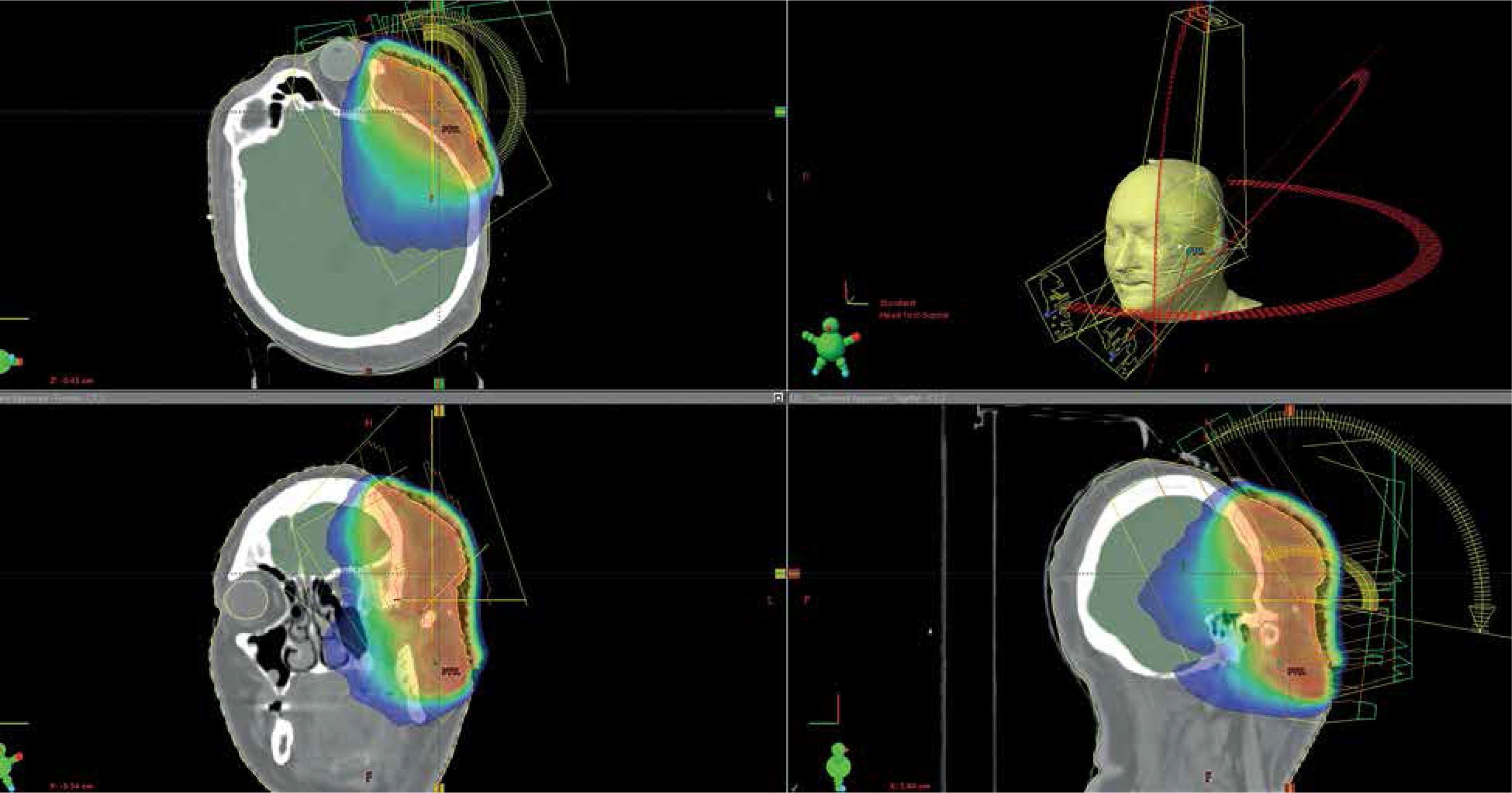
The total EBRT dose was reduced to decrease the toxicity risk in the free-flap region located in the proximity of radiation areas. Dose distribution was calculated according to dose constraints of our Radiotherapy Department with free-flap region treatment protocol. The patient was referred to the outpatient clinic for follow-up.
First CT scan was performed three months after adjuvant radiotherapy. The reconstructed area was recovered with no recurrence. Three months later (6 months after radiotherapy), the patient reported to a surgeon with a non-healing ulcer of the nasal bridge. Incisional biopsy revealed angiosarcoma. Wide local excision with skin graft reconstruction was planned. An intraoperative histopathologic report showed positive margins and another angiosarcoma spot close to left eyebrow. Due to multifocal disease and lack of possibility for further margin resection, the surgery was completed after skin graft reconstruction from right thigh.
The patient was not qualified for the second external beam radiotherapy, because the residual tumor was located too close to an eye, an area that was previously reconstructed and irradiated. Two months after the second surgery, the patient was referred to our Brachytherapy Department. Even though there was no data, surface IMM HDR was considered as an alternative option.
Brachytherapy treatment
On the baseline, the patient had a visual impairment in the left eye. Moreover, erythema of both left eyelids was reported with sclera injection and skin necrosis located close to the medial angle of the left eye. There were two main angiosarcoma foci on the face. The first one was localized on the right side from right nasolabial fold to the right medial eye angle, and the second included the left temple, left upper eyelid, and left nasolabial fold. The disease progressed rapidly as both lesions were fast-growing.
Due to lesions expansion and localization on both sides of the face, the treatment was split into two stages. Brachytherapy was performed with two individually designed custom moulds. These are made, based on the individual local imprint, with dental impression silicone covering visible tumor mass on the skin surface with 1 cm margin. The surface reflection is done in the plaster model to press the acrylic plate on it and to gain an individual mould (3 mm thick). Catheters (plastic tubes) are fixed to the opposite side of anatomic surface. The number and spatial configuration of the catheters are individually adjusted to achieve optimal conformity and homogeneity of the dose distribution and target coverage.
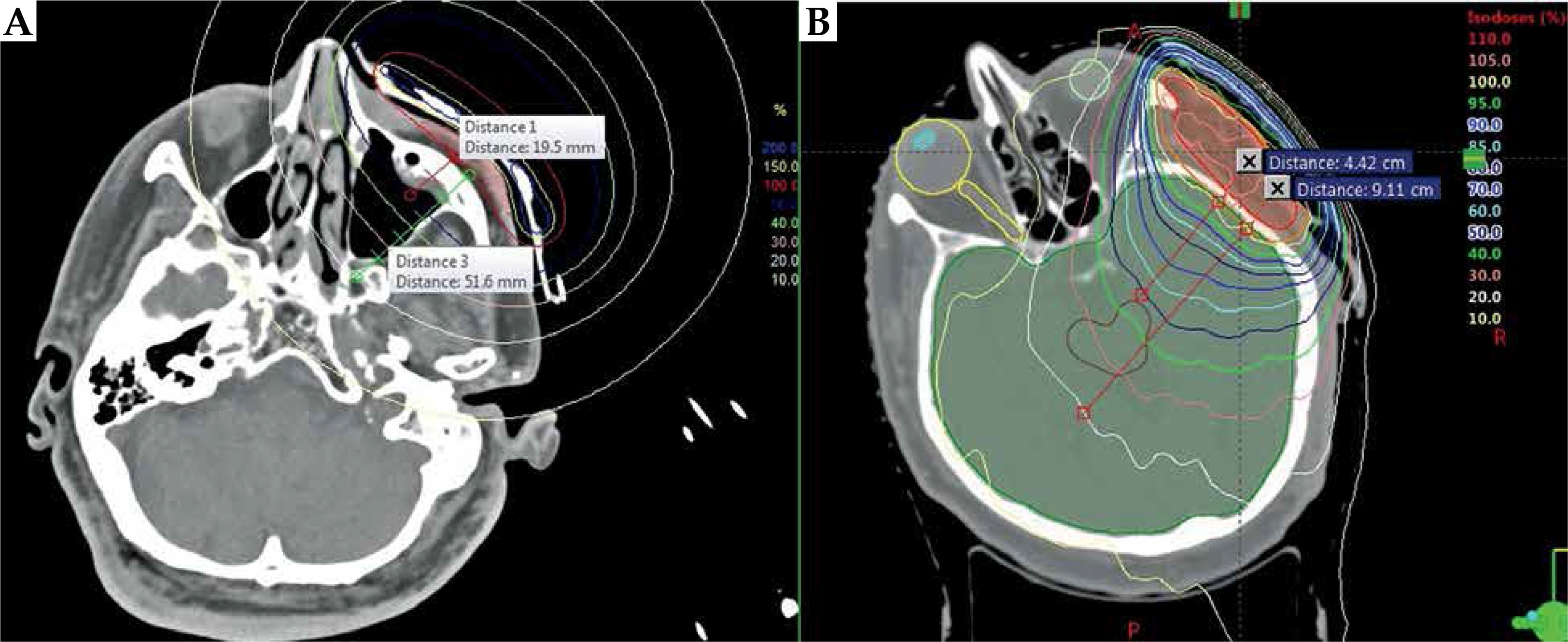
Dose distribution delivered with IMM HDR brachytherapy is a leading method for delivering high-dose to the target located on the surface of the skin, with simultaneous sparing of healthy tissue located below the treated region. In comparison to EBRT, relative dose delivered to the target is high, while steep dose gradient allows for reducing the dose in the healthy structures (Figure 2).
Fig. 2
Treatment plan calculated for (A) CTV2 (brachytherapy) and (B) PTV (EBRT). Dose distribution is presented in a relative mode as a percentage of the prescribed dose. Relative dose gradient was measured and compared between plans. Dose gradient was measured from the 100% to the 50% isodose and 20% isodose on the selected plane. Distance between isodoses differ for BT and EBRT treatment plan
CTV1 was designated for the right side, while CTV2 included left part of the face. Target volumes were specified based on the tumor mass observed on the patient skin. A margin for sub-clinical disease spread was added to the target volumes. According to the rules included in our internal, departmental protocol for non-melanoma skin cancer, clinical target volume (CTV) is a 10 mm margin added circumferentially to the target volume. CTV1 and CTV2 were planned to be treated separately with one-week gap. Two moulds were prepared. Eleven and twenty-one catheters were used for CTV1 and CTV2, respectively.
OncentraBrachy treatment planning system v. 4.5.2 (Elekta Instrument AB Stockholm) was used for planning 3D dose distribution. The dose was calculated on CT, with 1 mm slice thickness. The reference dose was initially specified as 5 mm (average) from the individual mould surface for both CTV1 and CTV2 (Figure 3). In our department, for contact brachytherapy, the reference dose is delivered according to rules included in departmental protocol, which was established based on the 20-year experience in treating non-melanoma skin cancer with superficial applicators. The 5 mm dose specification from the surface of the applicator (below the patient skin surface) has been recognized as a safe and efficient method of treating superficially located lesions. 3D treatment planning based on CT allows for CTV contouring and dose prescription based on the specified target volume. Introducing 3D treatment planning into clinical practice is a complex process. To fully benefit from 3D imaging, a clinical results collected from the 2D era should be considered as a starting point for prescribing and optimizing 3D dose distribution. Therefore, the reference dose was specified as 5 mm below the surface of the applicator, and then the optimization of dwell times was applied to improve CTV coverage in the region where the tumor penetrated deeper (more than 5 mm) into the tissue, but the dose of 200% of the reference dose should not exceed the maximal skin dose. Maximal dose point of 200% was the dose constraint for the skin. The depth and the range of the reference isodose was also compromised by the normal tissues as well as eyes and previously treated region located in the proximity of CTV1 and CTV2.
Fig. 3
Axial, coronal, and sagittal view of computed tomography (CT)-based treatment plan for CTV1 (A) and CTV2 (B). Contour of the patient body together with the reference isodose line (red line) and reconstructed catheters. Treatment plans were calculated with OncentraMasterPlan v. 4.5.2. Dose distribution was calculated based on the axial CT scans acquired with 1 mm thickness
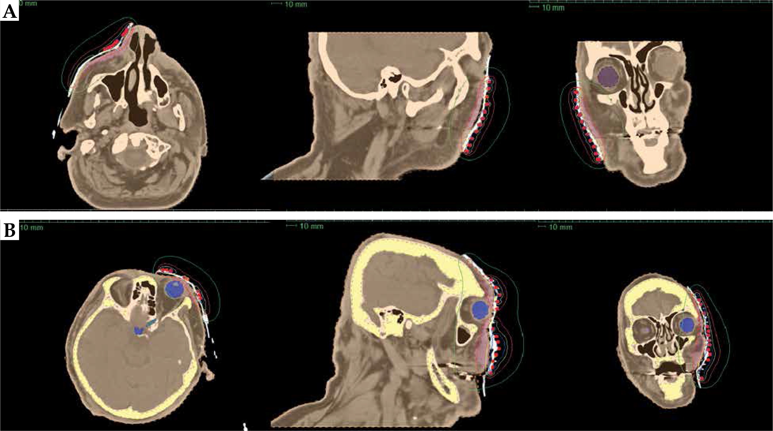
The total dose of 48 Gy (reference dose) in 12 fractions was planned. Four fractions per week were scheduled for both CTVs to three weeks of overall treatment time per side. The dose was specified on 100% isodose. The total dose was compromised by the dose delivered previously with external beam therapy and the extent of the lesion. Eye shielding for reducing the dose delivered to both lenses was used; round plates of lead (3 mm thick) were placed on the patient eyelids at every fraction.
Results
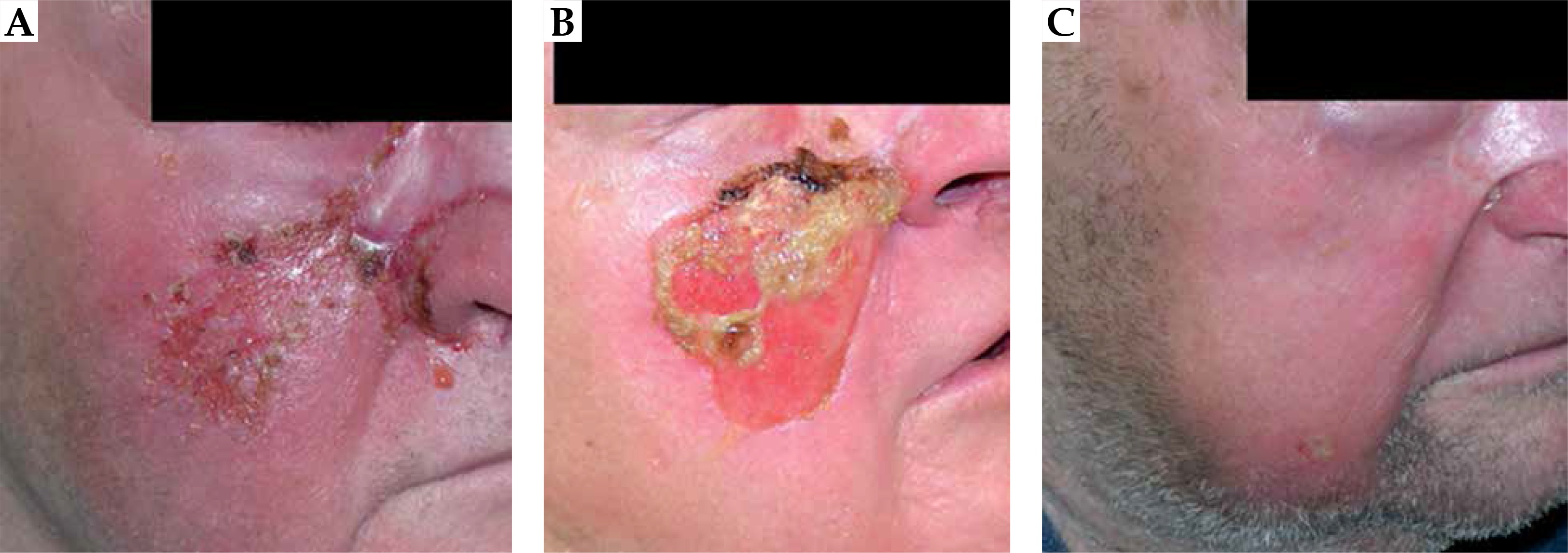
One month after surface IMM HDR, healing process of the skin was observed in treated regions. Skin erythema with diffused inflammation and desquamation was reported. Moreover, acute erythema of left eyelid with severe conjunctivitis including corneal drying and scleral atrophy was diagnosed by ophthalmologist, probably both after EBRT and HDR. In the next three months of the follow-up, the vision in the left eye improved (patient reacted to light), but conjunctivitis, corneal drying, and scleral atrophy were as before. Six months from HDR, skin erythema was still observed, but irradiated areas recovered (Figure 4). During the next eight months of follow-up, purulent dermatitis in the left nasolabial fold was diagnosed and cured in our center’s surgical ambulatory. The general condition of the patient has worsened two years after diagnosis and one year after HDR. Local recurrence had occurred 11/12 months after finishing the treatment. The patient was hospitalized to receive palliative care.
Discussion
Angiosarcoma accounts for no more than 2% of all soft tissue sarcomas, which is a very complex and heterogeneous group of tumors [2,3]. Survival rates of cutaneous angiosarcoma correlate with age, multifocal disease, anatomical site, and stage of disease [5]. Based on the analysis of the group of 50 patients, Bernstein et al. demonstrated worse survival of scalp angiosarcoma than face angiosarcoma [6]. Most of the published data on angiosarcoma treatment show small groups of patients collected throughout the decades [8,20,21]. Treatment procedures including surgery and radiotherapy have evolved in the last decades. Irradiation techniques used for the treatment of angiosarcoma have changed significantly, which may have an impact on the treatment results. Although published data on brachytherapy for angiosarcoma are mostly case reports, every individually approached patient and results reported are valuable because of the rarity of this disease [2,13,15,22,23].
In our case, high-dose-rate brachytherapy was used to treat multifocal angiosarcoma located on the face. The main advantage of this treatment was the possibility of the dose distribution tailoring due to an individual multi-catheter mould used. IMM HDR allowed optimizing dwell time and position of the source to deliver the optimal dose in the problematic locations (curved surface or proximity of the organs at risk). Moreover, the extent of the disease and its fast progression made the treatment complicated. The treatment was delivered with salvage intent, and it was achieved as a rapid decrease of the lesions and an acceptable cosmetic result shortly after brachytherapy. Additionally, patient’s psychological condition strengthened after treatment apart from left eye vision improvement.
Efficacy and safety of the surface brachytherapy in the skin lesions management, especially non-melanoma skin cancer, has been well documented. Different techniques and applicators can be used to deliver a dose to skin [16,17,24,25]. Stepping source (192Ir) has an optimal dose profile to deliver the dose to the skin surface efficiently with excellent and good cosmetic outcomes [24]. IMM HDR seems to be an optimal approach to treat lesions in the problematic areas, particularly in the head and neck region. Unfortunately, there is scarce data on surface HDR in the angiosarcoma patients, probably because angiosarcoma is a rare condition with an aggressive history [13,14,15]. The overall prognosis of head and neck angiosarcoma is poor [7,14,26,27]. Patel et al. analyzed retrospectively 56 patients with scalp/face angiosarcomas treated in Mayo Clinic [27]. Eighty per cent of patients underwent surgery as a wide local excision ± reconstruction. Although, radical resection including re-resection was planned, final histopathologic examination revealed positive margins in 12 (27%) patients. Majority of patients (65%) received adjuvant treatment (i.e., radiotherapy, chemotherapy or both). Other patients received single treatment (28%) or combination of radio- and chemotherapy (7%). Median follow-up was 25 months, with 38% and 16% of overall (OS) and recurrence-free survival (RFS) at 5 years, respectively. Univariate analysis showed survival benefit of 46% for combined modality therapy. Ogawa et al. analyzed failure patterns in the group of 48 patients suffering from head and neck angiosarcoma [3]. Median survival time was 13.4 months. Their data showed that combination of surgery and radiotherapy had a significantly better prognosis than sole surgery/radiotherapy or no surgery/no radiotherapy approaches (2-year OS: 46% vs. 11% vs. 0%; p < 0.0001). Sanada et al. presented a group of 9 patients with head and neck angiosarcoma treated with image-guided brachytherapy [14]. Only two patients received surgery before irradiation and seven received chemo- or immunotherapy. The 3-year OS and local control were 51% and 78%, respectively. Significant improvement in the local control was reported for patients treated with brachytherapy doses higher than 60 Gy in 20 fractions.
Wide local excision with negative margins is the treatment of choice for angiosarcoma [8,28,29,30], even though it may not be reachable due to an extensive subclinical tumor spread [8]. Adjuvant irradiation plays an important role in enhancing local control and overall survival. Combination of these two modalities is the most efficient way of treatment [6,8,11,12,14,23,26,27,28,30]. Due to its advantages, HDR should be considered as one of the useful techniques in angiosarcoma management, while IMM HDR could be beneficial in the individualized approach, especially with problematic treatment area. The latest NCCN guidelines for soft tissue sarcoma consider brachytherapy as an equivalent technique to external beam therapy [9].
Conclusions
High-dose-rate brachytherapy may be a valuable option for angiosarcoma treatment in cases where difficult lesion location may yield non-radical surgery (i.e., positive margins). Surface IMM HDR provides high conformity in respect to EBRT treatment. Catheters, which are individually placed in the mould, allow optimizing the dose to shape its coverage to anatomic structures and individual clinical situation. It enhances the dose inside target volumes and decrease the dose delivered to the organs at risk (OARs).


