
Although often asymptomatic, common bile duct (CBD) stones – choledocholithiasis – can result in life-threatening complications such as biliary pancreatitis, acute cholangitis, or obstructive jaundice if not promptly managed [1–3]. Interestingly, despite the minimal symptoms in many cases, their detection and removal remain a high priority in clinical practice. These stones are present in roughly 10–20% of patients undergoing cholecystectomy for gallstones, with or without clinical manifestations.
Cholangioscopy-guided lithotripsy, particularly with laser or electrohydraulic energy, offers a more direct and targeted alternative, especially in complex situations where standard methods fall short [3–6]. As endorsed by European Society for Gynaecological Endoscopy (ESGE) and other international bodies, early intervention is strongly advised to prevent complications, regardless of symptom presence. In some instances, particularly when the anatomy is unfavourable or stones are difficult in size or number, conventional endoscopic retrograde cholangiopancreatography (ERCP) becomes ineffective, failing in about 10% to 15% of attempts [1, 2].
Remarkably, a flexible ureteroscope – more common in urological procedures – was used successfully in the current case to manage an impacted CBD stone. The innovative use of laparoscopic cholangioscopy with laser lithotripsy is the focus of this report. It demonstrates how tools from one specialty can be repurposed effectively in another, especially in advanced centres dealing with difficult biliary cases.
Typically, ERCP – considered the standard of care – relies on endoscopic sphincterotomy followed by extraction using a balloon or basket catheter. When this method fails, clinicians must rely on alternative strategies to relieve the biliary obstruction and avoid emergency scenarios. This case illustrates the feasibility of such an approach when ERCP is either not feasible or unsuccessful and highlights its potential as a rescue therapy.
Six weeks of gradually worsening yellowish discoloration of the skin and sclera prompted a 44-year-old male to seek evaluation in the emergency department. He had no documented medical comorbidities. Notably, the patient had previously been diagnosed with obstructive jaundice and had undergone ERCP, during which a stent was placed in the CBD. The progression of jaundice occurred despite this prior intervention.
An abdominal ultrasound revealed a significantly dilated CBD, obstructed by a stone measuring approximately 3 cm in diameter. Interestingly, despite the high bilirubin levels, the patient remained haemodynamically stable upon admission. Laboratory tests showed a haemoglobin level of 15.9 g/dl and a creatinine level of 0.59 mg/dl, both within normal limits. However, marked hyperbilirubinaemia was evident, with total bilirubin at 11.6 mg/dl and direct bilirubin at 7.1 mg/dl.
Based on the clinical picture, the decision was made to proceed with diagnostic laparoscopy and cholecystectomy for both diagnostic clarification and therapeutic intervention.
The procedure began with diagnostic laparoscopy performed by the general surgery team. Intraoperative findings included a thickened gallbladder wall with evident oedema. Dissection of Calot’s triangle was carried out with identification of the cystic duct and cystic artery. The cystic artery was clipped and divided, followed by clipping of the cystic duct near the gallbladder. A standard laparoscopic cholecystectomy was then completed.
Subsequently, an incision was made in the cystic duct to perform intraoperative cholangiography. The cholangiogram demonstrated a large filling defect in the mid-CBD with upstream biliary tree dilatation. No contrast passage into the duodenum was noted, and the previously placed CBD stent was visualised.
Given the complexity of the case, the urology team was consulted intraoperatively. A single urologist performed laparoscopic cholangioscopy using a 9.5 Fr flexible ureteroscope introduced through the cystic duct. Direct visualisation confirmed the presence of a large stone occupying and obstructing the CBD lumen, positioned around and possibly compressing the previously placed stent.
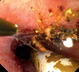
Laser lithotripsy was performed using a Holmium:YAG laser with energy settings of 0.3 J and 30 Hz frequency (9 Watts), optimised for stone dusting. The stone was successfully fragmented (Figure 1), and complete dusting was achieved (Figure 2). Residual fragments were retrieved using a closed-tip Dormia basket (Figure 3). The bile duct was examined from the common hepatic duct to the sphincter of Oddi to ensure full patency, and the stent was left in place.
Figure 1
Intraoperative image showing initiation of low-energy holmium:YAG laser dusting for common bile duct stone fragmentation
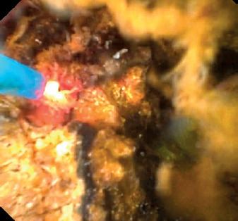
Figure 2
Intraoperative view demonstrating complete dusting of the common bile duct stone following holmium:YAG laser lithotripsy
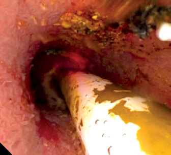
Figure 3
Retrieval of residual common bile duct stone fragments using a closed-tip dormia basket following laser lithotripsy
To conclude the procedure, the general surgery team resected the residual portion of the cystic duct, which was noted to be friable, and secured closure using an absorbable Vicryl suture.
Day 1 post-surgery: initial recovery: On the first postoperative day, the patient remained clinically stable. He reported tolerable abdominal discomfort, and his vital signs were within normal limits. The surgical drain placed intraoperatively showed minimal output with serous content, and no signs of infection or bile leakage were observed. Laboratory evaluation revealed a total bilirubin level of 9.5 mg/dl and a direct bilirubin of 7.0 mg/dl. These values, although still elevated, were expected in the early postoperative period given the recent manipulation of the biliary system. The patient was closely monitored for clinical and biochemical changes.
Day 2 post-surgery: biochemical paradox: Although the patient appeared visibly better – reporting improved well-being and showing reduced yellowing of the skin and conjunctiva – his biochemical profile told a different story. Surprisingly, by the second postoperative day, total bilirubin had climbed slightly to 11.7 mg/dl, with direct bilirubin rising to 7.5 mg/dl. Clinically, there were no alarming signs: drain output remained minimal, free of bile or signs of infection. This unexpected mismatch between the improving physical condition and worsening lab parameters raised concern. As a result, a decision was made to perform magnetic resonance cholangiopancreatography (MRCP) on the third postoperative day to reassess the biliary tree and evaluate the stent’s status.
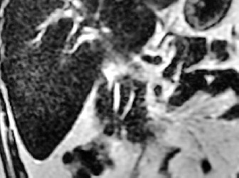
Day 3 post-surgery: MRCP evaluation: MRCP conducted on the third postoperative day provided further insights into the puzzling bilirubin elevation. While the main common bile duct was confirmed to be patent and not dilated, a notable finding was a defect at the distal end of the biliary stent – raising suspicion of partial blockage. The left hepatic duct was also dilated and showed a filling defect, pointing toward possible retained stones or biliary sludge. Interestingly, the right hepatic duct could not be clearly visualised on the imaging. There was no evidence of fluid collections or bile leakage. Figure 4 illustrates these findings, showing the stent in position along with mild inflammatory changes in the surrounding tissue. Mild oedema around the porta hepatis was also observed, which may have contributed to transient obstruction or impaired drainage.
Figure 4
MRCP image showing the stent in situ with no evidence of biliary leak or perihepatic collection; mild oedema noted around the porta hepatis
Day 3 post-surgery: clinical decision and discharge: Although imaging confirming complete biliary clearance was only performed months later, discharge was initiated early, based largely on the patient’s stable condition and lack of complaints. Interestingly, a 6-month MRCP had already been planned at this point, not due to active symptoms, but as a precautionary measure to assess long-term patency and rule out subclinical issues. The common bile duct, once examined again, showed a normalised diameter, and the previously observed porta hepatis oedema had entirely resolved. With no complications identified, the patient was ultimately discharged from all hepatobiliary follow-up.
One month post-surgery: clinical resolution: The patient’s procedure tolerance was notable – no complications followed the ERCP used to remove the stent. Biochemically, recovery was evident: total bilirubin dropped to 1.1 mg/dl and direct bilirubin to 0.2 mg/dl. Resolution of scleral icterus was clearly observed during physical examination, and the patient described a return to full well-being. No further jaundice, discomfort, or laboratory abnormalities were detected. Based on this positive progression, no additional endoscopic or imaging follow-up was deemed necessary at that time.
Six-month follow-up: radiological clearance: MRCP performed 6 months postoperatively, although clinically unnecessary by then, served to confirm the absence of complications. No signs of bile duct stricture, leak, or stone recurrence were observed. The porta hepatis oedema, which had contributed to the early biochemical-clinical mismatch, was entirely resolved. Ductal anatomy appeared normal, and the patient remained symptom-free. Official discharge from hepatobiliary care followed this reassuring imaging.
Standard treatment for choledocholithiasis continues to be ERCP, particularly in cases involving straightforward stone removal [1]. Nonetheless, around 10–15% of patients present with technical challenges such as large stones, strictures, or altered anatomy, rendering conventional ERCP inadequate [2–5].
Single-operator peroral cholangioscopy (SOPOC), a technology enabling direct intraductal visualisation, allows for precision-guided fragmentation through laser or electrohydraulic lithotripsy. In this case, SOPOC proved instrumental in targeting a large, impacted stone using Holmium:YAG laser, achieving clearance in a single session without further intervention.
Not all stones are created equal; some exceed 1.5 cm, become impacted, or reside in anatomically difficult locations. These “difficult stones” pose a major obstacle to routine ERCP [3, 7]. Laser lithotripsy, whether Holmium:YAG or Nd:YAG, offers focused energy delivery with minimal trauma to the ductal wall – an advantage when managing such cases [1, 3].
In this case, a flexible ureteroscope – typically a urological instrument – was innovatively repurposed for laparoscopic cholangioscopy, demonstrating how cross-disciplinary collaboration can deliver effective, minimally invasive solutions even in technically complex scenarios. The Holmium:YAG laser was selected for its well-documented safety profile and effectiveness in fragmenting hard, large, or impacted stones. Its pulsed energy delivery and shallow penetration (0.4 mm) minimise the risk of ductal wall injury while enabling precise “dusting” of the stone into retrievable fragments. Such properties make it preferable over other lithotripsy modalities in anatomically challenging or inflamed bile ducts [4–6].
An important feature of this case was the transient postoperative rise in bilirubin, despite clear clinical improvement. This phenomenon can be explained by multiple mechanisms. Manipulation of the biliary tree during surgery can cause temporary oedema at the porta hepatis or around the sphincter of Oddi, leading to partial outflow resistance. Additionally, the use of laser lithotripsy and fragmentation may result in transient microscopic sludge or debris migrating distally, briefly impairing drainage before clearance. Such transient hyperbilirubinaemia is well described in biliary surgery literature and generally resolves with conservative management as local inflammation subsides [3, 6–9].
This approach is not only novel but also feasible and potentially replicable in settings where advanced endoscopic resources are limited or where standard ERCP fails. By using a familiar urological tool via the cystic duct, the surgical team was able to achieve complete stone clearance in a single session without open surgery or repeated endoscopic procedures [1–3].
Compared with advanced techniques such as SpyGlass-guided cholangioscopy with laser or electrohydraulic lithotripsy, laparoscopic ureteroscope-guided laser lithotripsy offers comparable efficacy but with distinct advantages and limitations. Studies of single-operator cholangioscopy report high clearance rates – Buxbaum et al. [4] showed 93% clearance for cholangioscopy-guided lithotripsy vs. 67% for conventional ERCP, while Troncone et al. [3, 8] found an overall 94% pooled success rate. Electrohydraulic lithotripsy achieves similar efficacy but is associated with higher ductal wall injury risk due to less focused energy [5]. Holmium:YAG laser lithotripsy, used in this case, delivers precise energy with shallow penetration (~0.4 mm), reducing perforation risk [5, 9]. Cost analyses [6, 8] show early SpyGlass use reduces repeat procedures by ~27%, offsetting its higher unit cost. However, ureteroscope-guided laparoscopic access may be more feasible in centres lacking cholangioscopes, using existing urology equipment to achieve single-session clearance without the need for repeat ERCP, thus offering a cost-effective alternative in selected complex cases.
From a healthcare economics perspective, the advantages are equally compelling. Deprez et al. [5] reported a significant reduction in procedure volume (27%) and overall costs (11%) when SOPOC was used early. Sandha et al. [6] supported this, noting per-case savings between $1619 and $3210 CAD, with a clinical success rate of 93% and a low incidence (14%) of minor adverse events.
Altogether, these findings support the early integration of cholangioscopy-assisted lithotripsy into management algorithms for difficult bile duct stones. When ERCP fails or anatomy proves unfavourable, this minimally invasive technique offers a highly effective, safe, and economically sound alternative [1, 5, 8].
This case report highlights the successful use of cholangioscopy assisted by Holmium:YAG laser lithotripsy in managing a large, impacted common bile duct stone that was not amenable to conventional ERCP techniques. Beyond demonstrating technical feasibility, this cross-disciplinary approach offers a practical, replicable alternative that can be adopted in advanced surgical centres facing difficult biliary stones. As supported by literature, cholangioscopy-guided lithotripsy significantly improves stone clearance rates, reduces the need for repeat procedures and surgical intervention, and offers a cost-effective alternative to standard therapy. Although associated with longer operative times, the benefits in terms of efficacy and resource utilisation support its broader adoption in cases of complex biliary stone disease.














