
Introduction
Crohn’s disease (CD) and ulcerative colitis (UC) represent the primary forms of inflammatory bowel disease (IBD), characterised by a chronic, granulomatous inflammatory process that can affect any segment of the gastrointestinal tract and may be associated with a broad spectrum of systemic manifestations [1–5]. The clinical course of CD extends beyond gastrointestinal symptoms, often involving extraintestinal manifestations (EIM), which can occur in up to 50% of patients and may precede the diagnosis by more than 2 years [5]. Of particular importance are cutaneous and mucosal lesions, which are observed in 22% to as much as 75% of patients with IBD and pose significant diagnostic and therapeutic challenges [5–9].
Five main categories of mucocutaneous manifestations associated with inflammatory bowel disease (IBD) have been identified, based on their pathophysiological mechanisms, diagnostic approach, and treatment strategies:
1) specific lesions that share identical histopathological features with intestinal involvement;
2) cutaneous and mucosal lesions directly related to the disease process;
3) immune-mediated reactions triggered by shared intestinal and cutaneous antigens, resulting in reactive dermatoses;
4) therapy-related adverse cutaneous reactions secondary to IBD treatment;
5) lesions secondary to nutritional deficiencies caused by malabsorption [5, 6, 10–13].
Among the cutaneous manifestations of inflammatory bowel disease, metastatic Crohn’s disease (MCD) holds a particularly significant place, being considered a rare yet clinically important phenomenon [5, 7–9]. Due to its wide phenotypic variability and often nonspecific clinical presentation, the true incidence of MCD is probably underestimated in existing epidemiological data [9, 14]. First described by Parks et al. in 1965, MCD remains a diagnostic and therapeutic challenge even today [15]. Given the increasing number of reported cases and growing clinical interest in this condition, the development of standardised diagnostic and therapeutic frameworks is essential to support physicians in daily practice – particularly gastroenterologists and dermatologists.
In recent years, clinical guidelines for the diagnosis and treatment of MCD have been published for the first time, marking a significant milestone in the understanding of this rare manifestation of CD [9]. In 2014, the first recommendations were issued by the American Medical Association, and in 2025 detailed diagnostic criteria were proposed aimed at improving recognition and management of this clinical entity [14].
Pathogenesis of metastatic Crohn’s disease
MCD remains one of the most enigmatic aspects of IBD pathophysiology. Currently, it is recognised as a manifestation of a systemic immune response, in which immune activation surpasses the gut barrier and extends to the skin, leading to granulomatous inflammatory lesions [16–31].
ECCO guidelines and the latest expert consensus published in 2025 emphasise that the pathogenesis of EIM is complex and involves interactions between genetic, microbiotic, and immunological factors, as well as “epitope spreading” and T-cell “mis-homing” mechanisms [5, 9, 29–34]. There is a focus on the need for early recognition of these mechanisms and the development of personalised therapeutic strategies.
The classical concept of MCD pathogenesis was based on the hypothesis of deposition of circulating antigens and a Th1-type delayed hypersensitivity reaction, but studies have not confirmed a direct correlation between the presence of bacterial antigens in the intestine and in the skin [7, 17, 30]. However, more recently, greater importance has been attributed to vascular injury in the skin, leading to vasculitis and perivascular granulomas, which confirms the immunological basis of these lesions [7, 12, 17]. The most significant roles are played by immunological mechanisms, especially excessive activation of the Th17/IL-23/TNF-α axis and neutrophil infiltration, which are observed both in intestinal and cutaneous lesions [12, 17, 23–25, 29, 30]. High activity of these pathways promotes autoimmune processes and tissue destruction.
The phenomenon of epitope spreading, i.e. the expansion of the immune response to additional autoantigens, also appears important, explaining the coexistence of autoimmune dermatoses in IBD patients [12, 29, 30]. A breakthrough in understanding IBD was the identification of mutations in the NOD2/CARD15 genes, which regulate the response to intestinal bacteria. These mutations predispose to excessive immune activation and the development of autoimmune reactions in both the gut and the skin [12, 17, 26–30]. A significant mechanism is the abnormal migration of activated T lymphocytes from the gut to the skin (“mis-homing”), which leads to local inflammation and granuloma formation [7, 12, 17, 29–31]. A key element of this migration is the involvement of integrins, which mediate the adhesion and trafficking of lymphocytes from the circulation to specific target tissues, including the skin. Altered expression of integrins, particularly α4β7 and ligands such as MAdCAM-1, facilitates the adhesion and transmigration of activated immune cells from the blood to extraintestinal sites. In the pathogenesis of cutaneous IBD lesions, the role of integrins as mediators of T-lymphocyte ‘homing’ appears crucial, as confirmed by both experimental studies and the clinical effects of anti-integrin therapies in IBD [23, 24, 29]. Given the rarity of MCD and the complexity of its development, further molecular and immunological studies are needed to better understand its pathogenesis and more effectively treat patients with this form of CD, which may also help us better understand the pathogenesis of its intestinal form.
Clinical presentation
MCD is a rare clinical phenomenon; in most cases, it develops in patients with a previous diagnosis of IBD. However, MCD may also occur during remission of intestinal symptoms, and in some cases, it can even be the first manifestation of IBD, sometimes appearing months or even years before the diagnosis of IBD is established [9, 14, 35–40]. This form of the disease occurs more frequently in children, especially with isolated cutaneous or genital lesions [7, 15, 17, 41–46]. In children with MCD, compared to adults, the proportion of genital lesions is higher – about two-thirds of cases in children involve this region, while in adults it is around half [7, 17, 21].
The clinical manifestations of MCD are highly variable and may be generalised or limited to single sites [9, 14, 47–50]. The most characteristic lesions are oedematous, erythematous, and erythematous-infiltrative changes often with linear, “knife-like” fissures (arrow in the images), as well as papules and nodules that frequently break down, resulting in discharge of bloody-purulent material (asterisk in the images) [51–54]. There may also be isolated oedema of the external genitalia or labia, reflecting granulomatous inflammation (Figures 1 A–G) [9, 14, 55–61].
Figure 1
Heterogeneity of the clinical presentation of metastatic Crohn’s disease (asterisks mark discharge of bloody-purulent material, arrows indicate linear, “knife-like” fissures). Images from the authors’ own clinical archive. A – Erythematous-infiltrative lesion with erosion and discharge of bloody-purulent material in the abdominal skin. B – Erythematous-infiltrative change with knife-like fissure in the gluteal cleft area (arrow). Nodular lesion with discharge of bloody-purulent material (asterisk). C – Infiltrative lesion with knife-like fissure in the perioral area in a patient without oral cavity involvement, demonstrating extraoral manifestation. D – Knife-like ulceration in the thigh area. E – Infiltrative lesion with fissure and discharge of bloody-purulent material in the umbilical area in a patient after multiple intestinal surgeries. F – Asymmetric oedema and severe persistent granulomatous swelling of the labia majora in a patient with intestinal Crohn’s disease. G – Erythematous nodule in the abdominal skin area in a patient with Crohn’s disease
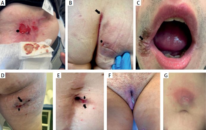
In cases of long-standing or deeply infiltrating lesions, scarring may occur, and in the anogenital region, secondary deformities may develop, which significantly impacts patients’ quality of life [9, 14, 62–68]. Notably, MCD dermal lesions are often painful at onset, but with chronic progression, pain typically subsides, and patients experience mainly discomfort or pruritus [69–71].
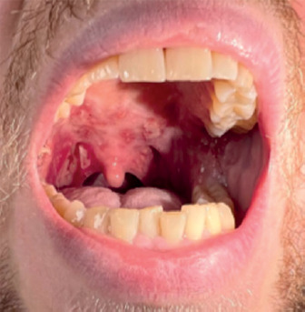
Due to the high frequency of involvement of the intimate areas, MCD is often classified as either genital or extragenital lesions [7, 12, 17, 72]. Depending on the location, the morphology of the lesions may differ – lesions on the limbs and buttocks often present as immobile, erythematous nodules, frequently with exudation (Figure 1); whereas, in the skin folds, linear erosions are typical (Figure 1). Lesions on the mucous membranes resemble those found in the gut, with numerous aphthae, erosions, or deep ulcers [7, 12, 29] (Figure 2).
Figure 2
Sudden onset of numerous confluent aphthae > 1 cm in a patient with diagnosed intestinal Crohn’s disease (visible aphthae > 1 cm); similar lesions were also observed in the oesophagus during endoscopic examination. Images from the authors’ own clinical archive
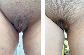
Genital lesions most often include erythema, oedema, ulcers, and fissures affecting the vulva, labia, clitoris, scrotum, penis, and perineum. Less commonly, there are wart-like lesions, skin nodules, or isolated lymphatic oedema, which can make diagnosis more difficult [7, 12, 17, 21, 29].
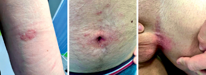
Given the diversity of clinical presentations and the lack of pathognomonic features, MCD should be considered in patients with a history of CD, especially those with colonic involvement, who present with persistent or atypical dermal lesions. In such cases, histopathological examination is recommended [7, 12, 17, 29, 31, 50–72] (Figures 3, 4).
Diagnostic criteria for metastatic Crohn’s disease
MCD is a rare but increasingly reported EIM of IBD [5–10]. The rising number of recognised cases reflects both enhanced clinical awareness and improved understanding of its diverse clinical presentation. In response, the first official diagnostic criteria for MCD were published in 2025 by an American expert panel through the Delphi consensus process [9]. Notably, the most recent guidelines do not require histopathological confirmation for diagnosis, emphasising the growing clinical awareness of MCD and the evolving clarity of its phenotype (Table I) [9]. The disease has been divided into two main groups: oral MCD and genital/other MCD. Oral MCD, previously referred to as orofacial Crohn’s disease, requires a prior diagnosis of intestinal Crohn’s disease and at least one minor clinical criterion. Genital/other MCD is primarily a clinical diagnosis, requiring either two major criteria, one major and one minor criterion, or three minor criteria. While histopathological confirmation is not mandatory as per the new consensus, it can offer strong supportive evidence in equivocal scenarios. In particular, the presence of naked granulomas after exclusion of other diagnoses, or at least one histopathological feature from the minor criteria together with one major or two minor clinical criteria, can be considered sufficient for diagnosis.
Table I
Clinical and histopathological diagnostic criteria for MCD (Modified by Delphi Consensus 2025)
| Category | Criteria necessary for diagnosis | Major criteria | Minor criteria |
|---|---|---|---|
| Oral MCD (Figure 2) | • Known diagnosis of luminal inflammatory bowel disease • 1 Minor criterion | *None* | • Linear ulceration of the mouth • Granulomatous cheilitis • Pyostomatitis vegetans • Sublingual soft-tissue swelling • Large (> 1 cm) oral aphthae |
| Genital/other MCD (Figure 3) | • 1 Major + 1 Minor criterion • 2 Major criteria alone • 3 Minor criteria alone | • Genital swelling • Knifelike ulcers of the genital, inguinal, or intertriginous skin • Lymphoedematous changes of the genital skin | • Perianal skin tags • Genital fissuring • Nonhealing genital ulcerations (negative HSV test result) • A cutaneous biopsy suggestive of MCD • A diagnosis of inflammatory bowel disease |
| Histologic criteria | Sufficient: 1 major criterion + no more likely alternative diagnosis Highly suggestive, but not sufficient: any number of major or minor criteria | • Naked granulomas with negative infectious staining • Loose granulomatous inflammation with lymphoplasmacytic inflammation • Epithelioid granulomas • Intravascular histiocytic aggregates + perivascular/perilymphatic lymphohistiocytic granulomatous | • Noncaseating granuloma with any of the following: ulceration, lymphocytes and eosinophils, or vasculitis |
The adoption of these criteria should facilitate earlier diagnosis and appropriate management of MCD [9]. Clinicians should maintain a high index of suspicion for MCD in any patient with IBD presenting with chronic, treatment-resistant skin or mucosal lesions.
Differential diagnosis of metastatic Crohn’s disease
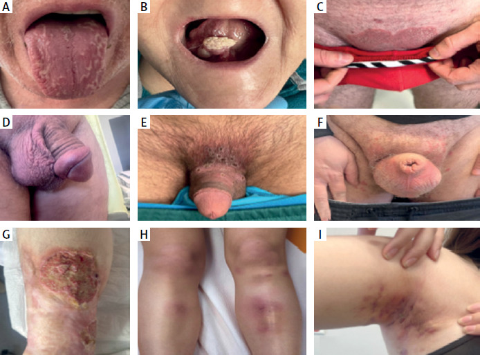
Due to the significant polymorphism of skin lesions, the differential diagnosis of MCD presents a considerable clinical challenge. The clinical classification of skin lesions proposed by American experts in 2025 divides MCD into several main groups, which has significantly facilitated the diagnostic process [9]. The differential diagnosis according to the type of lesion is summarised in Table II, along with practical tips and useful laboratory investigations (Figures 5).
Table II
The differential diagnosis according of metastatic Crohn’s disease to the type of lesion
| MCD location | Other possible conditions | Characteristic skin finding | Additional clinical features | Useful initial tests and consultation |
|---|---|---|---|---|
| Oral | Viral infections (HSV, Coxsackie, etc.) | • Erythema, oedema, small erosions or ulcers, often preceded by a vesicular stag | • Sudden onset • Other symptoms of infection | Inflammatory markers, consider serological/molecular tests |
| Mycoses (e.g., Candida spp.) (Figure 5 A) | • Aphthae that often can be scraped off with a spatula | • Often with other risk factors (immunosuppression, HIV, diabetes, antibiotic therapy) | In recurrent cases: OGTT, HIV Ag/Ab, consider measuring immunosuppressant drug levels | |
| Lichen planus, neoplasm, cancer (Figure 5 B) | • Often a single flat or exophytic lesion, often at sites of chronic trauma | • History of HPV/+ substance abuse | HBsAg, anti-HCV, HIV Ag/Ab, OGTT [89–91], consider ENT consultation in prolonged lack of improvement | |
| Behçet’s disease | • Erosions in the oral cavity and genital area, with a self-limiting and recurrent course | • Pathergy phenomenon • Frequently accompanied by non-specific limb lesions (e.g. EN) • Ocular and other symptoms | Inflammatory markers [63, 65], ophthalmology consultation | |
| Genital | Viral infections (HSV, monkeypox etc.) (Figure 5 E) | • Painful erosions on erythematous base • Erosions covered with crusts at a later stage | • Sudden onset • Other symptoms of infection | Inflammatory markers, HIV Ag/Ab, RPR, consider pathogen-specific tests and dermatology consultation |
| Syphilis (Figure 5 D) | • Painless ulcer with smooth margins and indurated base, usually solitary and < 2 cm | • Apparently self-limiting course* | Positive treponemal/nontreponemal serology (VDRL, RPR, FTA-ABS, TPHA, etc.) | |
| Mycoses (e.g. Candida spp.) | • Erythema, pruritus, burning sensation, whitish curd-like discharge, often accompanied by satellite papules or pustules | • Often with other risk factors (immunosuppression, HIV, diabetes, antibiotic therapy) | In recurrent cases: OGTT, HIV Ag/Ab, consider measuring immunosuppressant drug levels | |
| Lichen planus, neoplasm, cancer | • Often a single flat or exophytic lesion, -often at sites of chronic trauma | History of HPV/+ substance abuse | HBsAg, anti-HCV, HIV Ag/Ab, OGTT [89–91], consider dermatology consultation and histopathology in prolonged lack of improvement | |
| Scabies (Figure 5 F) | • May present as isolated genital swelling • Red papules, excoriations | • Accompanied by intense pruritus, particularly pronounced at night or after warming | Microscopic examination of scrapings, exceptionally histopathological examination | |
| Psoriasis (Figure 5 C) | • More monomorphic and infiltrated lesions • Diffuse or peripheral scaling • Rare bleeding cracks; | • Often accompanied by lesions elsewhere (including scalp, nails) • Risk factors include prolonged TNF-α inhibitor use, positive family history of psoriasis | Measurement of immunosuppressant drug levels, anti-anti-TNF antibodies | |
| Other (trunk and extremities) | Hidradenitis suppurativa (Figure 5 I) | • Often numerous lesions in skin folds, with comedones, fistulas, skin bridges and scarring • Lesions often with abundant purulent discharge | • Active acne or acne history • Features of metabolic syndrome | Pus culture usually sterile or skin flora; metabolic syndrome screening (often associated) [112–114] |
| Sarcoidosis | • Often clustered, painless lesions, without tendency to ulcerate | • Diascopy sign • Other manifestations of systemic sarcoidosis | Chest X-ray, inflammatory markers, ACE activity (elevated in many patients with systemic sarcoidosis) [62, 87, 88] | |
| Pyoderma gangrenosum (Figure 5 G) | • Crater-like ulcer covered with fibrin layer • Often arising at sites of minor trauma | • Positive pathergy phenomenon | Elevated inflammatory markers (usually with neutrophilia) [85, 86] | |
| Erythema nodosum (Figure 5 H) | Red, painful nodules without tendency to ulcerate, often symmetric on anterior lower legs | Often accompanies infections (signs of infection on physical examination) | Elevated inflammatory markers (ESR > 50 mm/h in many patients [83, 84]), consider chest X-ray (may accompany systemic sarcoidosis) | |
| Cutaneous tuberculosis/Mycobacterioses (Figure 5 G) | • Nodules and papules ranging in colour from red to brown, with a tendency to ulceration | Commonly on distal sites spread along lymphatic vessels (proximally) | Positive IGRA in up to 95% of TB cases and up to 73% of NTM skin involvement [58–61, 80–82] |
Figure 5
A – Oral candidiasis: easily scrapable plaques. B – Oral cancer: exophytic lesion suggestive of carcinoma. C – Psoriasis: well-demarcated erythematous-infiltrative lesion. D – Syphilis: preputial swelling over a regressing primary lesion. E – Monkeypox: erosions with crusts at the site of viral contact. F – Scabies: scrotal and perilesional swelling with papules. G – Pyoderma gangrenosum: ulcer with crater-like edge covered by fibrin. H – Erythema nodosum: painful erythematous-oedematous lesions. I – Hidradenitis suppurativa: nodules, scars, and comedones in the axilla. Images from the authors’ own clinical archive
In the initial evaluation of dermal lesions, a detailed personal and family history is essential – particularly regarding the presence of IBD, as well as rheumatologic and dermatologic disorders. Retrospective analyses involving more than 700 cases of CD have demonstrated that dermal lesions occur more frequently in patients with colonic involvement than in those with ileal-only disease activity. Nevertheless, most studies have not shown a clear correlation between the severity of intestinal disease and the occurrence of skin manifestations [7, 15, 17, 30]. However, certain extraintestinal manifestations (EIMs) of IBD exhibit a particularly strong association with disease activity. Erythema nodosum typically presents as tender, erythematous nodules – most often on the shins – and usually coincides with IBD flares. Pyoderma gangrenosum manifests as rapidly progressive, painful ulcers and may also worsen with increasing intestinal inflammation. In both conditions, the severity of cutaneous lesions often parallels the activity of the underlying bowel disease. In contrast, psoriasis that develops during anti-TNF therapy is unique in that it most commonly arises during periods of IBD remission rather than during active disease [5–7, 15, 17, 30].
Additional useful investigations in the preliminary workup include blood glucose measurement, serological testing for hepatitis B virus (HBV), hepatitis C virus (HCV), human immunodeficiency virus (HIV), rapid plasma reagin (RPR), interferon-gamma release assay (IGRA; e.g. QuantiFeron), chest X-ray, and abdominal ultrasound. In cases of chronic or long-standing lesions, histopathological examination is recommended; in selected cases, tissue sampling is particularly justified when the IGRA test is positive [1–5]. Furthermore, therapeutic drug monitoring and assessment for anti-drug antibodies may be helpful, especially in patients presenting with erosive or psoriasis-like lesions [6]. When differentiating MCD from other dermatoses, the fistulising form of CD – particularly enterocutaneous and perianal fistulas – should always be considered [1–4]. Enterocutaneous fistulas require evaluation with magnetic resonance enterography/enteroclysis or computed tomography to assess any potential communication with the intestinal tract. In cases where lesions are located in the anogenital region, pelvic magnetic resonance imaging (MRI) should be performed to determine whether there is a connection with the distal gastrointestinal tract [1–4]. Histopathologically, MCD is characterised by the presence of non-caseating granulomas resembling those observed in inflammatory lesions of the gastrointestinal tract obtained during endoscopic examinations [7, 9, 14]. These granulomas typically contain Langhans-type multinucleated giant cells and foreign body-type giant cells [14, 16–18].
The lesions are predominantly located in the deep dermis and often extend into the subcutaneous tissue [14, 16]. They are accompanied by inflammatory infiltrates composed of histiocytes, lymphocytes, and plasma cells, usually arranged in a perivascular or perifollicular pattern [7, 9, 14, 16–18]. The pathogenesis of MCD is hypothesised to involve multiple mechanisms, including type IV hypersensitivity reactions, dissemination of intestinal antigens to the skin, as well as genetic predisposition and enzymatic dysfunctions [9, 14, 16–18]. Histologically, MCD should primarily be differentiated from other granulomatous diseases such as sarcoidosis, mycobacterial infections, and other infectious granulomatous dermatoses. This workup may require supplementary histochemical staining (Ziehl-Neelsen, Giemsa, PAS) and examination under polarised light. In MCD, these tests typically do not reveal any specific abnormalities [56–67, 71–73] (Figure 6, Table III).
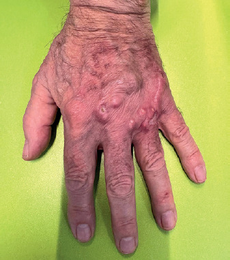
Table III
Differential diagnosis according to histopathological features
Management of metastatic Crohn’s disease
The treatment of MCD requires an individualised, multidisciplinary approach and the management of both dermal lesions and underlying CD activity [74–93]. Improvement or remission of dermal lesions is often only possible after the effective initiation of appropriate systemic therapy for the intestinal disease, which is why assessment of the underlying disease activity and adjustment of systemic treatment, if necessary, is always recommended [5, 14, 34, 48].
According to the 2014 American Academy of Dermatology guidelines, treatment should begin with topical therapy. The effectiveness of topical therapy is estimated at approximately 30% of patients [9, 14, 75]. Recommended topical treatments include glucocorticosteroids and calcineurin inhibitors, particularly for the face and anogenital area. However, high doses of topical calcineurin inhibitors should not be used on extensive lesions, especially those with epidermal loss, due to increased absorption through damaged skin and the risk of reaching even therapeutic plasma concentrations [75, 94–98]. If topical therapy is insufficient, oral metronidazole is recommended. Metronidazole is one of the best-documented adjuvant therapies for MCD, particularly in cases complicated by infection, abscesses, or fistulous lesions. Its effects go beyond antimicrobial action – it also demonstrates immunomodulatory and anti-inflammatory properties, inhibits neutrophil activation and migration to the inflammatory site, limits inflammatory infiltrate and local symptoms, improves intestinal barrier function, reduces bacterial antigen translocation, lowers the expression of pro-inflammatory cytokines (TNF-α, IL-1β, IL-6), and stabilises the gut microbiota [13, 14, 32–34, 99–105]. The effectiveness of metronidazole (in monotherapy or combination) is estimated at about 44%, but therapy should be carried out with caution due to the risk of peripheral neuropathy with long-term use [14, 34, 106].
In cases of extensive lesions or lack of response to topical and antibiotic therapy, systemic corticosteroids are recommended. If this also proves ineffective, immunosuppressive agents such as azathioprine, methotrexate, or cyclosporine are introduced. These drugs can achieve remission of both cutaneous and intestinal lesions, although their efficacy is moderate (approximately 37.5%) [5, 14, 98–110].
In refractory and extensive MCD, biological therapies are increasingly important. TNF-α inhibitors (infliximab, adalimumab) have shown the highest effectiveness among available treatments, ranging from 64.3% to 70.6% both as monotherapy and in combination with glucocorticosteroids or azathioprine [5, 14, 17, 37, 42, 66–70, 72, 75]. In cases of lack of efficacy, increasing the drug dose should be considered, but before dose escalation or changing therapy, it is advisable to assess serum drug levels and anti-drug antibodies [66–70, 106–107]. Moreover, similarly to the intestinal form, there are increasing reports on the efficacy and good safety profile of IL-12/23 and IL-23 inhibitors; the use of IL-23 inhibitors is pathophysiologically justified by histopathological observations: dendritic cells and macrophages in skin lesions express IL-23, suggesting that blocking this pathway may be beneficial in MCD therapy [17, 23–25, 29, 34, 38, 40, 43–45, 48, 69, 70, 111–120] (Figure 3).
There are also single case reports on the use of vedolizumab in patients with MCD. A case published in 2023 described complete remission of dermal lesions in a patient with refractory MCD after vedolizumab monotherapy [39]. However, in a 2020 ECCO/CONFER multicentre study, vedolizumab was effective in only 1 patient, whereas ustekinumab achieved remission in 5 patients [40]. It is concluded that vedolizumab’s mechanism of action, mainly targeting the gastrointestinal tract, may limit its effectiveness in the cutaneous manifestations of MCD, especially when these are independent of intestinal activity [39, 40]. Nonetheless, vedolizumab remains a potential therapeutic option, particularly in patients with concurrent intestinal disease activity and exhaustion of other pharmacological options [40].
The latest and most promising reports also document the efficacy of JAK inhibitors such as upadacitinib, especially in particularly difficult and refractory cases resistant to TNF-α and more selective IL-23p19 inhibitors [38, 73, 76].
As in the intestinal form of CD, combination therapy may be used in MCD due to its broader mechanism of action and the protective effect against anti-drug antibody formation (for example, adding azathioprine or methotrexate to monoclonal antibodies) [13, 14, 17, 37]. Although there are anecdotal reports of improvement with regimens such as IVIG, methotrexate, and hydroxychloroquine, or with thalidomide, these approaches are not routinely recommended [35, 68]. Adjunctive therapies include hyperbaric oxygen therapy (HBOT), which can accelerate healing of chronic or difficult ulcers, particularly in perianal and complicated cases [47, 97]. Surgical intervention is considered only for extensive lesions, complications like abscesses or fistulas, or significant post-traumatic or scar-related deformities – always after exhausting conservative options and with histopathological confirmation of the diagnosis [110]. Based on the latest reports and clinical experience, the authors propose a practical, case series-based treatment scheme, “DUET-MCD” (Dermatology–Gastroenterology Unified Escalation Treatment), presenting the longest therapeutic approach reported in the literature to date, spanning 24+ months (Figure 7). The horizontal arrow represents the timeline, with the upper pathway depicting the primary treatment scheme and the lower pathway presenting an alternative treatment approach. Both pathways follow a unified route until month 9, after which they diverge based on therapeutic response. Vertical arrows indicate critical timepoints requiring detailed patient evaluation.
Figure 7
A novel 24-month therapeutic scheme for metastatic Crohn’s disease: DUET-MCD (Dermatology– Gastroenterology Unified Escalation Treatment) proposed treatment pathway for metastatic Crohn’s disease, based on a series of clinical cases. The horizontal arrow represents a timeline spanning 24+ months. The upper pathway depicts the primary treatment scheme, while the lower pathway presents an alternative approach; both converge until month 9, and then diverge based on patient response. Vertical arrows mark evaluation timepoints requiring detailed assessment. The scheme follows stepwise escalation from topical corticosteroids (TCS) or calcineurin inhibitors, through metronidazole (MTZ), systemic corticosteroids (SCS), to conventional immunosuppressants (AZA, MTX, 6-MP). Subsequent options include IL-23 inhibitors, TNF-α inhibitors, dose escalation, hyperbaric oxygen therapy (HBOT), or switching to upadacitinib. Intralesional corticosteroid injections may be considered at any stage for localised lesions
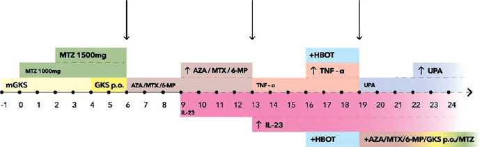
Treatment should be initiated with high-potency topical glucocorticosteroids (e.g. clobetasol propionate) or calcineurin inhibitors (for facial lesions), applied twice daily for 4 weeks. Systemic therapy is marked from month 0, with the note that topical corticosteroid therapy may be initiated earlier if clinically indicated.
If there is no improvement, metronidazole 2 × 500 mg for 8 weeks is added, and if necessary, 3 × 500 mg for another 8 weeks. If lesions persist, oral corticosteroids (e.g. prednisone 0.5 mg/kg of ideal body weight) are started for 8 weeks with a gradual dose reduction, while discontinuing topical glucocorticosteroids. After another 8 weeks, efficacy should be evaluated – if satisfactory improvement is not achieved, both dermatological symptoms and endoscopic findings of the gastrointestinal tract should be assessed. Next, azathioprine, methotrexate, or mercaptopurine is added and assessed after 12 weeks [9]. In the absence of improvement, dose escalation or a switch to alternative treatment may be considered, namely the implementation of an IL-23 inhibitor (ustekinumab, risankizumab, guselkumab). If there is still no effect after another 16 weeks, one may switch to a TNF-α inhibitor (infliximab, adalimumab) or increase the dose of the IL-23 inhibitor. If this is also ineffective, consider increasing the dose of the TNF-α inhibitor or adding hyperbaric oxygen therapy; if improvement still does not occur, reconsider reintroducing metronidazole 2 × 500 mg or systemic glucocorticosteroids or conventional immunosuppressants (AZA, MTX, 6-MP) or switching therapy to upadacitinib. In patients with single or few lesions, intralesional triamcinolone or methylprednisolone injections every 4–8 weeks may be considered at any stage of therapy.
It is important, in addition to pharmacological treatment, to provide psychological support to patients with MCD – particularly those with lesions on the genitalia. Chronic, disfiguring lesions may lead to body image disorders, depression, and difficulties in sexual and social functioning, especially among young individuals [14, 84, 87]. Untreated skin lesions in MCD may eventually become resistant to pharmacological and surgical treatment, and an accurate dermatological diagnosis with prompt referral to a gastroenterologist can significantly accelerate the diagnosis of Crohn’s disease and prevent serious and irreversible complications of this condition.
Summary
MCD remains one of the most enigmatic aspects of IBD, particularly due to its rare but highly problematic cutaneous symptoms. Despite significant advances in recent years, many questions regarding its pathogenesis, diagnosis, and treatment remain unanswered, and the number of uncertainties often exceeds the current state of clinical knowledge [1–18, 23–34, 39–40]. This makes MCD a particular challenge not only for gastroenterologists and dermatologists but also for psychologists and all specialists involved in patient care.
The manifestations of MCD can be extremely diverse – from oedema and ulceration of the genitalia to infiltrative and nodular lesions on the limbs or trunk. These lesions may appear in patients with newly diagnosed Crohn’s disease, during periods of remission, or – even more insidiously – long before the underlying disease is diagnosed [7, 15, 17, 49–55]. In children, lesions are more often limited to the genital area, further complicating diagnosis. The greatest diagnostic challenge remains the lack of pathognomonic features – morphology is so variable that MCD can easily be mistaken for other dermatoses, infections, or autoimmune conditions such as sarcoidosis, mycobacterial infections, Behçet’s disease, or hidradenitis suppurativa [56–67, 71–73].
Thus, a detailed personal and family history (including IBD, rheumatologic, and dermatologic diseases) is crucial, alongside comprehensive differential diagnostics: laboratory tests, imaging studies, and especially histopathological analysis in chronic or unclear cases [1–5, 9, 14, 31, 50–72]. In 2025, the first official diagnostic criteria for MCD (Delphi Consensus) were published, which do not require mandatory histopathological confirmation but emphasise fulfilment of clinical criteria and exclusion of other causes [9]. While this approach facilitates diagnosis in clinical practice, it demands vigilance and close interdisciplinary collaboration from the clinician.
Recent research points to the complexity of MCD pathogenesis – genetic factors (NOD2/CARD15 mutations), impaired T-cell migration (“mis-homing”), and abnormal integrin expression leading to the migration of immune cells beyond the gut barrier to the skin are all important [12, 17, 26–30]. Excessive activation of the Th17/IL-23/TNF-α axis, the phenomenon of epitope spreading, and immunological complications also play a significant role [12, 17, 23–25, 29, 30]. Still, there is no clear answer as to why only a small group of patients develop cutaneous lesions, despite similar genetic profiles and intestinal disease course.
Treatment of MCD requires an individualised approach and close cooperation between the gastroenterologist, dermatologist, and – increasingly – a psychologist. Cutaneous lesions, especially in the genital area, are not only chronic and recurrent but also resistant to standard dermatological therapies [61–65]. Guidelines suggest starting with topical glucocorticosteroids and calcineurin inhibitors, then introducing metronidazole, and, if ineffective, immunosuppressive drugs (azathioprine, methotrexate) or biological agents (TNF-α, IL-23, or IL-12/23 inhibitors) [9, 14, 17, 37, 42, 66–70, 72, 75, 111–120]. Increasingly, JAK inhibitors (upadacitinib) are reported to be effective, especially in refractory cases [38, 73, 76]. Recent reports highlight the effectiveness of ustekinumab and risankizumab and the limited efficacy of vedolizumab in cutaneous lesions – which underscores the different immunopathogenesis of extraintestinal versus intestinal manifestations [39, 40].
Surgical treatment and supportive therapies such as hyperbaric oxygen therapy are considered only in complicated or particularly refractory cases [47, 97, 110]. Monitoring therapeutic effects and safety parameters is essential, especially with long-term antibiotic or immunosuppressive therapy [14, 34, 106].
The impact of cutaneous lesions on the psychological well-being of patients cannot be underestimated – chronicity, recurrence, pain, and the location of lesions (especially in the genital area) can lead to serious body image disturbances, anxiety, depression, and difficulties in sexual and social functioning, particularly among young adults [9, 84, 87].
Patients often struggle with feelings of shame, social withdrawal, and stigmatisation, and the chronic nature of the disease demands long-term psychological and psychiatric support. Unfortunately, this problem is often underestimated, leading to reduced quality of life and growing frustration due to the lack of effective treatment methods. Psychological support, psychoeducation, and enabling patients to join support groups are indispensable elements of modern MCD therapy.
MCD presents a clinical challenge due to its rarity, extreme variability of symptoms, diagnostic difficulties, and limited treatment options. Each case requires a holistic approach, considering both clinical and psychological aspects. The still unclear pathogenesis and insufficient molecular research mean that treatment is based mainly on clinical experience and the latest, frequently changing, guidelines. In the future, further studies on immunological mechanisms and the search for new, targeted therapies are essential not only to more effectively treat cutaneous lesions but also to improve patients’ quality of life. Recognition and treatment of MCD demand constant clinical vigilance, interdisciplinary cooperation, and comprehensive support – both medical and psychological.














