en POLSKI
eISSN: 2083-8441
ISSN: 2081-237X
Pediatric Endocrinology Diabetes and Metabolism
Current issue Archive Manuscripts accepted About the journal Supplements Editorial board Reviewers Abstracting and indexing Subscription Contact Instructions for authors Ethical standards and procedures
Editorial System
Submit your Manuscript
SCImago Journal & Country Rank
3/2021
vol. 27
 
Share:
Share:
Review paper

Mucopolysaccharidosis III: Molecular basis and treatment

Lidvana Spahiu
1
,
Emir Behluli
1
,
Borut Peterlin
2
,
Hilada Nefic
3
,
Rifat Hadziselimovic
3
,
Thomas Liehr
4
,
Gazmend Temaj
5

1.
Pediatric Clinic, University Clinical Center of Kosovo, Prishtina, Kosovo
2.
Department of Genetics and Genomics, University Clinical Center Ljubljana, Slovenia
3.
Faculty of Science, University of Sarajevo, Sarajevo, Bosnia and Herzegovina, Bosnia and Herzegovina
4.
Institut für Humangenetik, Universitätsklinikum Jena, Friedrich Schiller Universität, Jena Germany, Germany
5.
Human Genetics, College UBT, Faculty of Pharmacy Prishtina, Kosovo
Pediatr Endocrinol Diabetes Metab 2021; 27 (3): 199–208
Online publish date: 2021/09/30
Article file
Get citation
 
PlumX metrics:
 

Introduction

Lysosomal storage diseases (LSDs) are inherited metabolic disorder diseases; these kinds of diseases are caused by enzyme deficiencies resulting in accumulation of un-degraded substrate. This storage process leads to clinical manifestations depending on the specific substrate and site of accumulation. LSDs are a heterogeneous group of diseases [1]. They are characterized by accumulation of macromolecules in lysosomes, like glycosaminoglycan (GAG). If GAG degradation cannot be performed normally, the resulting LSD is called mucopolysaccharidosis (MPS). Here the degradation of carbohydrate complex is hampered and thus, GAG cannot be processed normally [2]. Mucopolysaccharidoses constitute ~30% of all LSDs [3].

Mucopolysaccharidosis

Mucopolysaccharidoses are known as a group of metabolic diseases caused by absence or malfunction of an enzyme being necessary for breakdown of molecules long chains (carbohydrates) in the cells. In such patients deficiency of 11 different enzymes being required for breakdown of carbohydrates in small and simple molecules have been reported. These GAGs are accumulated in blood, brain, spinal corda and different connective tissue and are there responsible for local cell damage. Symptoms may be similar or may vary among different types of MPSs [4].

The prevalence of MPSs is different in different countries and varies between 0.07 in Australia and 4.05 per 100,000 life-births in Estonia; reported differences may be due to undiagnosed cases and/or different genetic prevalence: It must be suggested that MPSs are underdiagnosed in many countries due to lack of corresponding diagnostic means.

Mucopolysaccharidoses are known to be autosomal recessive disorder, meaning that only individuals with two mutated alleles are affected. When both parents are heterozygote for the disease causing allele in each pregnancy there is a one in four chance that the child will be affected. Accordingly, unaffected persons in a family may be either heterozygous for a disease causing allele, or may carry two non-disease causing ones. Only MPSII or Hunter syndrome is an X-linked recessive disorder [57].

Generally, following factors may increase the chance to have or pass an MPS-disease-causing allele:

  • family history of an MPS;

  • a couple is consanguineous or derive from same distinct ethnic or geographically clustered community;

  • a couple is heterozygous for MPS disease causing allels.

Among all types of MPS, MPS III (or Sanifilippo syndrome) is most frequent; it was first described ~50 years ago [8]. Here heparin sulfate (HS) plays a pivotal role, as it does also in neuronal development [9]. Accordingly, neurological pathology found in MPS III patients can be explained. Relevance of lysosomes in maintaining of neuron and brain homeostasis has been reported recently [10]. Impairment of HS metabolism, where also lysosomes are involved and accumulation of α-synuclein, connected MPS III with other neurological diseases such as Parkinson disease is reported [11]. On the other hand MPS III can be seen in one line with autism spectrum disorders (ASDs) which are associate with neurodevelopmental delay, including language, impaired social interactions and restrictive, repetitive and stereotyped behaviors [12, 13].

Mucopolysaccharidosis III can be divided into five subtypes [3]. These are caused by mutation of sulfamidase (MPS IIIA; OMIM#252900) [14], α-n-acetylglucosaminidase (NAGLU, MPS IIIB; OMIM#252920) [15], heparan acetyl CoA: α-glucosaminide N-acetyltransferase (HGSNAT, MPS IIIC; OMIM#252930) [16], and N-acetylglucosamine 6-sulfatase (GNS, MPS IIID; OMIM#252940) [17].

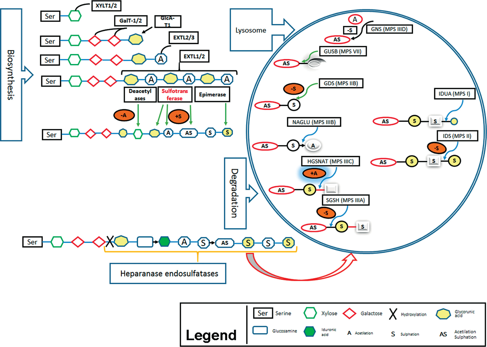
Here we describe the molecular basis of MPS III, and summarize therapeutic approaches. The process of heparin sulfate degradation and enzymes which are involved in this process are presented in Fig. 1.

Figure 1

Synthesis and degradation of HS (Heparan Sulfate) is presented schematically including cell organelles (Lysosome) and enzymes which are responsible for each MPSs; also modification of residues is shown

/f/fulltexts/PEDM/45203/PEDM-27-45203-g001_min.jpg

Genetics of mucopolysaccharidosis III

The known common mutations being responsible for MPS III are presented in Table IIa. Besides, other recently found genes involved in MPS III in Table IIb (only single case reports, yet). Approximate frequencies of genes involved in MPS IIIA, MPS IIIB, MPS IIIC, MPS IIID and MPS IIIE were determined based on reports in Varsome and ClinVar databases [99100].

Mucopolysaccharidosis IIIA

Mucpolysaccharidosis IIIA (~27% of MPS III) is caused by mutations in the gene SGSH localized in chromosome 17q25.3 (Table II, III). This gene is responsible for coding of sulfaminidase (heparin sulfate sulfatase or N-sulfoglucosamine sulfohydrolase) [18]. The first patients diagnosed with symptoms of MPS IIIA were described by Sanfilippo et al. [8].

Specific mutations in sulfaminidase are found in variant frequencies in different countries. The missense mutations p.R74C occurs at a frequency of 56% in Polish MPS III population [19]. In Australia amino acid change at p.R245H is most frequent with 31%; in Germany frequency of this mutation is 35%, and in Netherlands 58% [19, 20]. In Italian patients missense mutation for p.S66W was found with a frequency of 29% [21].

Table I

Incidences of MPS in different ethnicities

CountryPrevalence per 100,000 life-birthsReferences
Netherland1.89[75]
Australia0.07[76]
Portugal0.12[77]
Scandinavian countries0.67[78]
Japan1.53[79]
Switzerland1.56[79]
Taiwan2.04[80]
South Korea1.35[81]
Tunisia2.27[82]
British Columbia (Columbia)1.94[79]
Brazil1.04[79]
Czech Republic3.27[83]
Estonia4.05[84]
Germany3.51[79]
Poland1.80[85]

Mucopolysaccharidosis IIIB

Mutation in gene NAGLU (17q21.2) causes MPS IIIB (~36% of MPS III; Table II). This gene encodes N-acetyl-α-glucosaminidase, a lysosomal enzyme of 720 amino acids with six glucosylation site [22]. Most frequently observed are missense mutations p.F48L, p.G69S, p.S612G and p.R643C which are associated with late-onset phenotype [2325].

Table II

Summary of the phenotypic, enzymatic, and genetic classification of the MPS III

SubtypePhenotypeEnzymeGene locusCytogenetic locationType of mutationPosition of mutationReference
MPS III AOMIM
252900
SulfamidaseSGSH17q25.3Missensep.R245H
p.S66W
[19, 20, 21]
MPS III BOMIM
252920
a-n-acetyl- glucosaminidaseNAGLU17q21.1Missensep.F48L
p.G69S
p.R643O
[23, 24, 25]
MPS III COMIM
252930
Heparan acetyl CoA;
a-glucosamine
6-sulfatase
HGSNAT8p11.1Missensep.R344O
p.S518F
p.G262R
p.S539C
[27, 28, 29, 30]
MPS III DOMIM
252940
N-acetylglucosamine
6-sulfatase
GNS12q14.4Missensep.S94I
p.K340R
p.G418E
[43]
Nonsense Splice site mutationp.K388X
c.875 + 2delT
c.1097_1098
+ 1delAGGinsGGT
c.1309-2A>G
c.1420-2A>G
c.625-637del8
Frame shift mutationsc.109dupG
c.123dupG
Large deletionDel EX1 +
Del EX6,7
Del EX9-14
MPS III En.a.N-glucosamine
3-O-sulfatase
ARSG17q24.2n.a.[32, 33, 34]

Mucopolysaccharidosis IIIC

The third form of mucpolysaccharidosis, MPS IIIC (~30% of MPS III), is caused by lysosomal membrane acetyl-CoA α-glucosaminide N-acetyltransferase coded by HGSNAT gene (8p11.21) (Tables II, III) [26]. Missense mutations in position p.R344C (22%) and p.S518F (29%) [27] are most frequently observed. MPS IIIC can be associated with other diseases such as Retinitis pigmentosa, when HGSNAT missense mutations p.G262R and p.S539C in membrane domain of the enzyme are present [2830].

Mucopolysaccharidosis IIID

Mucopolysaccharidosis IIID is caused by GNS gene (12q14.3) mutations (~5% of MPS III, Tables II, III), which encodes lysosome N-acetylglucosamine-6-sulfatase. This enzyme contain 552 amino acids and 13 glucosylation (modification) sites [31]. Missense mutation occur in 13% of all MPS IIID cases.

Mucopolysaccharidosis IIIE

Mutations in ARSG gene (17q24.2) cause MPS IIIE (~2% of MPS-III, Tables II, III). The gene contains eleven exon, the protein N-glucosamine 3-O-sulfatse consist of 525 amino acids and four glucosylation (modification) sites in asparagine [324].

Table III

Summary of the MPS III associated with other diseases and gene mutation

Diseases which are found to be associated with MPS IIIBGeneGene encodedReference
Lung diseases, nephrotic syndrome, epidermolysis syndromeITGA3member of the integrin a chain family of proteins[86, 87, 88]
AutismHRH1histamine receptor H1[88, 89, 90]
Neurodevelopment disorders, development of learning and memoryeIF4A3 (eukaryote initiation factor 4A3)eIF4A3[88, 91, 92]
MPS types/subtypesID2transcription factor DNA binding 2[88]
Deafness, behavior problemHomer 2protein scaffold[88, 93, 94]
MPS I, MPS IIIA and MPS IIIBHomer 1Homer 1 protein[95]
Cancer diseasesB2M (-2-microglobulin)-2-microglobulin[88]
DiabetesINSR (insulin receptor)Insulin receptors[88]
Charcot-Marie Tooth, neurodegenerative diseasesMME (membrane metalloendopeptidase)Glycoproteins[96, 97]
Charcot-Marie Tooth, neurodegenerative diseasesCAPN2 (capelin 2, calcium dependent protease)Calpain-2 catalytic subunit[96, 97]
Neuropathy and neurodegenerationAPOEapolipoprotein E[98]
Neurodegenerative disordersUCHL1ubiquitin C-terminal hydrolase L1[88]
MPS IX, Rigid Spine Muscular Dystrophy 1 and Visceral HeterotaxySERPINE2SERPINE2 protein[88]
MPS IX, VLDLR-associated cerebellar hypoplasiaVLDLRlipoprotein receptor[88]

Laboratory diagnostic methods for mucopolysaccharidosis

Stepwise diagnostics of MPS is done biochemically, for example by detection of heparan sulfate levels in urine of patients. Dimethylmethylen blue is used for heparan sulfate detection at the wavelength of 520 nm in a spectrophotometer [35, 36]. Another method is high-resolution cellulose acetate electrophoresis of a urine sample which can separate and identify heparan sulfate [37]. Finally, tandem mass spectrometry can be applied for diagnosis of MPS [38]; identification of specific oligosaccharides that are accumulate as a results of enzyme deficiency can be picked up by that [3840]. Recently it was also suggested to measure protein amounts involved in MPS from dried blood spot using multiplex immune–quantification assay [41, 42].

The method to identify the underlying genetic cause and specific subtype of a suggested MPS is genomic DNA sequencing (NGS-next generation sequencing); also carrier status of parents can be assessed by this [43].

Postnatally, urine or peripheral blood can be studied. Prenatal genetic diagnostic is possible after invasive prenatal diagnostics, as chorionic villus sampling, amniocentesis or umbilical cord blood sampling [44].

Therapy for mucopolysaccharidosis patients

Most of the following treatment options are still experimental.

Enzyme replacement therapy

Enzyme replacement therapy is a possible approach to treat MPS patients. Injection of human sulfaminidase directly in brain [45] or cerebrospinal fluid via the cerebellomedullary cistern [46] has been shown to be principally possible in MPS IIIA mice [47] (Fig. 2A).
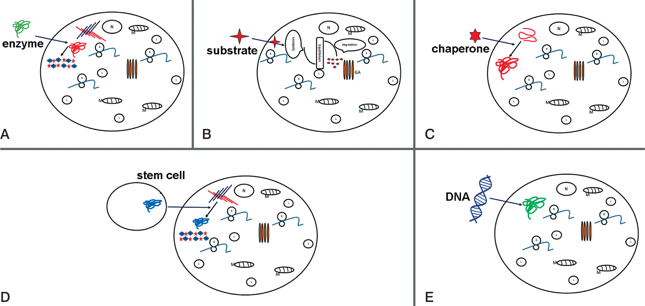
Figure 2

Potential therapy attempts to treat MPS. Schematic representations of: A) enzyme replacement therapy (ERT) to replace mutated form of protein; B) substrate reduction therapy (SRT), reduction of storage of undegraded substrate; C) pharmacological chaperone to correct substrate miss-folding; D) stem cell therapy (SCT) used for replacement of diseased cells; E) gene therapy to provide correct form of a mutated gene

/f/fulltexts/PEDM/45203/PEDM-27-45203-g002_min.jpg

Substrate reduction therapy

Substrate reduction therapy (STR) in another alternative therapy approach and outline in Figure 1B. Aim is here to approach molecular targets decreasing production of accumulated substrate and to restore balance between synthesis and degradation. This method is approved e.g. for treatment of lysosomal disorders with neurological and non-neurological symptoms [48]. For STR RNAi can be applied to key genes participating in GAG synthesis, such as EXTL other genes involved in linkage region formation. Dziedzic et al., applied siRNA to down-regulate XYLT1, XYTL2, GALTI and GALTII [49].

This strategy is successfully applied in treatment of MPS I and MPS IIIA, resulting in reduction of mRNA and protein levels of corresponding genes and a significant decrease in GAG synthesis. Using of shRNA (short hairpin RNA) has been shown to have a positive effect in downregulation of two other genes EXTL2 and EXTL3; both genes influence reduction of GAG synthesis [50] (Fig. 2B).

Pharmacological therapy

In many cases, mutation is shown to lead production of misfolded protein, which become rapidly degraded. The activation of chaperons (cellular proteins) may help functional correct protein folding. As shown in Fig. 2C, small compounds as actin-like chaperones (= amino and iminosugars) prevent protein misfolding by acting as enzyme inhibitors [51]. This approach is used to treat different LDS diseases such Farby, Morquio B, Pompe, Gaucher and Krabbe diseases [52].

The component is an isoflavine which is purified from soy and has the ability to reduce GAG (glucoaminglucans) level. The overall mechanism is not clarify. However, it seems likely that the mechanism involves protein kinase inhibitory, which in turn induces TFEB (transcription factor EB) transcriptional activity [53, 54].

Growth medium supplemented by genistein has been shown to reduce GAG level in skin fibroblast of MPS IIIA and MPS IIIB patients [55]. In murine model such treatment led to reduction of GAG storage [56]. Moreover, in a pilot study in patients with MPS IIIA and B administration of genistein reduced GAG levels in urine [57]. In mouse reduction of GAG levels was demonstrated after treatment with rhodamine in the liver and the brain [58].

Stem cell therapy

Stem cell therapy was suggested for treatment of neurological diseases in order to produce correct forms of enzyme (Figure 2D). Bone marrow transplantation can be used for treatment of neurological diseases, but in MPS III diseases this approach was not successful [59]. The treatment with hematopoietic stem cell in patients show that in brain cells microglia can be replaced and become enzyme secretion donor cells [60]. Recently genetic modification of hematopoietic stem cells carrying normal copies of the SGSH or NAGLU genes led in mouse model to enzyme production [6164].

iPSC (induced pluripotent stem cell) development technology gave new possibilities for generation of neural stem cell (NSC). After transplantation of NSC they could migrate over long distances within brain and were integrated in host network. This form of treatment recovered neurological pathology of MPS VII [65] and MPS IIIB [66].

Gene therapy

The gene therapy is used to enlargement ERT (enzyme replacement therapy) (Fig. 1E) so far is attempted to introduce the coding sequence of the protein. The cell manipulation will possess in enzyme activity but also will participate in enzyme secretion and circulation up to altered cell. The sulfamidase injection as a recombinant adeno-associated virus (AAV) will improve coding sequence and in this form will increase enzyme activity (sulfamidase) in brain of mice [67]. This strategy now is used in human clinical traits [68].

Gene therapy is most promising therapeutic option for treatment of MPS diseases, and 5–15% of enzyme activity can be recovered in affected patients [69]. Several viral vectors are used for treatment of MPS patients, such as retroviruses, lentiviruses, adenoviruses and adeno-associated viruses (AAV). Using of nonviral vector also led to increased enzyme activity and GAG reduction in lysosome [70]. Adeno-associated viruses treatment seems to be suited best for gene therapy in neurological disorders. AAV therapy could e.g. induce cell death in murine neural cells and hippocampus, suggesting that these approaches should be carefully evaluated [71]. AVV therapy and other viral gene therapies may be associated with significant side effects, particularly during development. Adeno-associated virus serotype 5 (AAV5) has been used for treatment of MPS IIIB patients, results shown improvement of neurological progression in patients inducing sustained enzyme production in the brain [72]. AAVrh10 (adeno-associated virus serotype rh10) was used to integrate intact SGSH gene in MPS IIIA model mice. This treatment reduced HS accumulation and microglia activity administration [73]. The AAV8 delivered effectively NAGLU gene in MPS IIIB animal model and also facilitated robust somatic transduction of the heart and liver [74].

Conclusions

In this review we provide an overview for molecular basis of MPS III, laboratory diagnostic methods such biochemical detection of heparan sulfate in patients with MPS III. Genes responsible for MPS III have been identified by characterizing the functional role of gene products in the metabolism. A number of genetic and biochemically methods have been adopted in laboratory for diagnosis. Although four types of MPS III diagnosis remain difficult, our recommendations for screening and diagnosis are as follows:

  • All patients with speech delay, deficit or hyperactivity disorders, autism should be screened for MPS III. DMB (1,2-diamino-4,5-methylenedioxybenzene-2HCl) assay is strongly recommended.

  • All positive samples from quantitative assay should be investigate by electrophoresis, GAG depolymerization followed by HPLC/MS/MS analysis.

  • An enzyme activity assay must be done to confirm the diagnosis.

  • Molecular genetic testing should be offered to all patients; this test is informative for the family when they making decision for family plan.

Therapeutic options for MPS III disease, once considered untreatable, are available for patients with these diseases. The iPSC have been established, and will be very useful in drug screening studies to identify the drugs which will be potential for human treatment.

Conflict of interests

none declared.

References

1 

Sun A. Lysosomal storage disease overview. Ann Transl Med.2018; 6: 476. 10.21037/atm.2018.11.39.

2 

Tomatsu S, Lavery C, Giugliani R, et al. Mucopolysaccharidoses Update; Nova Science Publishers: Hauppauge, NY, USA 2018.

3 

Neufeld EF, Muenzer J. The Mucopolysaccharidoses. In: The Metabolic and Molecular Bases of Inherited Disease. Scriver CR, Valle DL, Antonarakis S, et al. (eds.). McGraw-Hill, New York, NY, USA, 2001; 3421–3452.

4 

Malm G, Månsson JE. Mucopolysaccharidosis type III (Sanfilippo disease) in Sweden: clinical presentation of 22 children diagnosed during a 30-year period. Acta Paediatr 2010; 99: 1253-1257. 10.1111/j.1651-2227.2010.01800.x.

5 

Campos D, Monaga M. Mucopolysaccharidosis type I: current knowledge on its pathophysiological mechanisms. Metab Brain Dis. 2012; 27: 121-129. 10.1007/s11011-012-9302-1.

6 

Muenzer J. The mucopolysaccharidoses: a heterogeneous group of disorders with variable pediatric presentations. J Pediatr 2004; 144 (5 Suppl): S27-34. 10.1016/j.jpeds.2004.01.052.

7 

Clarke L.A. The mucopolysaccharidoses: a success of molecular medicine. Expert Rev Mol Med 2008; 18: 10: e1. 10.1017/S1462399408000550.

8 

Sanfilippo SJ, Good RA, Podosin R, et al. Mental Retardation Associated with Acid Mucopolysacchariduria (Heparitin Sulfate Type). J. Pediatr-Us. 1963; 63: 837–838.

9 

De Pasquale V, Pavone LM. Heparan sulfate proteoglycans: The sweet side of development turns sour in mucopolysaccharidoses. Biochim Biophys Acta Mol Basis Dis 2019; 1865: 165539. 10.1016/j.bbadis.2019.165539.

10 

Lloyd-Evans E, Haslett LJ. The lysosomal storage disease continuum with ageing-related neurodegenerative disease. Ageing Res Rev 2016; 32: 104–121. 10.1016/j.arr.2016.07.005.

11 

Winder-Rhodes SE, Garcia-Reitbock P, Ban M, et al. Genetic and pathological links between Parkinson’s disease and the lysosomal disorder Sanfilippo syndrome. Mov Disord 2012; 27: 312–315. 10.1002/mds.24029.

12 

Lau AA, Tamang SJ, Hemsley KM. MPS-IIIA mice acquire autistic behaviours with age. J Inherit Metab Dis.2018; 41: 669-77. 10.1007/s10545-018-0160-9.

13 

Çöp E, Yurtbasi P, Öner Ö, et al. Genetic testing in children with autism spectrum disorders. Anadolu Psikiyatri Derg 2015; 16: 426-32. 10.5455/apd.1414607917.

14 

Kresse H. Mucopolysaccharidosis 3 A (Sanfilippo A disease): deficiency of a heparin sulfamidase in skin fibroblasts and leucocytes. Biochem Biophys Res Commun. 1973; 54: 1111–1118. 10.1016/0006-291x(73)90807-3.

15 

von Figura K. Human α-N-acetylglucosaminidase. Purification and properties. Eur J Biochem 1977; 80: 523–533.

16 

Klein U, Kresse H, von Figura K. Sanfilippo syndrome type C: deficiency of acetyl-CoA: α-glucosaminide N-acetyltransferase in skin fibroblasts. Proc Natl Acad Sci U S A 1978; 75: 5185–5189. 10.1073/pnas.75.10.5185.

17 

Kresse H, Paschke E, von Figura K, et al. Sanfilippo disease type D: deficiency of N-acetylglucosamine-6-sulfate sulfatase required for heparan sulfate degradation. Proc Natl Acad Sci U S A 1980; 77: 6822–6826. 10.1073/pnas.77.11.6822.

18 

Scott HS, Blanch L, Guo XH, et al. Cloning of the sulphamidase gene and identification of mutations in Sanfilippo A syndrome. Nat Genet 1995; 11: 465–467. 10.1038/ng1295-465.

19 

Bunge S, Ince H, Steglich C, et al. Identification of 16 sulfamidase gene mutations including the common R74C in patients with mucopolysac-charidosis type IIIA (Sanfilippo A). Hum Mutat 1997; 10: 479–485. doi: 10.1002/(SICI)1098-1004(1997)10:6<479::AID-HUMU10>3.0.CO;2-X.

20 

Weber B, Guo XH, Wraith JE, et al. Novel mutations in Sanfilippo A syndrome: implications for enzyme function. Hum Mol Genet 1997; 6: 1573–1579. 10.1093/hmg/6.9.1573.

21 

Di Natale P, Balzano N, Esposito S, et al. Identification of molecular defects in Italian Sanfilippo A patients including 13 novel mutations. Hum Mutat 1998; 11: 313–320. 10.1002/(SICI)1098-1004(1998)11:4<313::AID-HUMU9>3.0.CO;2-P.

22 

Zhao HG, Li HH, Bach G, Schmidtchen A, Neufeld EF. The molecular basis of Sanfilippo syndrome type B. Proc Natl Acad Sci USA 1996; 93: 6101–6105.

23 

Zhao HG, Aronovich EL, Whitley CB. Genotype-phenotype correspondence in Sanfilippo syndrome type B. Am. J Hum Genet 1998; 62: 53–63.

24 

Weber B, Guo XH, Kleijer WJ, et al. Sanfilippo type B syndrome (mucopolysaccharidosis III B): allelic heterogeneity corresponds to the wide spectrum of clinical phenotypes. Eur J Hum Genet 1999; 7: 34–44. 10.1038/sj.ejhg.5200242.

25 

Yogalingam G, Hopwood JJ. Molecular genetics of mucopolysac-charidosis type IIIA and IIIB: diagnostic, clinical, and biological implications. Hum Mutat 2001; 18: 264–281. 10.1002/humu.1189.

26 

Zelei T, Csetneki K, Voko Z, et al. Epidemiology of Sanfilippo syndrome: Results of a systematic literature review. Orphanet J. Rare Dis. 2018; 13: 53. 10.1186/s13023-018-0796-4.

27 

Ruijter GJ, Valstar MJ, van de Kamp JM, et al. Clinical and genetic spectrum of Sanfilippo type C (MPS IIIC) disease in The Netherlands. Mol Genet Metab 2008; 93: 104–111. 10.1016/j.ymgme.2007.09.011.

28 

Fan X, Zhang H, Zhang S, et al. Identification of the gene encoding the enzyme deficient in mucopolysaccharidosis IIIC (Sanfilippo disease type C). Am J Hum Genet 2006; 79: 738–744. 10.1086/508068.

29 

Hřebíček M, Mrázová L, Seyrantepe V, et al. Mutations in TMEM76* cause mucopolysaccharidosis IIIC (Sanfilippo C syndrome). Am J Hum Genet 2006; 79: 807–819. 10.1086/508294.

30 

Fedele AO, Filocamo M, Di Rocco M, et al. Mutational analysis of the HGSNAT gene in Italian patients with mucopolysaccharidosis IIIC (Sanfilippo C syndrome). Hum Mutat 2007; 28: 523. 10.1002/humu.9488.

31 

Robertson DA, Freeman C, Nelson PV, et al. Human glucosamine-6-sulfatase cDNA reveals homology with steroid sulfatase. Biochem. Biophys. Res Commun 1988; 157: 218–224. 10.1016/s0006-291x(88)80035-4.

32 

Kowalewski B, Lamanna WC, Lawrence R, et al. Arylsulfatase G inactivation causes loss of heparan sulfate 3-O-sulfatase activity and mucopolysaccharidosis in mice. Proc Natl Acad Sci U S A 2012; 109: 10310–10315. 10.1073/pnas.1202071109.

33 

Ferrante P, Messali S, Meroni G, Ballabio A. Molecular and biochemical characterisation of a novel sulphatase gene: arylsulfatase G (ARSG). Eur J Hum Genet 2002; 10: 813–818. 10.1038/sj.ejhg.5200887.

34 

Frese MA, Schulz S, Dierks T. Arylsulfatase G, a novel lysosomal sulfatase. J Biol Chem 2008; 283: 11388–11395. 10.1074/jbc.M709917200.

35 

Farndale RW, Sayers CA, Barrett AJ. A direct spectrophotometric microassay for sulfated glycosaminoglycans in cartilage cultures. Connect Tissue Res 1982; 9: 247–248. 10.3109/03008208209160269.

36 

de Jong JG, Wevers RA, Liebrand-van Sambeek R. Measuring urinary glycosaminoglycans in the presence of protein: an improved screening procedure for mucopolysaccharidoses based on dimethylmethylene blue. Clin Chem 1992; 38: 803–807.

37 

Hopwood JJ, Harrison JR. High-resolution electrophoresis of urinary glycosaminoglycans: an improved screening test for the mucopolysaccharidoses. Anal Biochem 1982; 119: 120–127. 10.1016/0003-2697(82)90674-1.

38 

Fuller M, Rozaklis T, Ramsay SL, et al. Disease-specific markers for the mucopolysaccharidoses. Pediatr Res 2004; 56: 733–738. 10.1203/01.PDR.0000141987.69757.DD.

39 

Mason KE, Meikle PJ, Hopwood JJ, et al. Characterization of sulphated oligosaccharides in mucopolysaccharidosis type IIIA by electrospray ionization mass spectrometry. Anal Chem 2006; 78: 4534–4542. 10.1021/ac052083d.

40 

Mason K, Meikle P, Hopwood J, et al. Distribution of heparan sulfate oligosaccharides in murine mucopolysaccharidosis type IIIA. Metabolites 2014; 4: 1088–1100. 10.3390/metabo4041088.

41 

Meikle PJ, Grasby DJ, Dean CJ, et al. Newborn screening for lysosomal storage disorders. Mol Genet Metab 2006; 88: 307–314. 10.1016/j.ymgme.2006.02.013

42 

Fuller M, Tucker JN, Lang DL, et al. Screening patients referred to a metabolic clinic for lysosomal storage disorders. J Med Genet 2011; 48: 422–425. 10.1136/jmg.2010.088096.

43 

Valstar MJ, Bertoli-Avella AM, Wessels MW, et al. Mucopolysaccharidosis type IIID: 12 new patients and 15 novel mutations. Hum Mutat. 2010; 31: E1348–E1360. 10.1002/humu.21234.

44 

Hopwood JJ. Prenatal diagnosis of Sanfilippo syndrome. Prenat Diagn 2005; 25: 148–150. 10.1002/pd.1094.

45 

Savas PS, Hemsley KM, Hopwood JJ. Intracerebral injection of sulfamidase delays neuropathology in murine MPS-IIIA. Mol Genet Metab 2004; 82: 273–285. 10.1016/j.ymgme.2004.05.005.

46 

Hemsley KM, King B, Hopwood JJ. Injection of recombinant human sulfamidase into the CSF via the cerebellomedullary cistern in MPS IIIA mice. Mol Genet Metab 2007; 90: 313–328. 10.1016/j.ymgme.2006.10.005.

47 

Kishnani PS, Dickson PI, Muldowney L, et al. Immune response to enzyme replacement therapies in lysosomal storage diseases and the role of immune tolerance induction. Mol Genet Metab 2016; 117: 66–83. 10.1016/j.ymgme.2015.11.001.

48 

Venier RE, Igdoura SA. Miglustat as a therapeutic agent: Prospects and caveats. J Med Genet 2012; 49: 591–597. 10.1136/jmedgenet-2012-101070.

49 

Dziedzic D, Wegrzyn G, Jakobkiewicz-Banecka J. Impairment of glycosaminoglycan synthesis in mucopolysaccharidosis type IIIA cells by using siRNA: A potential therapeutic approach for Sanfilippo disease. Eur J Hum Genet 2010; 18: 200–205. 10.1038/ejhg.2009.144.

50 

Kaidonis X, Liaw WC, Roberts AD, et al. Gene silencing of EXTL2 and EXTL3 as a substrate deprivation therapy for heparan sulphate storing mucopolysaccharidoses. Eur J Hum Genet 2010; 18: 194–199. 10.1038/ejhg.2009.143.

51 

Parenti G, Andria G, Valenzano KJ. Pharmacological Chaperone Therapy: Preclinical Development, Clinical Translation, and Prospects for the Treatment of Lysosomal Storage Disorders. Mol Ther 2015; 23: 1138–1148. 10.1038/mt.2015.62.

52 

Suzuki Y. Emerging novel concept of chaperone therapies for protein misfolding diseases. Proc Jpn Acad Ser B 2014; 90: 145–162. 10.2183/pjab.90.145.

53 

Moskot M, Montefusco S, Jakóbkiewicz-Banecka J, et al. The phytoestrogen genistein modulates lysosomal metabolism and transcription factor EB (TFEB) activation. J Biol Chem 2014; 289: 17054–17069. 10.1074/jbc.M114.555300.

54 

Moskot M, Jakóbkiewicz-Banecka J, Kloska A, et al. Modulation of expression of genes involved in glycosaminoglycan metabolism and lysosome biogenesis by flavonoids. Sci Rep 2015; 5: 9378. 10.1038/srep09378.

55 

Piotrowska E, Jakóbkiewicz-Banecka J, Barańska S, et al. Genistein-mediated inhibition of glycosaminoglycan synthesis as a basis for gene expression-targeted isoflavone therapy for mucopolysaccharidoses. Eur J Hum Genet 2006; 14: 846–852. 10.1038/sj.ejhg.5201623.

56 

Malinowska M, Wilkinson FL, Bennett W, et al.Genistein reduces lysosomal storage in peripheral tissues of mucopolysaccharide IIIB mice. Mol Genet Metab 2009; 98: 235–242. 10.1016/j.ymgme.2009.06.013

57 

Piotrowska E, Jakóbkiewicz-Banecka J, Tylki-Szymanska A, et al. Genistin-rich soy isoflavone extract in substrate reduction therapy for Sanfilippo syndrome: An open-label, pilot study in 10 pediatric patients. Curr Ther Res Clin Exp 2008; 69: 166–179. 10.1016/j.curtheres.2008.04.002.

58 

Roberts AL, Fletcher JM, Moore L, et al. Trans-generational exposure to low levels of rhodamine B does not adversely affect litter size or liver function in murine mucopolysaccharidosis type IIIA. Mol Genet Metab 2010; 101: 208–213. 10.1016/j.ymgme.2010.06.008.

59 

Lau AA, Hannouche H, Rozaklis T, et al. Allogeneic stem cell transplantation does not improve neurological deficits in mucopolysaccharidosis type IIIA mice. Exp Neurol 2010; 225: 445–454. 10.1016/j.expneurol.2010.07.024.

60 

Krivit W, Sung JH, Shapiro EG, Lockman LA. Microglia: The elector cell for reconstitution of the central nervous system following bone marrow transplantation for lysosomal and peroxisomal storage diseases. Cell Transpl 1995; 4: 385–392. 10.1016/0963-6897(95)00021-o.

61 

Langford-Smith A, Wilkinson FL, Langford-Smith KJ, et al. Hematopoietic Stem Cell and Gene Therapy Corrects Primary Neuropathology and Behavior in Mucopolysaccharidosis IIIA Mice. Mol Ther 2012; 20: 1610–1621. 10.1038/mt.2012.82.

62 

Holley RJ, Ellison SM, Fil D, et al. Macrophage enzyme and reduced inflammation drive brain correction of mucopolysaccharidosis IIIB by stem cell gene therapy. Brain 2018; 141: 99–116. 10.1093/brain/awx311.

63 

Sergijenko A, Langford-Smith A, Liao AY, et al. Myeloid/Microglial driven autologous hematopoietic stem cell gene therapy corrects a neuronopathic lysosomal disease. Mol Ther 2013; 21: 1938–1949. 10.1038/mt.2013.141.

64 

Ellison SM, Liao A, Wood S, et al. Pre-clinical Safety and Efficacy of Lentiviral Vector-Mediated Ex Vivo Stem Cell Gene Therapy for the Treatment of Mucopolysaccharidosis IIIA. Mol Ther Methods Clin Dev 2019; 13: 399–413. 10.1016/j.omtm.2019.04.001.

65 

Grin TA, Anderson HC, Wolfe JH. Ex vivo gene therapy using patient iPSC-derived NSCs reverses pathology in the brain of a homologous mouse model. Stem Cell Rep 2015; 4: 835–846. 10.1016/j.stemcr.2015.02.022.

66 

Clarke D, Pearse Y, Kan SH, et al. Genetically Corrected iPSC-Derived Neural Stem Cell Grafts Deliver Enzyme Replacement to A_ect CNS Disease in Sanfilippo B Mice. Mol Ther Methods Clin Dev 2018; 10: 113–127. 10.1016/j.omtm.2018.06.005.

67 

Fraldi A, Hemsley K, Crawley A, et al. Functional correction of CNS lesions in an MPS-IIIA mouse model by intracerebral AAV-mediated delivery of sulfamidase and SUMF1 genes. Hum Mol Genet 2007; 16: 2693–2702. 10.1093/hmg/ddm223.

68 

Tardieu M, Zérah M, Husson B, et al. Intracerebral administration of adeno-associated viral vector serotype rh.10 carrying human SGSH and SUMF1 cDNAs in children with mucopolysaccharidosis type IIIA disease: results of a phase I/II trial. Hum Gene Ther 2014; 25: 506–516. 10.1089/hum.2013.238.

69 

Parenti G, Andria G, Ballabio A. Lysosomal storage diseases: From pathophysiology to therapy. Annu Rev Med 2015; 66: 471–486. 10.1146/annurev-med-122313-085916.

70 

Quiviger M, Arfi A, Mansard D, et al. High and prolonged sulfamidase secretion by the liver of MPS-IIIA mice following hydrodynamic tail vein delivery of antibiotic-free pFAR4 plasmid vector. Gene Ther 2014; 21: 1001–1007. 10.1038/gt.2014.75.

71 

Johnston S, Parylak S, Kim S, et al. AAV Ablates Neurogenesis in the Adult Murine Hippocampus. Elife 2021; 10:e59291. 10.7554/eLife.59291.

72 

Tardieu M, Zerah M, Gougeon ML, et al. Intracerebral gene therapy in children with mucopolysaccharidosis type IIIB syndrome: An uncontrolled phase 1/2 clinical trial. Lancet Neurol. 2017; 16: 712–720. 10.1016/S1474-4422(17)30169-2.

73 

Hocquemiller M, Hemsley KM, Douglass ML, et al. AAVrh10 Vector Corrects Disease Pathology in MPS IIIA Mice and Achieves Widespread Distribution of SGSH in Large Animal Brains. Mol Ther Methods Clin Dev 2020; 17: 174–187. 10.1016/j.omtm.2019.12.001.

74 

Gilkes JA, Bloom MD, Heldermon CD. Preferred transduction with AAV8 and AAV9 via thalamic administration in the MPS IIIB model: A comparison of four rAAV serotypes. Mol Genet Metab Rep 2016; 6: 48–54. 10.1016/j.ymgmr.2015.11.006.

75 

Poorthuis BJ, Wevers RA, Kleijer WJ, et al. The frequency of lysosomal storage diseases in The Netherlands. Hum Genet 1995; 105: 151–156. 10.1007/s004399900075.

76 

Meikle PJ, Hopwood JJ, Clague AE, Carey WF. Prevalence of lysosomal storage disorders. JAMA 1999; 281: 249–254. 10.1001/jama.281.3.249.

77 

Pinto R, Caseiro C, Lemos M, et al Prevalence of lysosomal storage diseases in Portugal. Eur J Hum Genet. 2004; 12: 87–92. 10.1038/sj.ejhg.5201044.

78 

Malm G, Lund AM, Mansson JE, Heiberg A. Mucopolysaccharidoses in the Scandinavian countries: incidence and prevalence. Acta Paediatr 2008; 97: 1577–1581. 10.1111/j.1651-2227.2008.00965.x.

79 

Khan SA, Peracha H, Ballhausen D, et al. Epidemiology of mucopolysaccharidoses. Mol Genet Metab 2017; 121: 227–240. 10.1016/j.ymgme.2017.05.016.

80 

Lin HY, Lin SP, Chuang CK, et al. Incidence of the mucopolysaccharidoses in Taiwan, 1984–2004. Am J Med Genetics Part A 2009; 149A: 960–964. 10.1002/ajmg.a.32781.

81 

Cho SY, Sohn YB, Jin DK. An overview of Korean patients with mucopolysaccharidosis and collaboration through the Asia Pacific MPS Network. Intractable Rare Dis Res 2014; 3: 79–86. 10.5582/irdr.2014.01013.

82 

Ben Turkia H, Tebib N, Azzouz H, et al. Incidence of mucopolysaccharidoses in Tunisia La. Tunis Med 2009; 87: 782–785.

83 

Poupetova H, Ledvinova J, Berna L, et al. The birth prevalence of lysosomal storage disorders in the Czech Republic: comparison with data in different populations. J Inherit Metab Dis 2010; 33: 387–396. 10.1007/s10545-010-9093-7.

84 

Krabbi K, Joost K, Zordania R, et al. The live-birth prevalence of mucopolysaccharidoses in Estonia. Genet Test Mol Biomarkers 2012; 16: 846–849. 10.1089/gtmb.2011.0307.

85 

Jurecka A, Lugowska A, Golda A, et al. Prevalence rates of mucopolysaccharidoses in Poland. J Appl Genet 2015; 56: 205–210. 10.1007/s13353-014-0262-5.

86 

McGrath JA. Recently Identified Forms of Epidermolysis Bullosa. Ann Dermatol 2015; 6: 658–666. 10.5021/ad.2015.27.6.658.

87 

Hamill KJ, McLean WH. The alpha-3 polypeptide chain of laminin 5: Insight into wound healing responses from the study of genodermatoses. Clin Exp Dermatol 2005; 30: 398–404. 10.1111/j.1365-2230.2005.01842.x.

88 

Pierzynowska K, Gaffke L, Podlacha M, Węgrzyn G. Genetic Base of Behavioral Disorders in Mucopolysaccharidoses: Transcriptomic Studies. Int J Mol Sci 2020; 21: 1156. 10.3390/ijms21031156.

89 

Wright C, Shin JH, Rajpurohit A, et al. Altered expression of histamine signaling genes in autism spectrum disorder. Transl Psychiatry 2017; 7: 1126. 10.1038/tp.2017.87.

90 

Wijburg FA, Wegrzyn G, Burton BK, et al. Mucopolysaccharidosis type III (Sanfilippo syndrome) and misdiagnosis of idiopathic developmental delay, attention deficit/hyperactivity disorder or autism spectrum disorder. Acta Paediatr 2013; 102: 462–470. 10.1111/apa.12169.

91 

Bartkowska K, Tepper B, Turlejski K, et al. Roles of the exon junction complex components in the central nervous system: A mini review. Rev Neurosci 2018; 29: 817–824. 10.1515/revneuro-2017-0113.

92 

McMahon JJ, Miller EE, Silver DL. The exon junction complex in neural development and neurodevelopmental disease. Int J Dev Neurosci 2016; 55: 117–123. 10.1016/j.ijdevneu.2016.03.006.

93 

Worley PF, Zeng W, Huang G, et al. Homer proteins in Ca2+ signaling by excitable and non-excitable cells. Cell Calcium 2007; 42: 363–371. 10.1016/j.ceca.2007.05.007.

94 

Lee KM, Coelho MA, Sern KR, Szumlinski KK. Homer2 within the central nucleus of the amygdala modulates withdrawal-induced anxiety in a mouse model of binge-drinking. Neuropharmacology 2018; 128: 448–459. 10.1016/j.neuropharm.2017.11.001.

95 

Wilkinson FL, Holley RJ, Langford-Smith KJ, et al. Neuropathology in mouse models of mucopolysaccharidosis type I, IIIA and IIIB. PLoS One 2012; 7: e35787. 10.1371/journal.pone.0035787.

96 

Pareyson D, Saveri P, Pisciotta C. New developments in Charcot-Marie-Tooth neuropathy and related diseases. Curr Opin Neurol 2017; 30: 471–480. 10.1097/WCO.0000000000000474.

97 

Wang Y, Bi X, Baudry M. Calpain-2 as a therapeutic target for acute neuronal injury. Expert Opin Ther Targets 2018; 22: 19–29. 10.1080/14728222.2018.1409723.

98 

Marais AD. Apolipoprotein E in lipoprotein metabolism, health and cardiovascular disease. Pathology 2019; 51: 165–176. 10.1016/j.pathol.2018.11.002.

 
Quick links
© 2024 Termedia Sp. z o.o.
Developed by Bentus.